. 2013 Feb;62(2):285-97.
doi: 10.1007/s00262-012-1334-1. Epub 2012 Aug 19.

Improving dendritic cell vaccine immunogenicity by silencing PD-1 ligands using siRNA-lipid nanoparticles combined with antigen mRNA electroporation

Affiliations

Improving dendritic cell vaccine immunogenicity by silencing PD-1 ligands using siRNA-lipid nanoparticles combined with antigen mRNA electroporation

Willemijn Hobo et al. Cancer Immunol Immunother. 2013 Feb.

Abstract

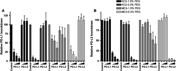
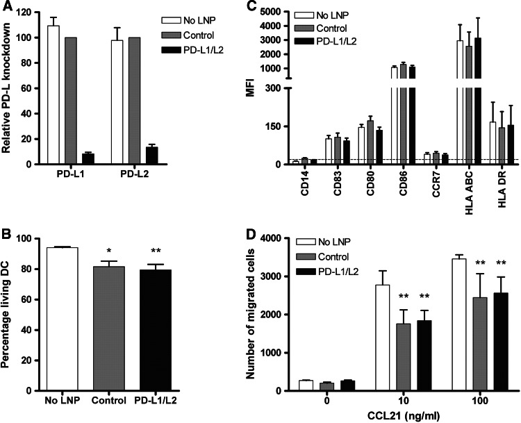
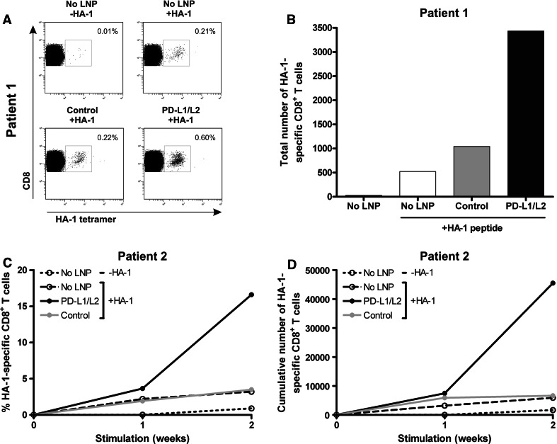
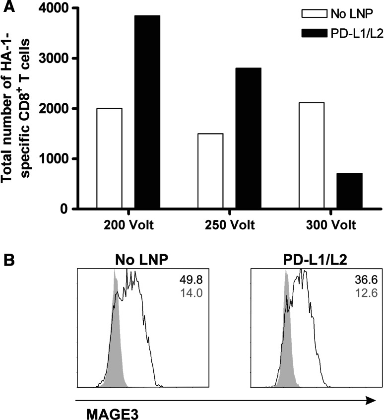
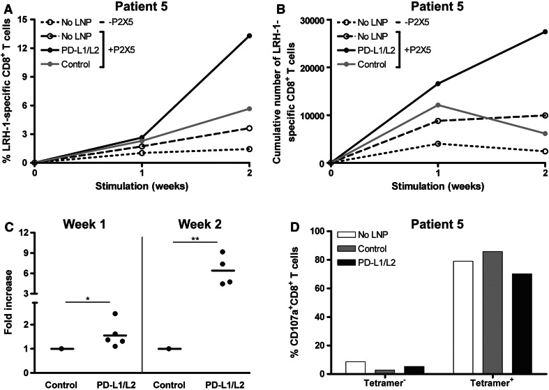
Dendritic cell (DC)-based vaccination boosting antigen-specific immunity is being explored for the treatment of cancer and chronic viral infections. Although DC-based immunotherapy can induce immunological responses, its clinical benefit has been limited, indicating that further improvement of DC vaccine potency is essential. In this study, we explored the generation of a clinical-grade applicable DC vaccine with improved immunogenic potential by combining PD-1 ligand siRNA and target antigen mRNA delivery. We demonstrated that PD-L1 and PD-L2 siRNA delivery using DLin-KC2-DMA-containing lipid nanoparticles (LNP) mediated efficient and specific knockdown of PD-L expression on human monocyte-derived DC. The established siRNA-LNP transfection method did not affect DC phenotype or migratory capacity and resulted in acceptable DC viability. Furthermore, we showed that siRNA-LNP transfection can be successfully combined with both target antigen peptide loading and mRNA electroporation. Finally, we demonstrated that these PD-L-silenced DC loaded with antigen mRNA superiorly boost ex vivo antigen-specific CD8(+) T cell responses from transplanted cancer patients. Together, these findings indicate that our PD-L siRNA-LNP-modified DC are attractive cells for clinical-grade production and in vivo application to induce and boost immune responses not only in transplanted cancer patients, but likely also in other settings.


Conflict of interest statement

Tatiana I. Novobrantseva, Jamie Wong, Stuart Milstein, Hila Epstein-Barash and Ju Liu are employees of Alnylam Pharmaceuticals. The other authors have no conflicting financial interests.

Figures

Fig. 1
Fig. 1
LNP-mediated siRNA delivery results in efficient PD-L1 and PD-L2 silencing on monocyte-derived DC. At days 3 and 7 of culture, siRNA-LNP were added at concentrations of 125, 250 or 500 nM for PD-L1 and PD-L2 compared to 500 nM of the control. Subsequently, DC were matured for 2 days, and PD-L expression levels were analyzed using flow cytometry. The relative knockdown of a PD-L1 and b PD-L2 was determined for four different siRNA-containing LNP formulations composed of either cationic lipid KC2 or MC3 in combination with 1.5 or 0.3 % PEG. Data of 3 donors are shown. The relative PD-L knockdown efficiency was calculated as follows: (ΔMFI PD-L LNP-treated DC/ΔMFI Control LNP-treated DC) × 100
Fig. 2
Fig. 2
PD-L-silenced DC show a mature phenotype and good migratory capacity toward CCL21. At days 3 and 7 of culture, 250 nM PD-L1 + 125 nM PD-L2 or 375 nM negative control siRNA-containing KC2-1.5 % PEG-LNP was added. Subsequently, DC were matured for 2 days, and DC viability, phenotype and migratory capacity were analyzed. a Relative PD-L1 and PD-L2 knockdown was examined using flow cytometry and calculated as follows: (ΔMFI PD-L LNP-treated DC/ΔMFI Control LNP-treated DC) × 100. b Viability was determined by trypan blue exclusion. Data are expressed as mean + SEM of 9 independent experiments. c Expression of maturation and co-stimulatory molecules by DC was analyzed using flow cytometry. Data are expressed as mean + SEM of 9 donors. The dotted line indicates the MFI of the isotype controls. d CCR7-mediated migratory capacity of DC cultured with or without LNP was determined toward increasing concentrations of chemokine CCL21. Depicted is the mean number of migrated cells + SD from triplo measurements. Data of one representative donor are shown. Statistical analysis was performed using a one-way ANOVA (c) or two-way ANOVA (d) followed by a Bonferroni post hoc test. *P < .05, **P < .01
Fig. 3
Fig. 3
PD-L knockdown DC loaded with MiHA peptide enhance MiHA-specific CD8+ T cell proliferation. At days 3 and 7 of DC culture, 250 nM PD-L1 + 125 nM PD-L2 or 375 nM negative control KC2-1.5 % PEG-LNP was added if indicated. Mature DC were loaded with 5 μM HA-1 peptide and cultured at a ratio of 0.1:1 with patient PBMC containing low numbers of MiHA-specific CD8+ T cells for 1–2 weeks. a After 1 week, cells were analyzed for tetramer-positive CD8+ T cells using flow cytometry. The numbers in the FACS plots represent the percentage of HA-1-specific CD8+ T cells within the total CD3+CD8+ T cell population. Data of one representative patient (patient 1) are shown. b Total number of HA-1-specific CD8+ T cells of patient 1 after 1 week of stimulation with PD-L-silenced DC loaded with or without HA-1 peptide. c and d The percentage (c) and cumulative numbers (d) of HA-1-specific CD8+ T cells of patient 2 obtained after stimulation with PD-L-silenced DC loaded with or without HA-1 peptide
Fig. 4
Fig. 4
siRNA-LNP-treated DC can be efficiently loaded with antigen-specific mRNA using electroporation. At days 3 and 7 of culture, 250 nM PD-L1 + 125 nM PD-L2 siRNA-containing KC2-1.5 % PEG-LNP was added. Mature DC treated with or without siPDL-LNP were electroporated with antigen-specific mRNA. a 1 h after loading with 20 μg HMHA1 mRNA using different electroporation voltages, DC were added at a ratio of 0.1:1 to patient PBMC containing low numbers of HA-1-specific CD8+ T cells. After 1 week, the expansion of HA-1-specific CD8+ T cells was analyzed using flow cytometry. b One hour after electroporation with 10 μg MAGE3 mRNA at 200 V, MAGE3 protein expression (black lines) as compared to isotype control (gray peaks) was analyzed using flow cytometry. Data of one representative donor out of 3 experiments are shown
Fig. 5
Fig. 5
The expansion of highly functional MiHA-specific CD8+ T cells is boosted by MiHA mRNA-loaded PD-L-silenced DC. At days 3 and 7 of culture, 250 nM PD-L1 + 125 nM PD-L2 or 375 nM negative control siRNA-containing KC2-1.5 % PEG-LNP was added when indicated. Mature DC were electroporated at 200 Volt with 20 μg MiHA-encoding mRNA and cultured at a ratio of 0.1:1 with patient PBMC containing low numbers of MiHA-specific CD8+ T cells for 1-2 weeks. a One week after each stimulation, cells were analyzed for tetramer-positive CD8+ T cells using flow cytometry. Data of one representative patient (patient 5) having a LRH-1-specific CD8+ T cell response are shown. b Total number of LRH-1-specific CD8+ T cells of patient 5 after 1-2 consecutive weeks of stimulation with PD-L-silenced DC loaded with P2X5 mRNA. c To combine data of all patients, the increase in absolute number of MiHA-specific CD8+ T cells after stimulation with PD-L-silenced DC was calculated relative to control siRNA-LNP DC stimulation. Data of 5 and 4 patients are shown for week 1 and 2, respectively. Lines indicate the mean. d Degranulation of LRH-1-tetramer-negative and positive CD8+ T cells of patient 5, stimulated for 2 weeks with P2X5 mRNA-loaded PD-L knockdown DC, was measured by staining for CD107a during overnight restimulation with 5 μM LRH-1 peptide. Statistical analysis was performed using a two-sample two-tailed t test assuming independent samples. *P < 0.05, **P < 0.01

References

    1. Palucka AK, Ueno H, Fay JW, Banchereau J. Taming cancer by inducing immunity via dendritic cells. Immunol Rev. 2007;220:129–150. doi: 10.1111/j.1600-065X.2007.00575.x. - DOI - PubMed
    1. Kalinski P, Urban J, Narang R, Berk E, Wieckowski E, Muthuswamy R. Dendritic cell-based therapeutic cancer vaccines: what we have and what we need. Future Oncol. 2009;5:379–390. doi: 10.2217/fon.09.6. - DOI - PMC - PubMed
    1. Garcia F, Routy JP. Challenges in dendritic cells-based therapeutic vaccination in HIV-1 infection workshop in dendritic cell-based vaccine clinical trials in HIV-1. Vaccine. 2011;29:6454–6463. doi: 10.1016/j.vaccine.2011.07.043. - DOI - PubMed
    1. Schuler G, Schuler-Thurner B, Steinman RM. The use of dendritic cells in cancer immunotherapy. Curr Opin Immunol. 2003;15:138–147. doi: 10.1016/S0952-7915(03)00015-3. - DOI - PubMed
    1. Banchereau J, Palucka AK. Dendritic cells as therapeutic vaccines against cancer. Nat Rev Immunol. 2005;5:296–306. doi: 10.1038/nri1592. - DOI - PubMed

Publication types

MeSH terms