. 2012 Dec;90(12):2259-71.
doi: 10.1002/jnr.23116. Epub 2012 Aug 18.

Comparison of nerve growth factor receptor binding models using heterodimeric muteins

Affiliations

Comparison of nerve growth factor receptor binding models using heterodimeric muteins

Hrishikesh M Mehta et al. J Neurosci Res. 2012 Dec.

Abstract

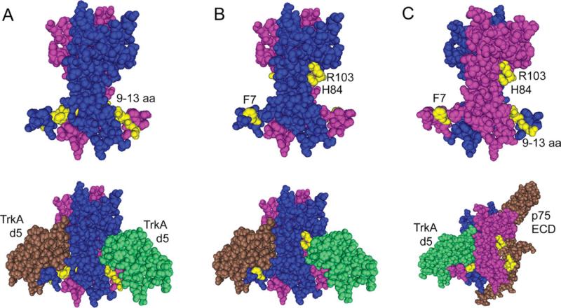
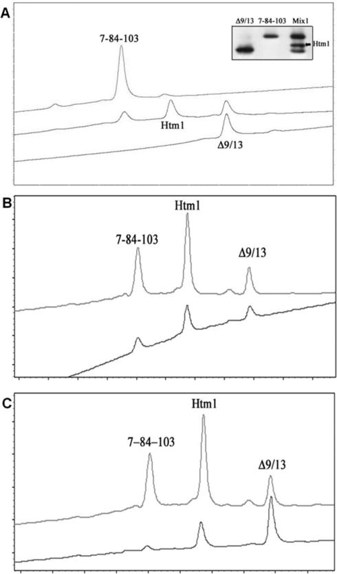
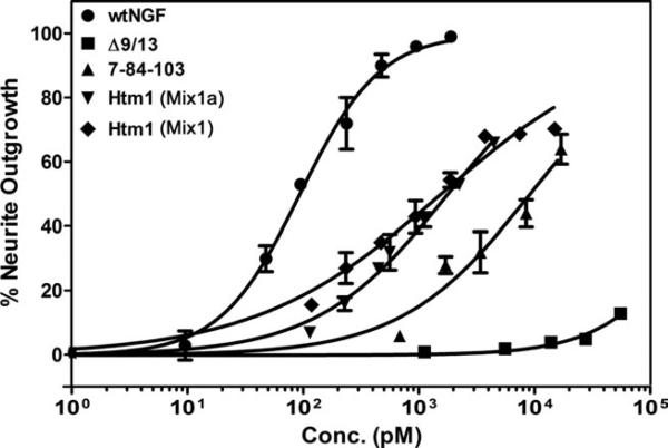
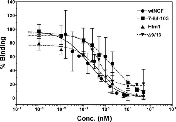
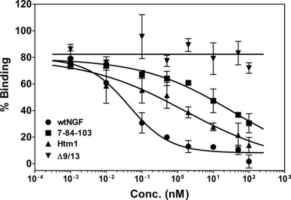
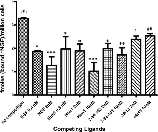
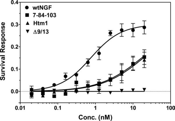
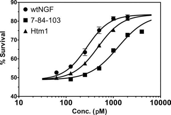
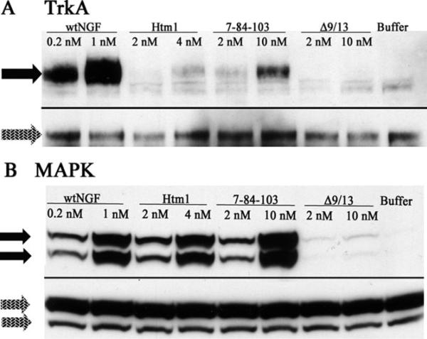
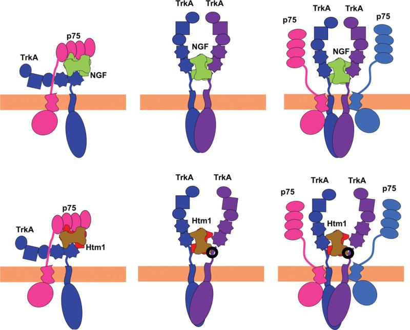
Nerve growth factor (NGF) is a homodimer that binds to two distinct receptor types, TrkA and p75, to support survival and differentiation of neurons. The high-affinity binding on the cell surface is believed to involve a heteroreceptor complex, but its exact nature is unclear. We developed a heterodimer (heteromutein) of two NGF muteins that can bind p75 and TrkA on opposite sides of the heterodimer, but not two TrkA receptors. Previously described muteins are Δ9/13 that is TrkA negative and 7-84-103 that is signal selective through TrkA. The heteromutein (Htm1) was used to study the heteroreceptor complex formation and function, in the putative absence of NGF-induced TrkA dimerization. Cellular binding assays indicated that Htm1 does not bind TrkA as efficiently as wild-type (wt) NGF but has better affinity than either homodimeric mutein. Htm1, 7-84-103, and Δ9/13 were each able to compete for cold-temperature, cold-chase stable binding on PC12 cells, indicating that binding to p75 was required for a portion of this high-affinity binding. Survival, neurite outgrowth, and MAPK signaling in PC12 cells also showed a reduced response for Htm1, compared with wtNGF, but was better than the parent muteins in the order wtNGF > Htm1 > 7-84-103 >> Δ9/13. Htm1 and 7-84-103 demonstrated similar levels of survival on cells expressing only TrkA. In the longstanding debate on the NGF receptor binding mechanism, our data support the ligand passing of NGF from p75 to TrkA involving a transient heteroreceptor complex of p75-NGF-TrkA.


Figures

Fig. 1
Fig. 1
Design rationale for an NGF heteromutein capable of forming a TrkA-NGF-p75 complex but not a TrkA-NGF-TrkA complex. A: The 9–13 amino acids on the N-terminus of NGF are highlighted in yellow on each protomer (pink, blue) of NGF dimer (upper structure). These amino acids are deleted in the mutein Δ9/13 that is unable to bind TrkA. The lower structure shows the interaction of residues 9–13 with TrkA d5 domain (brown, green). B: The F7, H84, and R103 amino acids are highlighted in the NGF dimer (upper structure), and their interaction with TrkA d5 domains (brown, green) is shown in the lower structure. F7A/H84A/R103A (7-84-103) mutein is an NGF mutein binding weakly to TrkA. C: A dimer of NGF in which 9–13 amino acids are highlighted on one protomer (pink) and F7, H84, and R103 are highlighted on the other protomer (blue) of NGF shows that, if these highlighted residues are mutated or deleted, it would abolish TrkA binding on one side (upper structure). Therefore, a heterodimer of Δ9/13 mutein and the 7-84-103 mutein would lose TrkA binding on one side but still have the ability to form a heteroreceptor complex (p75 ECD, brown; TrkA d5, green; lower structure). PDB IDs for NGFTrkA(d5) complex and NGF-p75 complex are 1WWW and 1SG1, respectively.
Fig. 2
Fig. 2
Separation, purification, and stability of Htm1. The parent muteins 7-84-103 and Δ9/13 were mixed 1:1 in 0.4% acetic acid (traditional storage buffer) and incubated at 4°C for at least 24 hr to form Htm1. Separation was achieved using HPIEC on a weak cation exchange column (polyCAT A), using a step gradient protocol. A: Separation profile for Htm1 from the parent muteins (middle trace). The upper trace is for 7-84-103 alone, and the bottom trace is for Δ9/13 alone. Inset: Separation pattern on an IEF gel; lane 1: Δ9/13; lane 2: 7-84-103; lane 3: Htm1 (Mix1a). B: Eluted Htm1 (from the central peak, upper trace), separated from the parent muteins in the Mix1a, was dialyzed out of the elution buffer for 8 hr and reloaded on the column (lower trace). Shorter times of stability could not be tested because a 4-hr dialysis did not sufficiently remove the salts to allow proper chromatography. C: Two different mixtures of the parent muteins were analyzed for the ratio of the three species of NGF muteins at equilibrium. Mix1a: 1:1 mixture of 7-84-103 and Δ9/13 was mixed and incubated for at least 24 hr at 4°C (upper trace). Mix1: 1:2 mixture of 7-84-103 and Δ9/13 was mixed and incubated for 24 hr at 4°C (lower trace).
Fig. 3
Fig. 3
Neurite outgrowth in PC12 cells. PC12 cells were used to measure the number of neurite-bearing cells after treatment with neurotrophins. Different concentrations of wtNGF, Δ9/13, 7-84-103, Htm1 (Mix1a), and Htm1 (Mix1) were added to PC12 cells in defined medium to stimulate neurite outgrowth for 48 hr. In this and subsequent figures, the concentration of Htm1 in each mixture was calculated from the total protein concentration and the percentage of Htm1 area under the peaks of the corresponding trace in Figure 2C and then plotted as the concentration of Htm1. Data are presented as mean ± SEM with n = 3. The dose–response curves were fit to the data points in GraphPad Prism 5.02. Data for wtNGF and 7-84-103 are from Mahapatra et al. (2009).
Fig. 4
Fig. 4
PC12 cell (TrkA+/p75+) competitive binding assay. Increasing concentrations of muteins were used in competition with labeled wtNGF to generate competition binding curves in cells expressing both p75 and TrkA receptors. PC12 cells were incubated in the presence of 0.1 nM 125I-NGF in the presence or absence of competing ligands for 30 min at room temperature, followed by determination of the bound fraction in 100-μl aliquots. Bound 125I-NGF in the absence of a competing ligand was set at 100% binding to determine relative binding. The data are presented as mean ± range, n = 2. Competitive binding curves were fit to the data points in GraphPad Prism. The IC50s determined from these individual binding curves (wtNGF = 0.22 nM, 7-84-103 = 1.9 nM, Htm1 = 0.53 nM, Δ9/13 = 0.51 nM) are similar to those listed for averages in Table I.
Fig. 5
Fig. 5
MG139 cell (TrkA+/p75) competitive binding assay. Increasing concentrations of muteins and wtNGF were used to compete with labeled wtNGF to generate competition binding curves in cells expressing TrkA only. MG139 cells were incubated with 0.2 nM 125I-NGF in the presence or absence of competing ligands for 45 min at room temperature, followed by determination of the bound fraction in 100-ll aliquots. Bound 125I-NGF in the absence of a competitor was set at 100% binding to determine relative binding. The data are presented as mean ± range, n = 2. Competitive binding curves were fit to the data points in GraphPad Prism. The IC50s determined from these binding curves (wtNGF = 0.04 nM, 7-84-103 = 15 nM, Htm1 = 0.64 nM, Δ9/13 = no binding) are similar to those listed for averages in Table I.
Fig. 6
Fig. 6
Cold-temperature, cold-chase stable competition binding. Wild-type NGF (0.2 nM 125I-NGF) was incubated with PC12 cells (106 cells/ml) in the absence or presence of a competitor for 45 min at room temperature, followed by 5 min of incubation on ice (0.5°C). A 1,000-fold excess of cold wtNGF was added to initiate dissociation of bound 125I-NGF and allowed to incubate on ice for an additional 30 min. The bound fraction was separated from free and represents the amount of cold-chase stable specific binding remaining after competition with the muteins. Data are presented as mean ± SEM, n = 3, and statistical significance was computed by using Dunnett's multiple-comparisons test as a posttest following a significant one-way ANOVA test (P = 0.0012). *P < 0.05, **P < 0.01, and ***P < 0.001 represent significant difference in the amount of cold cold-chase stable binding remaining after competition in comparison with no competition. #P < 0.05, ##P < 0.01, and ###P < 0.001 represent significant difference in the amount of cold cold-chase stable binding remaining compared with 10 nM Htm1.
Fig. 7
Fig. 7
XTT assay of cell survival response in MG139 cells (TrkA+/p75). Increasing concentrations of wtNGF or its muteins were added to log-phase MG139 cells in 96-well plates for 72 hr at 37°C. The XTT reagent was added for another 4 hr, and the absorbance at 450 nm was measured on a multiwell plate reader (Tecan). Data are presented as mean ± SEM for three separate experiments.
Fig. 8
Fig. 8
Trypan blue assay of cell survival response in PC12 cells (TrkA+/p75+). Increasing concentrations of either wtNGF or its muteins were added to log-phase PC12 cells, and cell survival was determined after 72 hr by counting cells under a phase-contrast microscope. The data are presented as mean ± SD, n = 3. Dose–response curves were generated in GraphPad Prism.
Fig. 9
Fig. 9
TrkA and MAPK phosphorylation in PC12 cells. A: TrkA phosphorylation was determined by immunoprecipitating TrkA, followed by immunoblotting with a pan-phosphotyrosine antibody. Once phosphoprotein bands (black arrows) were identified and scanned, the blot was stripped and reprobed with an anti-TrkA antibody to determine total TrkA (gray arrows). B: Phospho-MAPK was detected by running the lysate on the gel, followed by immunoblotting using a phospho-MAPK antibody (black arrows). The blot was then stripped and reprobed with anti-MAPK antibody to detect total MAPK (gray arrows). Horseradish peroxidase-conjugated secondary antibody was used for band detection by chemiluminescence.
Fig. 10
Fig. 10
Model of ligand passing and TrkA presentation for NGF binding. Upper row: NGF. Lower row: Htm1. Left column: Putative complex of NGF or Htm1 with TrkA and p75 that represents an intermediate in the ligand-passing model. Note the antiparallel orientation of the two receptors resulting from the binding mode of each ligand relative to the membrane (Barker, 2007). The formation of this complex would be preceded by the binding of NGF or Htm1 to the p75 receptor. Middle column: NGF or Htm1 binding to a homodimeric TrkA receptor. The binding pathway for NGF or Htm1 to form this complex can occur by directly binding to TrkA or via the two-step ligand-passing process, which involves formation of the complex shown in the left column. Right column: Putative heteroreceptor complex of undefined stoichiometry in the Trk presentation model. This complex represents the TrkA presentation model. A pre-existing TrkA-p75 heteroreceptor complex can bind to NGF with greater affinity than TrkA alone. In the lower middle and lower right models, the area in the circle designates additional modes of interaction among Htm1 loop L-I, L-II, and L-IV residues and the extracellular, juxtamembrane region of TrkA to explain the cellular and signaling data (see Discussion).

Similar articles

Cited by

References

    1. Barker PA. High affinity not in the vicinity? Neuron. 2007;53:1–4. - PubMed
    1. Barker PA, Shooter EM. Disruption of NGF binding to the low affinity neurotrophin receptor p75LNTR reduces NGF binding to TrkA on PC12 cells. Neuron. 1994;13:203–215. - PubMed
    1. Barker PA, Lomen-Hoerth C, Gensch EM, Meakin SO, Glass DJ, Shooter EM. Tissue-specific alternative splicing generates two isoforms of the trkA receptor. J Biol Chem. 1993;268:15150–15157. - PubMed
    1. Barker PA, Hussain NK, McPherson PS. Retrograde signaling by the neurotrophins follows a well-worn trk. Trends Neurosci. 2002;25:379–381. - PubMed
    1. Bernd P, Greene LA. Association of 125I-nerve growth factor with PC12 pheochromocytoma cells. Evidence for internalization via high-affinity receptors only and for long-term regulation by nerve growth factor of both high- and low-affinity receptors. J Biol Chem. 1984;259:15509–15516. - PubMed

Publication types

MeSH terms