. 2012 Aug 17;337(6096):857-60.
doi: 10.1126/science.1221698.

A TOG:αβ-tubulin complex structure reveals conformation-based mechanisms for a microtubule polymerase

Affiliations

A TOG:αβ-tubulin complex structure reveals conformation-based mechanisms for a microtubule polymerase

Pelin Ayaz et al. Science. .

Abstract

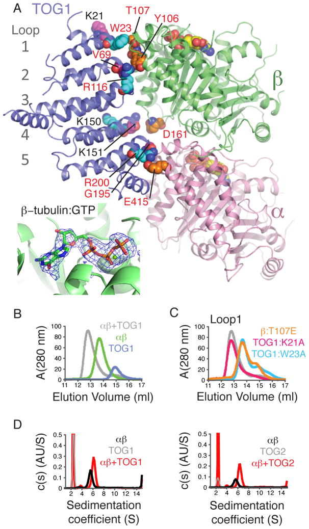
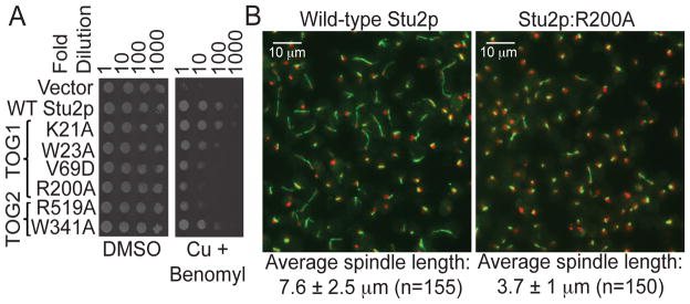
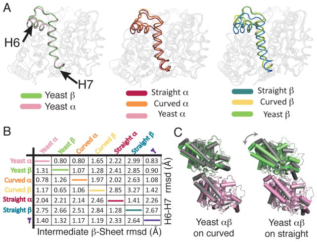
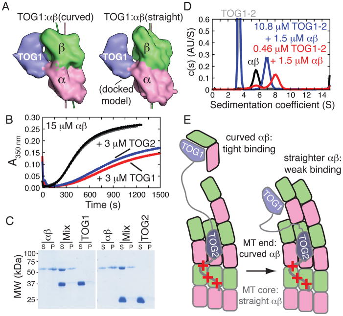
Stu2p/XMAP215/Dis1 family proteins are evolutionarily conserved regulatory factors that use αβ-tubulin-interacting tumor overexpressed gene (TOG) domains to catalyze fast microtubule growth. Catalysis requires that these polymerases discriminate between unpolymerized and polymerized forms of αβ-tubulin, but the mechanism by which they do so has remained unclear. Here, we report the structure of the TOG1 domain from Stu2p bound to yeast αβ-tubulin. TOG1 binds αβ-tubulin in a way that excludes equivalent binding of a second TOG domain. Furthermore, TOG1 preferentially binds a curved conformation of αβ-tubulin that cannot be incorporated into microtubules, contacting α- and β-tubulin surfaces that do not participate in microtubule assembly. Conformation-selective interactions with αβ-tubulin explain how TOG-containing polymerases discriminate between unpolymerized and polymerized forms of αβ-tubulin and how they selectively recognize the growing end of the microtubule.


Figures

Figure 1
Figure 1. Structure of a TOG1:αβ-tubulin complex, revealing significant contacts with α- and β-tubulin
A Cartoon representation of the complex (pink: α-tubulin, lime: β-tubulin, slate: TOG1). Contacts probed by mutagenesis are represented as spheres (colored to match panel C and Fig. S3), as are GTPs. (inset) mF0-DFc omit electron density map contoured at 3.5 σ and computed from a model without nucleotides: β-tubulin is bound to GTP. B Size-exclusion chromatography assay for TOG1:αβ-tubulin interactions. C TOG1:αβ-tubulin binding assay using interface mutants on Loop 1 (see Fig. S3). D TOG1 (left) and TOG2 (right) (grey) each form a 1:1 complex (red) with αβ-tubulin (black) as detected by analytical ultracentrifugation (Table S4). Curves are shown from S=1 to eliminate a slowly sedimenting contaminant in the TOG2 run.
Figure 2
Figure 2. Disruptive point mutations on the tubulin-binding interfaces of TOG1 or TOG2 affect Stu2p function in vivo
A Yeast carrying plasmid-based rescue constructs of Stu2p were plated at serial dilutions on media containing DMSO (control) or 500 μM CuSO4 (to deplete endogenous Stu2p) plus 20 μg/ml benomyl (to cause microtubule stress). TOG1 or TOG2 impaired for αβ-tubulin interactions only partially compensate for the depletion of endogenous Stu2p. B Fluorescence images one hour after release from hydroxyurea arrest (green: αβ-tubulin; red: DNA) of yeast depleted of endogenous Stu2p and rescued with wild-type (left) or R200A (right) Stu2p. Spindle elongation is compromised when TOG1:αβ-tubulin interactions are impaired (R200A).
Figure 3
Figure 3. αβ-tubulin-GTP adopts a curved conformation
A Superposition of yeast α (pink) and β-tubulin (lime) shows similar positioning of the H6-H7 segment (represented with darker colors). B Superposition of yeast α- and β-tubulin onto curved (α: orange, β: yellow) and straight (α: maroon, β: dark blue) structures shows the H6-H7 segments of yeast tubulins arranged as in prior curved structures. C Pairwise Cα r.m.s. coordinate deviations between yeast α- and β-tubulin and prior structures, computed for the subdomains indicated (Table S3). D The quaternary structure of yeast αβ-tubulin (pink and green) resembles that of the “curved” form (grey, left), and differs from the straight form (grey, right).
Figure 4
Figure 4. TOG1:αβ-tubulin interactions are conformation-selective
A The structure of the TOG1:αβ-tubulin complex (left) and a docked model with straight αβ-tubulin (right) illustrates how TOG1-contacting epitopes on α- and β-tubulin move relative to each other in the two conformations. B Microtubule assembly reactions (15 μM animal αβ-tubulin) containing 3 μM TOG1 (red) or TOG2 (blue) are inhibited relative to control reactions (black and grey) that received only buffer. C Microtubule co-sedimentation showing that TOG1 or TOG2 do not appreciably bind microtubules, even though the TOG-interacting epitopes are accessible on the outside of the microtubule (Fig. S7). S: supernatant, P: pellet. D Size distributions showing that substoichiometric concentrations of TOG1-TOG2 mixed with αβ-tubulin (red) behave as a complex that sediments faster than αβ-tubulin alone (black) and the TOG1-TOG2:(αβ)1 complex that results when TOGs are in molar excess over αβ-tubulin (blue). E Minimal cartoon model illustrating how conformation-selective TOG:αβ-tubulin interactions contribute to function. ‘+++’ denotes a basic region that provides microtubule affinity. TOG1 can efficiently capture unpolymerized αβ-tubulin in its naturally curved state (left). The straight/straighter conformation of αβ-tubulin in the MT has lower affinity interactions with TOG1 (right) and may be recognized by TOG2.

Similar articles

Cited by

References

    1. Desai A, Mitchison TJ. Microtubule polymerization dynamics. Annu Rev Cell Dev Biol. 1997;13:83. - PubMed
    1. Gard DL, Kirschner MW. A microtubule-associated protein from Xenopus eggs that specifically promotes assembly at the plus-end. J Cell Biol. 1987;105:2203. - PMC - PubMed
    1. Ohkura H, et al. Cold-sensitive and caffeine-supersensitive mutants of the Schizosaccharomyces pombe dis genes implicated in sister chromatid separation during mitosis. EMBO J. 1988;7:1465. - PMC - PubMed
    1. Wang PJ, Huffaker TC. Stu2p: A microtubule-binding protein that is an essential component of the yeast spindle pole body. J Cell Biol. 1997;139:1271. - PMC - PubMed
    1. Al-Bassam J, van Breugel M, Harrison SC, Hyman A. Stu2p binds tubulin and undergoes an open-to-closed conformational change. J Cell Biol. 2006;172:1009. - PMC - PubMed

Publication types

MeSH terms

Substances

Associated data