. 2012 Dec;194(23):6382-6.
doi: 10.1128/JB.00505-12. Epub 2012 Aug 17.

The helical MreB cytoskeleton in Escherichia coli MC1000/pLE7 is an artifact of the N-Terminal yellow fluorescent protein tag

Affiliations

The helical MreB cytoskeleton in Escherichia coli MC1000/pLE7 is an artifact of the N-Terminal yellow fluorescent protein tag

Matthew T Swulius et al. J Bacteriol. 2012 Dec.

Abstract

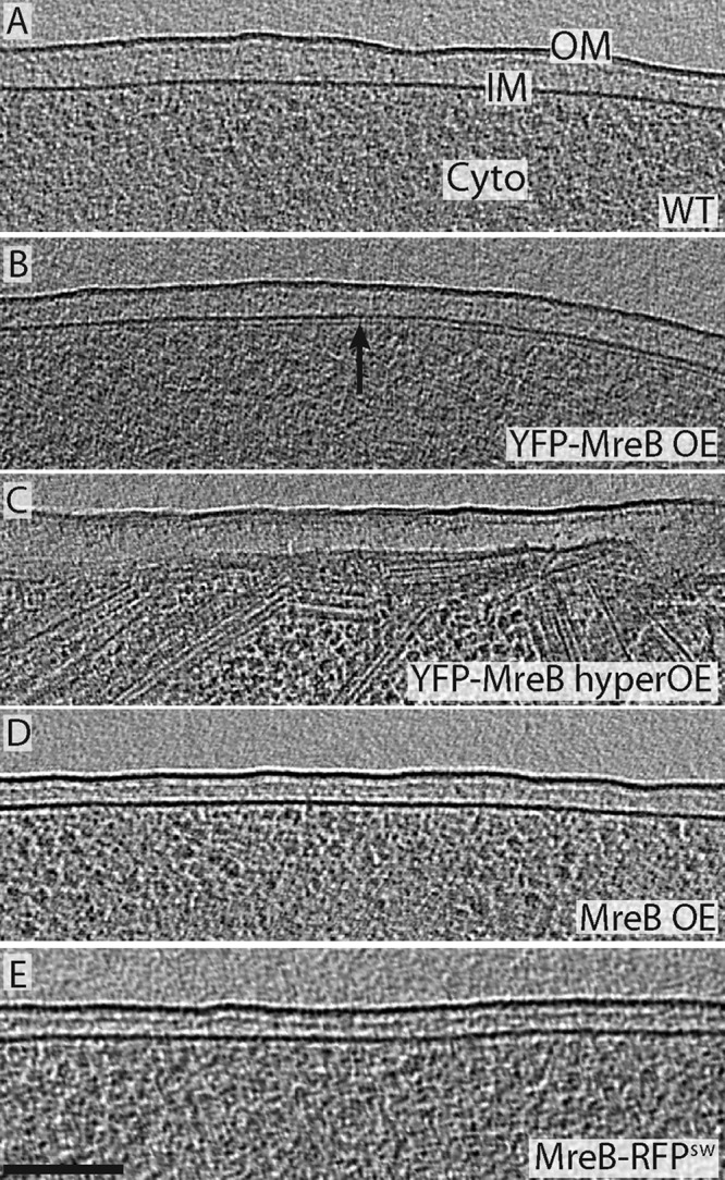
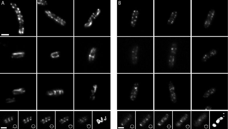
Based on fluorescence microscopy, the actin homolog MreB has been thought to form extended helices surrounding the cytoplasm of rod-shaped bacterial cells. The presence of these and other putative helices has come to dominate models of bacterial cell shape regulation, chromosome segregation, polarity, and motility. Here we use electron cryotomography to show that MreB does in fact form extended helices and filaments in Escherichia coli when yellow fluorescent protein (YFP) is fused to its N terminus but native (untagged) MreB expressed to the same levels does not. In contrast, mCherry fused to an internal loop (MreB-RFP(SW)) does not induce helices. The helices are therefore an artifact of the placement of the fluorescent protein tag. YFP-MreB helices were also clearly distinguishable from the punctate, "patchy" localization patterns of MreB-RFP(SW), even by standard light microscopy. The many interpretations in the literature of such punctate patterns as helices should therefore be reconsidered.


Figures

Fig 1
Fig 1
MreB helices are an artifact of the YFP tag. (A to E) Tomographic slices through the edge of five representative E. coli cells. No filaments were seen near the membranes of wild-type cells (A), cells overexpressing native MreB (D), or cells harboring MreB-RFPSW (E), but many helical and other filaments were found just inside the membranes of cells overexpressing YFP-MreB (strain pLE7) (B). We confirmed that these filaments are YFP-MreB by their dramatically increased abundance in cells overexpressing YFP-MreB at high levels (C). The outer membrane (OM), inner membrane (IM), and cytoplasm (Cyto) are labeled in panel A. Bar, 100 nm.
Fig 2
Fig 2
YFP-MreB filaments exhibit a range of lengths and helical pitches. (Top) 3D model of a pLE7 cell's inner membrane (blue) and YFP-MreB filaments (white) (1 to 3). Tomographic slices through the cell modeled in the top panel. The slice numbers correspond to the filaments of the same number in the top panel. Insets depict the angle of the slice through the cell and arrowheads point to the visible ends of the filament. The outer membrane (OM), inner membrane (IM), and cytoplasm (Cyto) are labeled in slice 1. The scale bar represents 100 nm.
Fig 3
Fig 3
Comparison of fluorescence patterns in strains MC1000/pLE7 and FB76. (A) Epifluorescence images of various MC1000/pLE7 cells overexpressing YFP-MreB. The top row shows the familiar punctate and helical streaks commonly shown in the literature. The two middle rows show a variety of other patterns seen. The bottom row shows the individual images of a focal stack through a cell with an extended helical fluorescent pattern. The diagrams illustrate where the focal plane was in the cell for each image. The rightmost panel is a 3D isosurface of the fluorescent signal in the focal stack, which is clearly helical. (B) Epifluorescence images of E. coli strain FB76. Individual cells in the top rows illustrate the puncta along the membrane that often look similar to cells in the top row of panel A. The middle two rows show a variety of patchy distributions seen in epifluorescence images of FB76. The patchy signal is seen consistently, and no long filamentous structures are visible as seen in MC1000/pLE7. For further comparison, the bottom row shows a focal stack through one cell and its corresponding isosurface, which is obviously not helical. Scale bars represent 1 μm.

Comment in

References

    1. Bendezú FO, Hale CA, Bernhardt TG, de Boer PAJ. 2009. RodZ (YfgA) is required for proper assembly of the MreB actin cytoskeleton and cell shape in E. coli. EMBO J. 28:193–204 - PMC - PubMed
    1. Briegel A, et al. 2006. Multiple large filament bundles observed in Caulobacter crescentus by electron cryotomography. Mol. Microbiol. 62:5–14 - PubMed
    1. Chastanet A, Carballido-Lopéz R. 2012. The actin-like MreB proteins in Bacillus subtilis: a new turn. Front. Biosci. (Schol. Ed.) 4:1582–1606 - PubMed
    1. Dempwolff F, Reimold C, Reth M, Graumann PL. 2011. Bacillus subtilis MreB orthologs self-organize into filamentous structures underneath the cell membrane in a heterologous cell system. PLoS One 6:e27035 doi: - DOI - PMC - PubMed
    1. Divakaruni AV, Baida C, White CL, Gober JW. 2007. The cell shape proteins MreB and MreC control cell morphogenesis by positioning cell wall synthetic complexes. Mol. Microbiol. 66:174–188 - PubMed

Publication types

MeSH terms