. 2012 Oct 5;287(41):34400-9.
doi: 10.1074/jbc.M112.390294. Epub 2012 Aug 17.

Spg5 protein regulates the proteasome in quiescence

Affiliations

Spg5 protein regulates the proteasome in quiescence

John Hanna et al. J Biol Chem. .

Abstract

The ubiquitin-proteasome system is the major pathway for selective protein degradation in eukaryotes. Despite extensive study of this system, the mechanisms by which proteasome function and cell growth are coordinated remain unclear. Here, we identify Spg5 as a novel component of the ubiquitin-proteasome system. Spg5 binds the regulatory particle of the proteasome and the base subassembly in particular, but it is excluded from mature proteasomes. The SPG5 gene is strongly induced in the stationary phase of budding yeast, and spg5Δ mutants show a progressive loss of viability under these conditions. Accordingly, during logarithmic growth, Spg5 appears largely dispensable for proteasome function, but during stationary phase the proteasomes of spg5Δ mutants show both structural and functional defects. This loss of proteasome function is reflected in the accumulation of oxidized proteins preferentially in stationary phase in spg5Δ mutants. Thus, Spg5 is a positive regulator of the proteasome that is critical for survival of cells that have ceased to proliferate due to nutrient limitation.


Figures
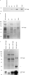
FIGURE 1.
FIGURE 1.
Spg5 binds to the RP and base but not mature proteasomes. A, binding of bacterially expressed GST-Spg5 (pJH101) to purified proteasome or proteasome subcomplexes. Visualization is by anti-GST antibody. Input represents ∼3% of the total GST-Spg5 present in each binding assay. GST-Spg5 was added in excess of the proteasome species. B, electrophoretic profile of the proteasome and proteasome subcomplexes from A, as visualized by Coomassie staining. (26 S, RP, and lid were generated from strain SDL133; base from SY36; CP from SDL135). Note that our CP preparations have a minor contaminant that comigrates with GST-Spg5, which may account for the weak band of this mobility in the 2nd lane, because this band is not detected by antibody to GST (A). C, binding of purified GST-Spg5 expressed in bacteria to the base subcomplex, as visualized by Coomassie stain (upper panel) or Western blot with ant-GST antibody (lower panel). The asterisk indicates a fragment of GST-Spg5 that comigrates with bona fide GST. The double asterisk indicates a nonspecific band.
FIGURE 2.
FIGURE 2.
spg5Δ proteasomes are similar to those of wild type in logarithmic or late-logarithmic phase cultures. A, electrophoretic profile of proteasomes purified from wild-type (SJH303) and spg5Δ strains (SJH304), visualized by Coomassie staining. B, suc-LLVY-AMC hydrolyzing activity of proteasomes (5 μg) from A. Control, buffer only. C, native gel electrophoresis coupled with in-gel suc-LLVY-AMC assay highlights proteolytically active proteasome species, as indicated, for the purified proteasomes from A. D, electrophoretic profile of the proteasome species, separated by native gel electrophoresis as in C, and visualized by Coomassie staining.
FIGURE 3.
FIGURE 3.
Stationary phase transcriptional induction of SPG5. A, quantitative real time PCR analysis of SPG5 transcription after 2 and 5 days of culture, expressed as fold-induction relative to exponential phase growth. The 18 S ribosomal RNA serves as a control. Error bars indicate standard deviations of experiments carried out in triplicate. RNA was generated from strain SUB62. White bars, 18 S RNA; black bars, SPG5 RNA. B, growth of wild-type (SJH92) and spg5Δ (SJH301) strains on rich medium at the indicated temperatures after growth for 28 or 60 days in rich medium at 30 °C.
FIGURE 4.
FIGURE 4.
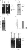
spg5Δ proteasomes from stationary phase cells are defective. A, native gel electrophoresis coupled with in-gel suc-LLVY-AMC assay highlights proteolytically active proteasome species, as indicated, for wild-type (SJH303) and spg5Δ (SJH304) proteasomes. B, electrophoretic profile of the proteasome species, separated by native gel electrophoresis as in A, and visualized by Coomassie staining. C, denaturing gel electrophoresis of the proteasome species as in A, visualized by Coomassie staining. D, suc-LLVY-AMC hydrolyzing activity of proteasomes (3.5 μg) from A. Control, buffer only. E, native gel electrophoresis coupled with in-gel suc-LLVY-AMC assay shows that restoration of SPG5 expression corrects the proteasome defects of the spg5Δ mutant. Strains: wild type with empty vector (SJH386), spg5Δ with empty vector (SJH387), and spg5Δ with SPG5 CEN plasmid (SJH388).
FIGURE 5.
FIGURE 5.
spg5Δ proteasomes from stationary phase cells show an altered proteasome subcomplex profile. A, wild-type and spg5Δ proteasomes as in Fig. 4, analyzed by native gel electrophoresis and visualized by suc-LLVY-AMC in-gel activity assay or immunoblotting with antibodies to the CP subunit α7, the base subunit Rpt4, and the lid subunit Rpn8, as indicated. B, denaturing gel electrophoresis of the proteasome species, visualized by Coomassie staining.
FIGURE 6.
FIGURE 6.
Regulation of SPG5 expression is distinct from that of known RP chaperones. Quantitative real time PCR analysis of transcription of the indicated genes after 5 days of culture, expressed as fold-induction relative to exponential phase growth. The 18 S ribosomal RNA serves as an endogenous control. Error bars indicate standard deviations of experiments carried out in triplicate. RNA was generated from strain SUB62.
FIGURE 7.
FIGURE 7.
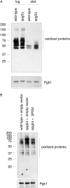
Accumulation of oxidized proteins in stationary phase in spg5Δ mutants. A, total cellular abundance of oxidized proteins in wild-type (SJH92) and spg5Δ (SJH301) strains in logarithmic and stationary phase growth, as indicated. Pgk1, loading control. Comparable results were obtained in four independent experiments. B, total cellular abundance of oxidized protein in stationary phase. Strains: wild-type with empty vector (sJH370), spg5Δ with empty vector (sJH371), and spg5Δ complemented with a plasmid-borne SPG5 gene (sJH372). Pgk1, loading control.

References

    1. Gray J. V., Petsko G. A., Johnston G. C., Ringe D., Singer R. A., Werner-Washburne M. (2004) “Sleeping beauty.” Quiescence in Saccharomyces cerevisiae. Microbiol. Mol. Biol. Rev. 68, 187–206 - PMC - PubMed
    1. Devenish R. J., Klionsky D. J. (2012) Autophagy: Mechanism and physiological relevance “brewed” from yeast studies. Front. Biosci. 4, 1354–1363 - PMC - PubMed
    1. Takeshige K., Baba M., Tsuboi S., Noda T., Ohsumi Y. (1992) Autophagy in yeast demonstrated with proteinase-deficient mutants and conditions for its induction. J. Cell Biol. 119, 301–311 - PMC - PubMed
    1. Teichert U., Mechler B., Müller H., Wolf D. H. (1989) Lysosomal (vacuolar) proteinases of yeast are essential catalysts for protein degradation, differentiation, and cell survival. J. Biol. Chem. 264, 16037–16045 - PubMed
    1. Gresham D., Boer V. M., Caudy A., Ziv N., Brandt N. J., Storey J. D., Botstein D. (2011) System-level analysis of genes and functions affecting survival during nutrient starvation in Saccharomyces cerevisiae. Genetics 187, 299–317 - PMC - PubMed

Publication types

MeSH terms