. 2012 Oct;23(10):1682-90.
doi: 10.1681/ASN.2012030283. Epub 2012 Aug 16.

Invasion of distal nephron precursors associates with tubular interconnection during nephrogenesis

Affiliations

Invasion of distal nephron precursors associates with tubular interconnection during nephrogenesis

Robert M Kao et al. J Am Soc Nephrol. 2012 Oct.

Abstract

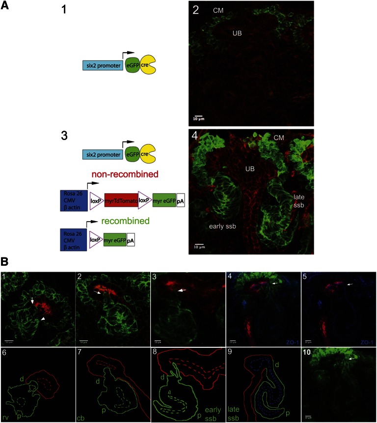
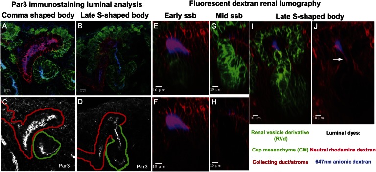
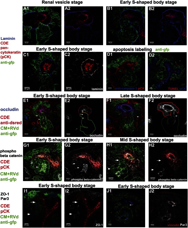
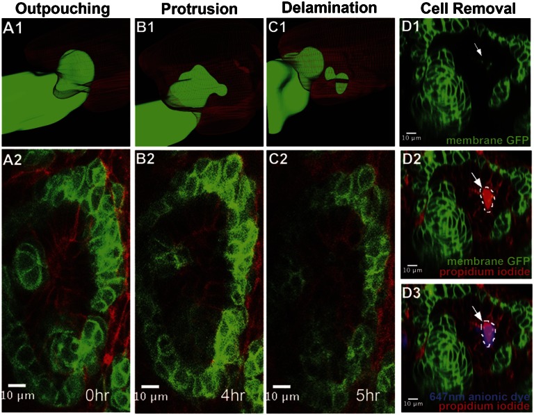
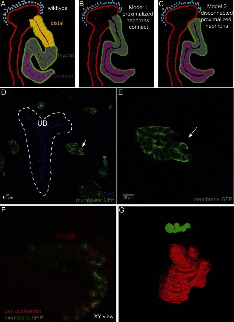
Formation of a functional renal network requires the interconnection of two epithelial tubes: the nephron, which arises from kidney mesenchyme, and the collecting system, which originates from the branching ureteric epithelium. How this connection occurs, however, is incompletely understood. Here, we used high-resolution image analysis in conjunction with genetic labeling of epithelia to visualize and characterize this process. Although the focal absence of basal lamina from renal vesicle stages ensures that both epithelial networks are closely apposed, we found that a patent luminal interconnection is not established until S-shaped body stages. Precursor cells of the distal nephron in the interconnection zone exhibit a characteristic morphology consisting of ill-defined epithelial junctional complexes but without expression of mesenchymal markers such as vimentin and Snai2. Live-cell imaging revealed that before luminal interconnection, distal cells break into the lumen of the collecting duct epithelium, suggesting that an invasive behavior is a key step in the interconnection process. Furthermore, loss of distal cell identity, which we induced by activating the Notch pathway, prevented luminal interconnection. Taken together, these data support a model in which establishing the distal identity of nephron precursor cells closest to the nascent collecting duct epithelium leads to an active cell invasion, which in turn contributes to a patent tubular interconnection between the nephron and collecting duct epithelia.


Figures

Figure 1.
Figure 1.
A dual fluorescent membrane labeling system to visualize interactions between nephron and collecting duct networks during mouse development. (A1 and A3) Schematic representation of transgenic alleles and reporter systems. (A2 and A4) GFP and TdTomato activity before and after recombination in sections of E15.5 kidney. (B1–10) Analysis of the interaction between RV derivatives and the ureteric epithelium at the indicated stages of nephrogenesis. Upper panels in B1–B5 and lower panel in B10 show fluorescent labeling of kidney sections at E15.5. The merged image in B4 is also labeled with antibodies to ZO-1 for labeling tight junction complexes (blue), anti-GFP to label Six2+ progenitor cells and RV derivatives (green), and antipan-cytokeratin to label collecting duct epithelium (red). Arrow points to late ssb connected to tip of collecting duct epithelium. (B5) Separated ZO-1 (blue) and pan-cytokeratin (red) from merged image in B4. (B10) Separated GFP-labeled image from B4. Lower panels in B6–B9 are the schematic representations of interactions between the developing nephron and collecting duct networks. Distal, d; proximal, p; rv, RV; cb, comma-shaped body; ssb, S-shaped body.
Figure 2.
Figure 2.
Luminal interconnection between the nephron and collecting duct systems. (A–J) Stage-specific analysis of connection events using epithelial markers and dye filling; stages and analysis are indicated on the panels. (A–D) Par3 highlights the apical organization of epithelia; continuous labeling between the RV derivative and ureteric epithelium indicates that a continuous epithelium exists at the late S-shaped body stage (compare A and C with B and D). Backfilling with dye injection into the bladder of an intact urogenital system at stage E16.5 shows that dye labels the distal arm of the developing nephron at the late S-shaped body stage (compare E–H with I and J). ssb, S-shaped body.
Figure 3.
Figure 3.
Analysis of cellular organization, cell behavior, and cell death at the site of epithelial interconnection in the mammalian kidney. (A1–C2) Anti-laminin immunostaining shows that the basal lamina is discontinuous at the RV to ureteric epithelial interface at RV (A1 and A2) and early S-shaped body (B1 and B2) stages. Pan-cytokeratin labels the collecting duct epithelium (CDE), whereas anti-GFP labels the cap mesenchyme (CM) and RV derivatives (RVd). At the early S-shaped body stage (C1 and C2), a distal cell invasion of an early S-body–derived cell is observed into the ureteric epithelium in an area displaying a punctate laminin distribution (arrow). (D1 and D2) Anticaspase-3 (D1) and TUNEL (D2) labeling fails to detect cell apoptosis at the S-shaped body/ureter junction although apoptotic figures are observed elsewhere (Supplemental Figure 3). (E1–F2) Occludin labeling of tight junctional complexes supports ZO-1 and Par3 analysis of a patent luminal connection between nephron and ureteric epithelium at late S-shaped body stages. (E2) Arrow indicates distal-most cells of RV derivative do not express occludin, whereas the neighboring RV derivative cells express occludin (arrowhead). (F2) Arrow indicates continuous occludin labeling between two epithelial networks. (G1–I2) The absence of phospho-β-catenin (G1–H2) and ZO-1 (I1 and I2), markers of adherens and tight junctional complexes, respectively, highlights the absence of an epithelial organization to the most distal cells of early and mid S-shaped body stages where they interface with the ureteric epithelium (white arrow). Proximal cells of RV derivative have more mature epithelial junctions (white arrowhead). (J1 and J2) Invading distal cells (arrow) do not express Par3.
Figure 4.
Figure 4.
Distal cell invasion of RV-derived cells into the lumen of the CDE. (A1– C2) 3D reconstruction (A1–C1) and still confocal optical cross-sectional images (A2–C2) from live imaging of the E15.5 stage mouse kidney (Supplemental Movie 2) highlighting the invasion of the ureteric epithelium by distal cells from the developing RV derivative. (D1–D3) Optical section analysis shows that a Six2-derived cell (arrowed in D1, green) within the lumen of the developing CDE (highlighted by anionic-dye backfilling in D3, blue) is focally labeled by propidium iodide (D2 and D3, magenta), indicating that the cell membrane is not intact and the cell is likely dead or dying.
Figure 5.
Figure 5.
Proximalized nephrons fail to connect to collecting duct network. (A–C) Shows a schematic representation of possible outcomes in an experimental test whether interconnection is dependent on distal nephrons fates. Ectopic activation of the NICD activates Notch signaling throughout the developing nephron leading to the loss of distal cell fates. (D) Cryosections from E13.5 Notch gain-of-function kidneys show developing RV derivative (arrow) that are not closely juxtaposed to the collecting duct (dotted white line, UB). (E) Close-up view of highlighted RV derivative in panel D. (F) ScaleA2 cleared kidneys at E18.5 stained in whole mount with pan-cytokeratin to visualize the ureteric epithelium. GFP highlights developing proximalized nephrons. (G) Example of a nephron reconstruction, no interconnection is observed between the nephron and ureteric epithelium.

Comment in

  • Connecting the segments.
    Zent R, Pozzi A. Zent R, et al. J Am Soc Nephrol. 2012 Oct;23(10):1603-5. doi: 10.1681/ASN.2012080850. Epub 2012 Sep 13. J Am Soc Nephrol. 2012. PMID: 22975669 No abstract available.

References

    1. Samakovlis C, Manning G, Steneberg P, Hacohen N, Cantera R, Krasnow MA: Genetic control of epithelial tube fusion during Drosophila tracheal development. Development 122: 3531–3536, 1996 - PubMed
    1. Sherwood DR: Cell invasion through basement membranes: An anchor of understanding. Trends Cell Biol 16: 250–256, 2006 - PubMed
    1. Sherwood DR, Butler JA, Kramer JM, Sternberg PW: FOS-1 promotes basement-membrane removal during anchor-cell invasion in C. elegans. Cell 121: 951–962, 2005 - PubMed
    1. Sherwood DR, Sternberg PW: Anchor cell invasion into the vulval epithelium in C. elegans. Dev Cell 5: 21–31, 2003 - PubMed
    1. Ziel JW, Hagedorn EJ, Audhya A, Sherwood DR: UNC-6 (netrin) orients the invasive membrane of the anchor cell in C. elegans. Nat Cell Biol 11: 183–189, 2009 - PMC - PubMed

Publication types

MeSH terms