. 2012;7(8):e43106.
doi: 10.1371/journal.pone.0043106. Epub 2012 Aug 14.

Isolation and characterization of three mammalian orthoreoviruses from European bats

Affiliations

Isolation and characterization of three mammalian orthoreoviruses from European bats

Claudia Kohl et al. PLoS One. 2012.

Abstract

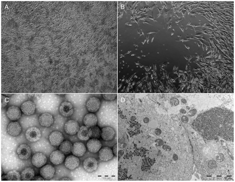
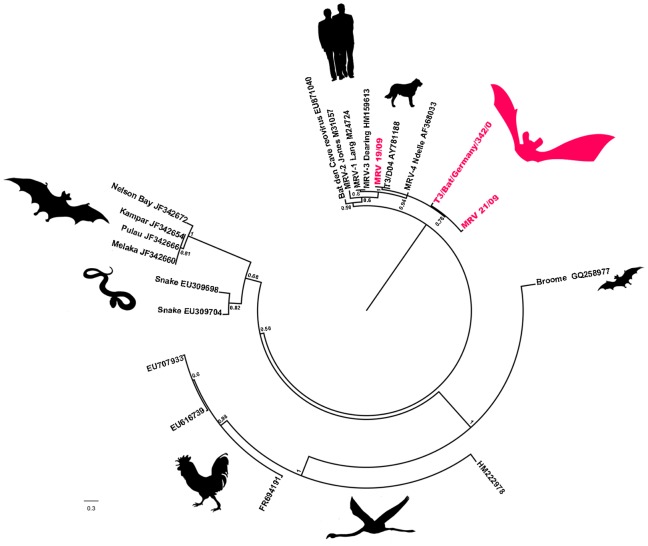
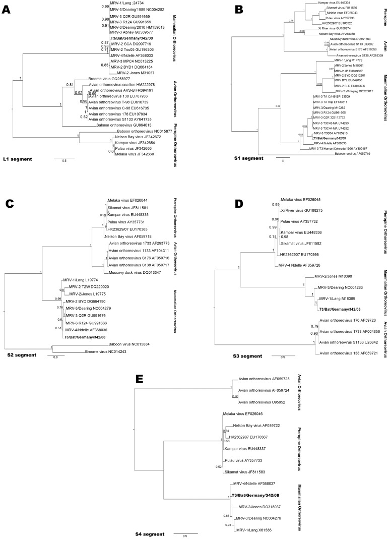
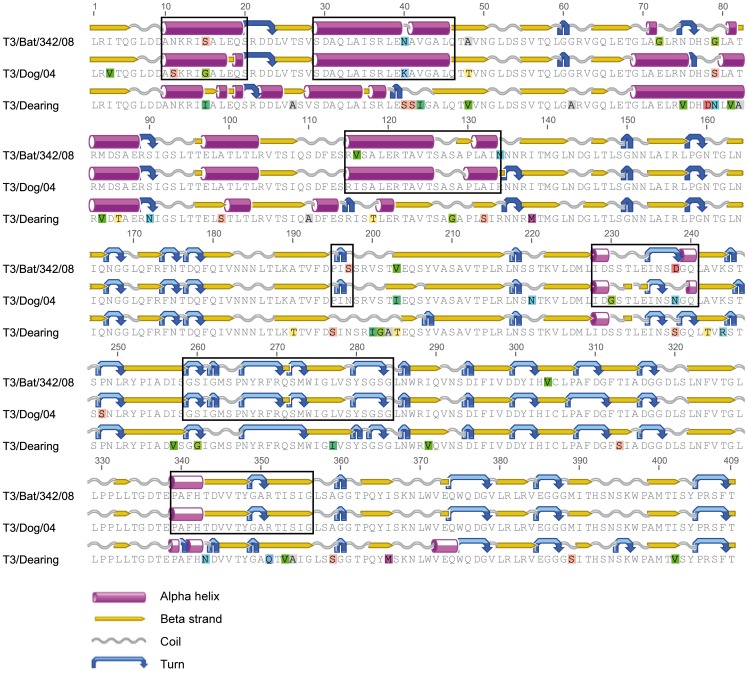
In recent years novel human respiratory disease agents have been described in South East Asia and Australia. The causative pathogens were classified as pteropine orthoreoviruses with strong phylogenetic relationship to orthoreoviruses of flying foxes inhabiting these regions. Subsequently, a zoonotic bat-to-human transmission has been assumed. We report the isolation of three novel mammalian orthoreoviruses (MRVs) from European bats, comprising bat-borne orthoreovirus outside of South East Asia and Australia and moreover detected in insectivorous bats (Microchiroptera). MRVs are well known to infect a broad range of mammals including man. Although they are associated with rather mild and clinically unapparent infections in their hosts, there is growing evidence of their ability to also induce more severe illness in dogs and man. In this study, eight out of 120 vespertilionid bats proved to be infected with one out of three novel MRV isolates, with a distinct organ tropism for the intestine. One isolate was analyzed by 454 genome sequencing. The obtained strain T3/Bat/Germany/342/08 had closest phylogenetic relationship to MRV strain T3D/04, isolated from a dog. These novel reoviruses provide a rare chance of gaining insight into possible transmission events and of tracing the evolution of bat viruses.


Conflict of interest statement

Competing Interests: The authors have declared that no competing interests exist.

Figures

Figure 1
Figure 1. Light microscopy images of Vero E6 cells.
A: control cells. B: cytopathogenic effect of infected cells 3 dpi with strain T3/Bat/Germany/342/08 (see also life cell imaging video Fig S1). Electron microscopy images of strain T3/Bat/Germany/342/08. C: Negative staining of cell-culture supernatant. Non-enveloped reoviral particles with double-layered capsid structure were observed (diameter = approximately 70 nm). Bar is indicating 100 nm. D: Ultra-thin sections of infected Vero E6 cells displayed typical contrast-rich virus particles, organized as paracrystalline structures within the cytosol. Bar is indicating 2 µm.
Figure 2
Figure 2. Phylogenetic tree containing the three novel isolates.
Phylogenetic (Bayesian) analysis of T3/Bat/Germany/342/08, Bat MRV 019/09 and Bat MRV 021/09, using free-end alignments of nucleotide sequences of the partial L1 segments of multiple orthoreovirus species. Posterior probability values are depicted. Strains T3/Bat/Germany/342/08, Bat MRV019/09 and Bat MRV021/09 are shown in magenta and bold. Scale bar shows the evolutionary distance of 0.3 nt substitutions per position. The calculations were unrooted, but for visualization the species reptilian orthoreovirus was used as an outgroup.
Figure 3
Figure 3. Phylogenetic trees of five particular segments of isolate Bat/342/08.
Bayesian analysis of T3/Bat/Germany/342/08 using free-end alignments of nucleotide sequences of five segments of multiple orthoreovirus species. In genome order: A: complete L1 segment. B: S1 segment, complete cds, C: complete S2 segment, D: complete S3 segment, E: complete S4 segment, Posterior probability values are depicted. Strain T3/Bat/Germany/342/08 is shown in bold. Scale bar shows the evolutionary distance of nt substitutions per position. The calculations were unrooted, but for visualization mid-point root was applied.
Figure 4
Figure 4. Free-end protein alignment of the partial σ1 protein.
Its abridged length is due to the availability of only a shorter T3D/04 sequence. The protein structure (α-helix, β-strand, coil and turn) was predicted by Geneious Pro for all strains, and rectangles indicate structure similarity between strains T3/Bat/Germany/342/08 and T3D/04 compared to prototype strain T3/Dearing.

Similar articles

Cited by

References

    1. Cutler SJ, Fooks AR, van der Poel WH (2010) Public health threat of new, reemerging, and neglected zoonoses in the industrialized world. Emerg. Infect. Dis. 16: 1–7. - PMC - PubMed
    1. Jones KE, Patel NG, Levy MA, Storeygard A, Balk D, et al. (2008) Global trends in emerging infectious diseases. Nature 451: 990–993. - PMC - PubMed
    1. Kuzmin IV, Bozick B, Guagliardo SA, Kunkel R, Shak JR, et al. (2011) Bats, emerging infectious diseases, and the rabies paradigm revisited. Emerg. Health Threats J. 4: 7159. - PMC - PubMed
    1. Gard GP, Marshall ID (1973) Nelson Bay virus. A novel reovirus. Arch. Gesamte Virusforsch. 43: 34–42. - PubMed
    1. Pritchard L, Chua K, Cummins D, Hyatt A, Crameri G, et al. (2006) Pulau virus; a new member of the Nelson Bay orthoreovirus species isolated from fruit bats in Malaysia. Arch. Virol. 151: 229–239. - PubMed

Publication types