. 2012;7(8):e43255.
doi: 10.1371/journal.pone.0043255. Epub 2012 Aug 14.

GMP-compliant isolation and large-scale expansion of bone marrow-derived MSC

Affiliations

GMP-compliant isolation and large-scale expansion of bone marrow-derived MSC

Natalie Fekete et al. PLoS One. 2012.

Abstract

Background: Mesenchymal stromal cells (MSC) have gained importance in tissue repair, tissue engineering and in immunosupressive therapy during the last years. Due to the limited availability of MSC in the bone marrow, ex vivo amplification prior to clinical application is requisite to obtain therapeutic applicable cell doses. Translation of preclinical into clinical-grade large-scale MSC expansion necessitates precise definition and standardization of all procedural parameters including cell seeding density, culture medium and cultivation devices. While xenogeneic additives such as fetal calf serum are still widely used for cell culture, its use in the clinical context is associated with many risks, such as prion and viral transmission or adverse immunological reactions against xenogeneic components.

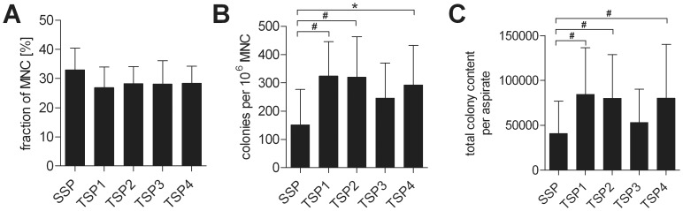
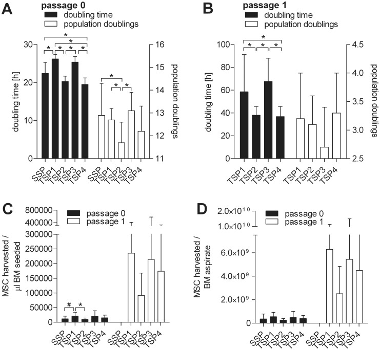
Methods and findings: We established animal-free expansion protocols using platelet lysate as medium supplement and thereby could confirm its safety and feasibility for large-scale MSC isolation and expansion. Five different GMP-compliant standardized protocols designed for the safe, reliable, efficient and economical isolation and expansion of MSC was performed and MSC obtained were analyzed for differentiation capacity by qPCR and histochemistry. Expression of standard MSC markers as defined by the International Society for Cellular Therapy as well as expression of additional MSC markers and of various chemokine and cytokine receptors was analysed by flow cytometry. Changes of metabolic markers and cytokines in the medium were addressed using the LUMINEX platform.

Conclusions: The five different systems for isolation and expansion of MSC described in this study are all suitable to produce at least 100 millions of MSC, which is commonly regarded as a single clinical dose. Final products are equal according to the minimal criteria for MSC defined by the ISCT. We showed that chemokine and integrin receptors analyzed had the same expression pattern, suggesting that MSC from either of the systems show equal characteristics of homing and adhesion.


Conflict of interest statement

Competing Interests: The authors have the following interest: NF, MTR, DF, MW, and HS are working for the German Red Cross Blood Donor Service, Institute for Clinical Transfusion Medicine and Immunogenetics, which is a non-profit organization producing and marketing platelet lysate. There are no patents or products in development to declare. This does not alter the authors' adherence to all the PLoS ONE policies on sharing data and materials, as detailed online in the guide for authors.

Figures

Figure 1
Figure 1. Schematic summary of the GMP-compliant single-step and two-step protocols used for clinical-scale isolation and expansion of MSCs from BM.
Figure 2
Figure 2. Analysis of starting material.
A. Fraction of MNC. B. Colonies per million MNC C. Total colony content per BM aspirate or the starting material (unprocessed bone marrow) used for expansion according to the single-step (SSP) or the two-step protocols (TSP1 - 4). *indicates statistically highly significant (p<0.01). #indicates statistically significant (p<0.05).
Figure 3
Figure 3. Comparative analysis of MSC yields. Shown are mean and SD values of A. doubling time and populations doublings of passage 0, B. doubling time and populations doublings of passage 1, C.
MSCs harvested per ml BM seeded and D. MSCs harvested per total BM aspirate for all of the established GMP-compliant expansion protocols at passage 0 and passage 1. *indicates statistically highly significant (p<0.01). #indicates statistically significant (p<0.05).
Figure 4
Figure 4. Flow cytometric analysis of MSC isolated and expanded according to a GMP-grade single-step or two-step protocol options.
Results of single-step MSC passage 0 (n = 15), two-step protocol 1 (TSP1), p0 (n = 8), p1 (n = 8), protocol 2 (TSP2), p0 (n = 10), p1 (n = 10), protocol 3 (TSP3), p0 (n = 6), p1 (n = 6), protocol 4 (TSP4), p0 (n = 11), p1 (n = 12) are shown as mean and SD values.
Figure 5
Figure 5. Flow cytometric analysis of MSC expanded according to a GMP-grade single-step or two-step protocol.
Results of single-step MSC passage 0 (n = 6–13), p1 (n = 7–12) and p>1 (n = 6–12) as well as two-step MSC at p0 (n = 6) and p1 (n = 8) are shown as percent positive in Tukey’s Whisker Plots.
Figure 6
Figure 6. Differentiation capacity of representative MSC batches isolated and expanded using the five different cell expansion systems.
Adipogenic (Oil RedO/haematoxylin staining), chondrogenic (methylene blue staining) and osteogenic (detection of alkaline phosphatase) differentiation assays are shown. Control assays were performed in aMEM supplemented with 10% FCS. Black bar indicates 1000 µm for adipogenic and 100 µm for chondrogenic and osteogenic differentiation.
Figure 7
Figure 7. Quantitative cytokine analysis and acquisition of metabolic parameters of single-step and two-step MSC expansion media.
Samples of cell culture supernatants were collected at the time points indicated during single-step and two-step (TSP1; 14+7-5-8) GMP-grade large-scale cell expansion runs and analyzed for their cytokine content. Depicted are results of glucose, lactate, bFGF, IL-6, GRO (CXCL1/2/3), sCD40L, RANTES/CCL5, PDGF-AA, VEGF (n = 4 single step runs; n = 3 two-step runs), sVCAM-1, PDGF-AB/BB (n = 3 single step runs; n = 3 two-step runs), sICAM-1, and TGF-β1 (n = 2 single step runs; n = 3 two-step runs) contents as mean and SD values.

References

    1. Dominici M, Le BK, Mueller I, Slaper-Cortenbach I, Marini F, et al. (2006) Minimal criteria for defining multipotent mesenchymal stromal cells. The International Society for Cellular Therapy position statement. Cytotherapy 8: 315–317. - PubMed
    1. Caplan AI (1994) The mesengenic process. ClinPlastSurg 21: 429–435. - PubMed
    1. Subbanna PK (2007) Mesenchymal stem cells for treating GVHD: in-vivo fate and optimal dose. MedHypotheses 69: 469–470. - PubMed
    1. Tolar J, Le BK, Keating A, Blazar BR (2010) Concise review: hitting the right spot with mesenchymal stromal cells. Stem Cells 28: 1446–1455. - PMC - PubMed
    1. EuropeanCommission (2007) REGULATION (EC) No 1394/2007 OF THE EUROPEAN PARLIAMENT AND OF THE COUNCIL of 13 November 2007 on advanced therapy medicinal products and amending Directive 2001/83/EC and Regulation (EC) No 726/2004. Official Journal of the European Union 50: 121–137.

Publication types