

Resection of ictal high-frequency oscillations leads to favorable surgical outcome in pediatric epilepsy
- PMID: 22905734
- PMCID: PMC3520059
- DOI: 10.1111/j.1528-1167.2012.03629.x
Resection of ictal high-frequency oscillations leads to favorable surgical outcome in pediatric epilepsy
Abstract
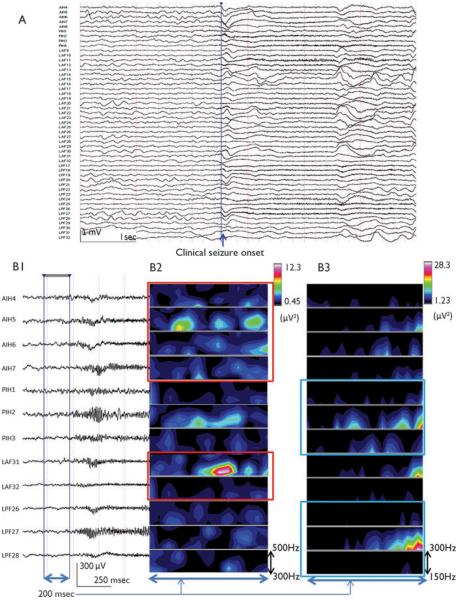
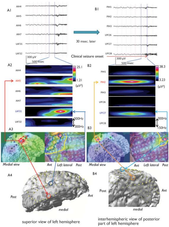
Purpose: Intracranial electroencephalography (EEG) is performed as part of an epilepsy surgery evaluation when noninvasive tests are incongruent or the putative seizure-onset zone is near eloquent cortex. Determining the seizure-onset zone using intracranial EEG has been conventionally based on identification of specific ictal patterns with visual inspection. High-frequency oscillations (HFOs, >80 Hz) have been recognized recently as highly correlated with the epileptogenic zone. However, HFOs can be difficult to detect because of their low amplitude. Therefore, the prevalence of ictal HFOs and their role in localization of epileptogenic zone on intracranial EEG are unknown.
Methods: We identified 48 patients who underwent surgical treatment after the surgical evaluation with intracranial EEG, and 44 patients met criteria for this retrospective study. Results were not used in surgical decision making. Intracranial EEG recordings were collected with a sampling rate of 2,000 Hz. Recordings were first inspected visually to determine ictal onset and then analyzed further with time-frequency analysis. Forty-one (93%) of 44 patients had ictal HFOs determined with time-frequency analysis of intracranial EEG.
Key findings: Twenty-two (54%) of the 41 patients with ictal HFOs had complete resection of HFO regions, regardless of frequency bands. Complete resection of HFOs (n = 22) resulted in a seizure-free outcome in 18 (82%) of 22 patients, significantly higher than the seizure-free outcome with incomplete HFO resection (4/19, 21%).
Significance: Our study shows that ictal HFOs are commonly found with intracranial EEG in our population largely of children with cortical dysplasia, and have localizing value. The use of ictal HFOs may add more promising information compared to interictal HFOs because of the evidence of ictal propagation and followed by clinical aspect of seizures. Complete resection of HFOs is a favorable prognostic indicator for surgical outcome.
Wiley Periodicals, Inc. © 2012 International League Against Epilepsy.
Figures
References
-
- Akiyama T, Otsubo H, Ochi A, Ishiguro T, Kadokura G, Ramachandrannair R, Weiss SK, Rutka JT, Carter Snead O., 3rd Focal cortical high-frequency oscillations trigger epileptic spasms: confirmation by digital video subdural EEG. Clin Neurophysiol. 2005;116:2819–2825. - PubMed
-
- Akiyama T, Otsubo H, Ochi A, Galicia EZ, Weiss SK, Donner EJ, Rutka JT, Snead OC., 3rd Topographic movie of ictal high-frequency oscillations on the brain surface using subdural EEG in neocortical epilepsy. Epilepsia. 2006;47:1953–1957. - PubMed
-
- Akiyama T, McCoy B, Go CY, Ochi A, Elliott IM, Akiyama M, Donner EJ, Weiss SK, Snead OC, III, Rutka JT. Focal resection of fast ripples on extraoperative intracranial EEG improves seizure outcome in pediatric epilepsy. Epilepsia. 2011;52:1802–1811. - PubMed
-
- Axmacher N, Elger CE, Fell J. Ripples in the medial temporal lobe are relevant for human memory consolidation. Brain. 2008;131:1806–1817. - PubMed
MeSH terms
Grants and funding
LinkOut - more resources
Full Text Sources
Medical
Molecular Biology Databases
Miscellaneous
