

Peritumoral lymphangiogenesis induced by vascular endothelial growth factor C and D promotes lymph node metastasis in breast cancer patients
- PMID: 22906075
- PMCID: PMC3499230
- DOI: 10.1186/1477-7819-10-165
Peritumoral lymphangiogenesis induced by vascular endothelial growth factor C and D promotes lymph node metastasis in breast cancer patients
Abstract
Background: Mounting clinical and experimental data suggest that the migration of tumor cells into lymph nodes is greatly facilitated by lymphangiogenesis. Vascular endothelial growth factor (VEGF)-C and D have been identified as lymphangiogenic growth factors and play an important role in tumor lymphangiogenesis. The purpose of this study was to investigate the location of lymphangiogenesis driven by tumor-derived VEGF-C/D in breast cancer, and to determine the role of intratumoral and peritumoral lymphatic vessel density (LVD) in lymphangiogenesis in breast cancer.
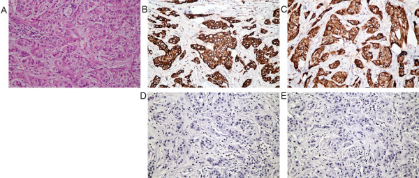
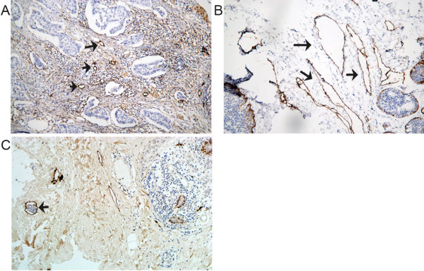
Methods: The expression levels of VEGF-C/D were determined by immunohistochemistry, and intratumoral LVD and peritumoral LVD were assessed using immunohistochemistry and the D2-40 antibody in 73 patients with primary breast cancer. The associations of intratumoral LVD and peritumoral LVD with VEGF-C/D expression, clinicopathological features and prognosis were assessed.
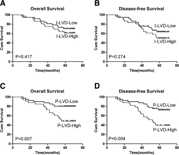
Results: VEGF-C and D expression were significantly higher in breast cancer than benign disease (P < 0.01). VEGF-C (P < 0.001) and VEGF-D (P = 0.005) expression were significantly associated with peritumoral LVD, but not intratumoral LVD. Intratumoral LVD was associated with tumor size (P = 0.01). Peritumoral LVD was significantly associated with lymph node metastasis (LNM; P = 0.005), lymphatic vessel invasion (LVI; P = 0.017) and late tumor,node, metastasis (TNM) stage (P = 0.011). Moreover, peritumoral LVD was an independent risk factor for axillary lymph node metastasis, overall survival and disease-free survival in multivariate analysis.
Conclusions: This study suggests that tumor-derived VEGF-C/D induce peritumoral lymphangiogenesis, which may be one mechanism that leads to lymphatic invasion and metastatic spread. Peritumoral LVD has potential as an independent prognostic factor in breast cancer patients.
Figures
Similar articles
-
Different significance between intratumoral and peritumoral lymphatic vessel density in gastric cancer: a retrospective study of 123 cases.BMC Cancer. 2010 Jun 17;10:299. doi: 10.1186/1471-2407-10-299. BMC Cancer. 2010. PMID: 20565772 Free PMC article.
-
Peritumoral lymphatic vessel density and vascular endothelial growth factor C expression in early-stage squamous cell carcinoma of the uterine cervix.Clin Cancer Res. 2005 Dec 1;11(23):8364-71. doi: 10.1158/1078-0432.CCR-05-1238. Clin Cancer Res. 2005. PMID: 16322297
-
Vascular endothelial growth factor (VEGF)-C, VEGF-D, VEGFR-3 and D2-40 expressions in primary breast cancer: Association with lymph node metastasis.Adv Clin Exp Med. 2017 Mar-Apr;26(2):245-249. doi: 10.17219/acem/58784. Adv Clin Exp Med. 2017. PMID: 28791841
-
Lymphatic microvessel density and vascular endothelial growth factor-C and -D as prognostic factors in breast cancer: a systematic review and meta-analysis of the literature.Mol Biol Rep. 2012 Dec;39(12):11153-65. doi: 10.1007/s11033-012-2024-y. Epub 2012 Oct 11. Mol Biol Rep. 2012. PMID: 23054001
-
Lymphangiogenesis in Breast Cancer: From Molecular Mechanisms to Clinical Implications.FASEB J. 2025 May 15;39(9):e70590. doi: 10.1096/fj.202500024R. FASEB J. 2025. PMID: 40320983 Review.
Cited by
-
Dysregulation of Lymphatic Endothelial VEGFR3 Signaling in Disease.Cells. 2023 Dec 28;13(1):68. doi: 10.3390/cells13010068. Cells. 2023. PMID: 38201272 Free PMC article. Review.
-
Peritumoral Lymphatic Vessels Associated with Resistance to Neoadjuvant Chemotherapy and Unfavorable Survival in Esophageal Cancer.Ann Surg Oncol. 2020 Oct;27(10):3762-3769. doi: 10.1245/s10434-020-08474-x. Epub 2020 Apr 23. Ann Surg Oncol. 2020. PMID: 32328984
-
Quantification of STAT3 and VEGF expression for molecular diagnosis of lymph node metastasis in breast cancer.Medicine (Baltimore). 2017 Nov;96(45):e8488. doi: 10.1097/MD.0000000000008488. Medicine (Baltimore). 2017. PMID: 29137038 Free PMC article.
-
The global landscape and research trend of lymphangiogenesis in breast cancer: a bibliometric analysis and visualization.Front Oncol. 2024 Mar 14;14:1337124. doi: 10.3389/fonc.2024.1337124. eCollection 2024. Front Oncol. 2024. PMID: 38549934 Free PMC article. Review.
-
Lymphangiogenesis in Classical Hodgkin Lymphoma - Preliminary Study with Clinicopathological Correlations.J Cancer. 2016 Oct 23;7(14):2117-2123. doi: 10.7150/jca.16389. eCollection 2016. J Cancer. 2016. PMID: 27877228 Free PMC article.
References
-
- Pepper MS. Lymphangiogenesis and tumor metastasis: myth or reality? Clin Cancer Res. 2001;7:462–468. - PubMed
-
- Ji RC. Lymphatic endothelial cells, tumor lymphangiogenesis and metastasis: new insights into intratumoral and peritumoral lymphatics. Cancer Metastasis Rev. 2006;25:677–694. - PubMed
-
- Mandriota SJ, Jussila L, Jeltsch M, Compagni A, Baetens D, Prevo R, Banerji S, Huarte J, Montesano R, Jackson DG, Orci L, Alitalo K, Christofori G, Pepper MS. Vascular endothelial growth factor-C-mediated lymphangiogenesis promotes tumour metastasis. EMBO J. 2001;20:672–682. doi: 10.1093/emboj/20.4.672. - DOI - PMC - PubMed
-
- Van Trappen PO, Steele D, Lowe DG, Baithun S, Beasley N, Thiele W, Weich H, Krishnan J, Shepherd JH, Pepper MS, Jackson DG, Sleeman JP, Jacobs U. Expression of vascular endothelial growth factor (VEGF)-C and VEGF-D, and their receptor VEGFR-3, during different stages of cervical carcinogenesis. J Pathol. 2003;201:544–554. doi: 10.1002/path.1467. - DOI - PubMed
Publication types
MeSH terms
Substances
LinkOut - more resources
Full Text Sources
Medical
