. 2012 Aug 20:7:28.
doi: 10.1186/1749-8104-7-28.

Neurog1 and Neurog2 coordinately regulate development of the olfactory system

Affiliations

Neurog1 and Neurog2 coordinately regulate development of the olfactory system

Tarek Shaker et al. Neural Dev. .

Abstract

Background: Proneural genes encode basic helix-loop-helix transcription factors that specify distinct neuronal identities in different regions of the nervous system. In the embryonic telencephalon, the proneural genes Neurog1 and Neurog2 specify a dorsal regional identity and glutamatergic projection neuron phenotype in the presumptive neocortex, but their roles in cell fate specification in the olfactory bulb, which is also partly derived from dorsal telencephalic progenitors, have yet to be assessed. Given that olfactory bulb development is guided by interactions with the olfactory epithelium in the periphery, where proneural genes are also expressed, we investigated the roles of Neurog1 and Neurog2 in the coordinated development of these two olfactory structures.

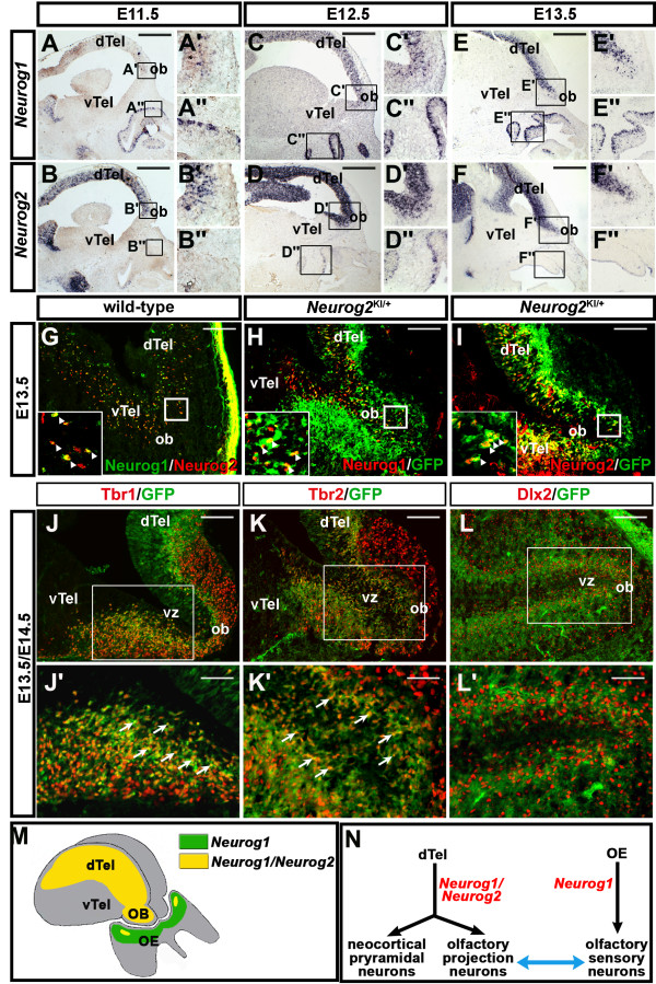
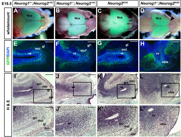
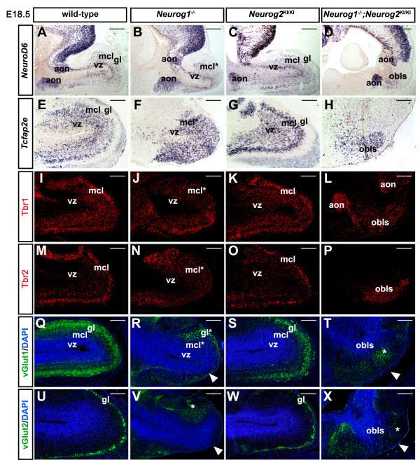
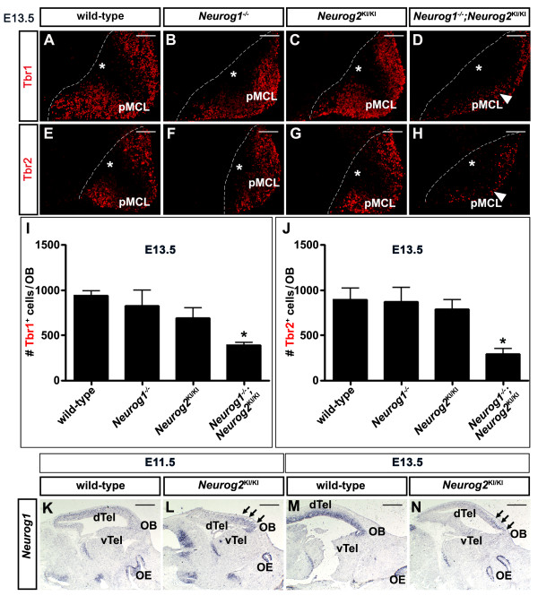
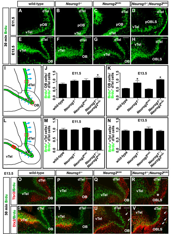
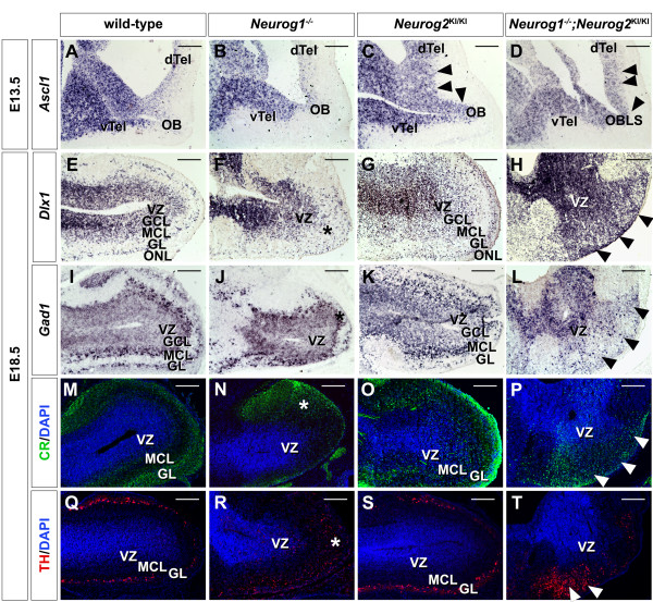
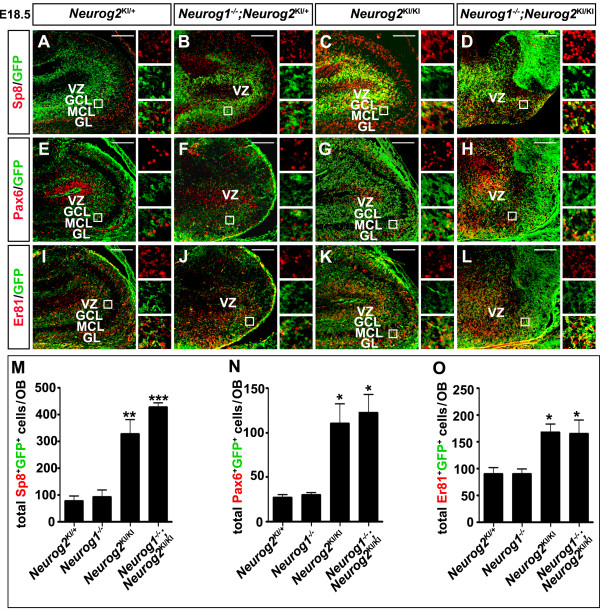
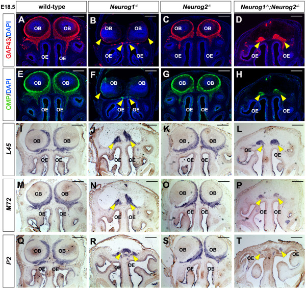
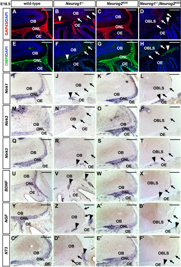
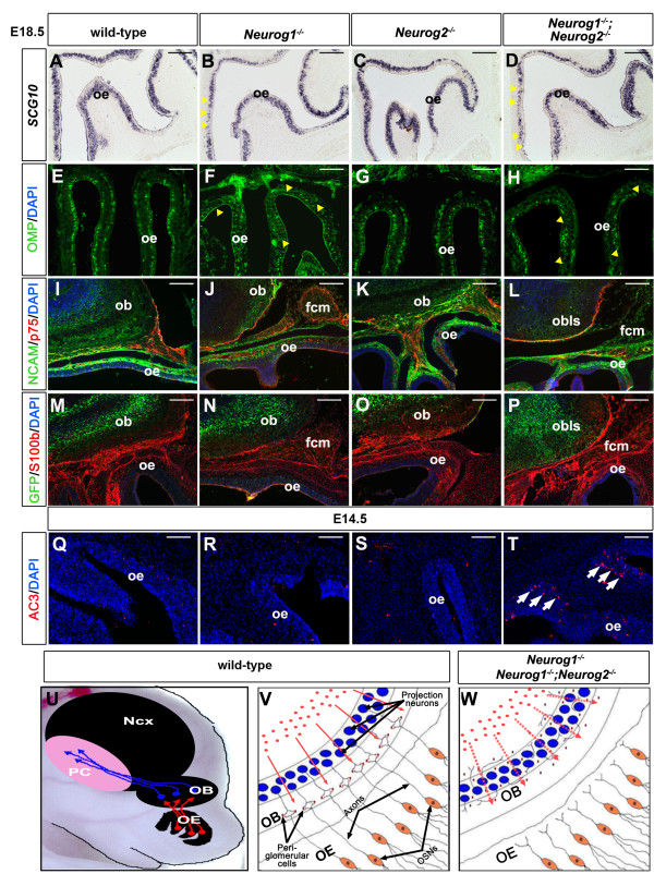
Results: Neurog1/2 are co-expressed in olfactory bulb progenitors, while only Neurog1 is widely expressed in progenitors for olfactory sensory neurons in the olfactory epithelium. Strikingly, only a remnant of an olfactory bulb forms in Neurog1-/-;Neurog2-/- double mutants, while this structure is smaller but distinguishable in Neurog1-/- single mutants and morphologically normal in Neurog2-/- single mutants. At the cellular level, fewer glutamatergic mitral and juxtaglomerular cells differentiate in Neurog1-/-;Neurog2-/- double-mutant olfactory bulbs. Instead, ectopic olfactory bulb interneurons are derived from dorsal telencephalic lineages in Neurog1-/-;Neurog2-/- double mutants and to a lesser extent in Neurog2-/- single mutants. Conversely, cell fate specification is normal in Neurog1-/- olfactory bulbs, but aberrant patterns of cell proliferation and neuronal migration are observed in Neurog1-/- single and Neurog1-/-;Neurog2-/- double mutants, probably contributing to their altered morphologies. Finally, in Neurog1-/- and Neurog1-/-;Neurog2-/- embryos, olfactory sensory neurons in the epithelium, which normally project to the olfactory bulb to guide its morphogenesis, fail to innervate the olfactory bulb.

Conclusions: We have identified a cell autonomous role for Neurog1/2 in specifying the glutamatergic identity of olfactory bulb neurons. Furthermore, Neurog1 (and not Neurog2) is required to guide olfactory sensory neuron innervation of the olfactory bulb, the loss of which results in defects in olfactory bulb proliferation and tissue morphogenesis. We thus conclude that Neurog1/2 together coordinate development of the olfactory system, which depends on tissue interactions between the olfactory bulb and epithelium.


Figures

Figure 1
Figure 1
Neurog1 and Neurog2 expression in the embryonic olfactory system. (A) to (F) Sagittal sections of embryonic day (E) 11.5, E12.5 and E13.5 embryos, showing the distribution of Neurog1 (A-A", C-C", E-E") and Neurog2 (B-B", D-D", F-F") transcripts. Insets in A to F are fourfold magnifications of the boxed areas in the OB (A' to F') and OE (A" to F"). (G) to (I) Co-immunolabeling of E13.5 Neurog2 KI/+ brains with antibodies to Neurog1 and Neurog2 (G), Neurog1 and GFP (H), and Neurog2 and GFP (I). Insets in (G) to (I) are fourfold magnifications of the boxed areas. (J) to (L) Co-immunolabeling of the E13.5 OB with Tbr1 and GFP (J,J') and with Tbr2 and GFP (K,K'), and labeling of the E14.5 OB with Dlx2 and GFP (L,L'). Boxed areas in (J) to (L) are magnified twofold in J', K', L' respectively. (M) Schematic representation of Neurog1 single-positive OE progenitors (green) and Neurog1/Neurog2 double-positive progenitors (yellow) in the OB and a small region of the OE. (N) Schematic illustration of the objectives of this study; to determine the roles of Neurog1 and Neurog2 in olfactory system development. dTel, dorsal telencephalon; OB, olfactory bulb; OE, olfactory epithelium; vTel, ventral telencephalon. Scale bars: 1 mm (A) to (F), 250 μm (G) to (L), 125 μm (J') to (L').
Figure 2
Figure 2
Defects in olfactory bulb morphogenesis and lamination in Neurog1 −/−and Neurog1 −/−;Neurog2 KI/KImutant embryos. (A) to (D) Whole-mount dorsal views of partially dissected embryonic day (E) 18.5 wild-type (A), Neurog1−/− (B), Neurog2 KI/KI (C), and Neurog1/2 −/− (D) brains, all heterozygous or homozygous for a Neurog2GFPKI allele. Brains were left in the cranium and imaged by merging bright-field and GFP fluorescent images. Arrowheads mark the reduction in OB size in Neurog1−/− embryos (B), and apparent loss of the OB in Neurog1/2 −/− embryos (D). (E) to (H) GFP epifluorescence (green) and nuclear DAPI staining (blue) of sagittal sections through E18.5 wild-type (E), Neurog1−/− (F), Neurog2KI/KI (G), and Neurog1/2−/− (H) OBs. (I) to (L) H & E histological analysis of E18.5 wild-type (I,I'), Neurog1−/− (J,J'), Neurog2KI/KI (K,K'), and Neurog1/2−/− (L,L') OBs. (I') to (L') are twofold magnifications of the boxed areas in (I) to (L), respectively. GCL, granule cell layer; Gl, glomerular layer; MCL, mitral cell layer; NCX, neocortex; OB, olfactory bulb; OBLS, olfactory bulb-like structure; ONL, outer nerve layer; VZ, ventricular zone. Scale bars: 2 mm (A) to (D), 500 μm (E) to (L), 250 μm (I') to (L').
Figure 3
Figure 3
Impaired lamination and differentiation of excitatory neurons in Neurog1/2 −/− single and double mutant olfactory bulbs. (A) to (X) Expression of NeuroD6 (A) to (D), Tcfap2e (E) to (H), Tbr1 (I) to (L), Tbr2 (M) to (P), vGlut1 (green)/DAPI (blue) (Q) to (T) and vGlut2 (green)/DAPI (blue) (U) to (X) in embryonic day 18.5 wild-type (A,E,I,M,Q,U), Neurog1−/− (B,F,J,N,R,V), Neurog2KI/KI (C,G,K,O,S,W) and Neurog1/2−/− (D,H,L,P,T,X) olfactory bulbs (OBs). White arrowheads in R,V,T,X mark diminished vGlut1/2 protein expression in the peripheral glomerular layer (GL). AON, anterior olfactory nucleus; MCL, mitral cell layer; OBLS, olfactory bulb-like structure; VZ, ventricular zone. Scale bars: 1 mm (A) to (D), 500 μm (E) to (X).
Figure 4
Figure 4
Defects in the differentiation of glutamatergic olfactory bulb neurons inNeurog1/2 double mutants. (A) to (H) Expression of Tbr1 (A) to (D) and Tbr2 (E) to (H) in the presumptive MCL of embryonic day (E) 13.5 wild-type (A,E), Neurog1−/− (B,F), Neurog2 KI/KI (C,G) and Neurog1/2 −/− (D,H) OBs. White arrowheads in D,H mark the reduction in glutamatergic neurons in Neurog1/2−/− OBs. White asterisks in (A) to (H) mark the ventricular zone. (I), (J) Quantitation of total numbers of Tbr1+ cells (I) and Tbr2+ cells (J) in E13.5 wild-type Neurog1−/−, Neurog2KI/KI and Neurog1/2−/− OBs. Asterisks denote P <0.05. (K) to (N) Analysis of Neurog1 expression in E11.5 (K,L) and E13.5 (M,N) wild-type and Neurog2−/− telencephalons. Arrows mark ectopic Neurog1 expression in (L) and (N). Scale bars: 250 μm (A) to (H), 1 mm (K) to (N).
Figure 5
Figure 5
Aberrant patterns of cell proliferation in Neurog1/2 −/− mutants. (A) to (H) BrdU-labeled S-phase progenitors at embryonic day (E) 11.5 (A) to (D), and E13.5 (E) to (H) in wild-type (A,E), Neurog1−/− (B,F), Neurog2 KI/KI (C,G), and Neurog1/2−/− (D,H) OBs. (I) to (K) Illustration schematizing areas where counts of BrdU-labeled S-phase progenitors were performed (I). Quantitation of the ratio of BrdU+ cells in the OB compared with the dorsal telencephalon at E11.5 (J) and E13.5 (K). Asterisks denote P <0.05. (L) to (N) Illustration schematizing areas where counts of BrdU-labeled S-phase progenitors were performed (L). Quantitation of the ratio of BrdU+ cells in the ventral compared with the dorsal telencephalon at E11.5 (M) and E13.5 (N). (O) to (V) Co-labeling of BrdU+ S-phase progenitors (green) with either Tbr2 (red, O to R) or Dlx2 (red, S to V) in E13.5 wild-type (O,S), Neurog1−/− (P,T), Neurog2KI/KI (Q,U), and Neurog1/2−/− (R,V) embryos. dTel, dorsal telencephalon; OB, olfactory bulb; pOB, presumptive olfactory bulb; pOBLS, presumptive olfactory bulb like structure; vTel, ventral telencephalon. Scale bars: 250 μm (A) to (H), (O) to (V).
Figure 6
Figure 6
Ectopic differentiation of olfactory bulb interneurons in Neurog2 KI/KI and Neurog1/2 −/− embryos. (A) to (D) Expression of Ascl1 in embryonic day (E) 13.5 wild-type (A), Neurog1−/− (B), Neurog2KI/KI (C), and Neurog1/2−/− (D) OBs. (E) to (T) Expression of Dlx1 (E) to (H), Gad1 (I) to (L), calretinin (CR) (M) to (P) and TH (Q) to (T) in E18.5 wild-type, (E,I,M,Q), Neurog1−/− (F,J,N,R), Neurog2KI/KI (G,K,O,S), and Neurog1/2−/− (H,L,P,T) OBs. Blue is DAPI nuclear stain in (M) to (T). Arrowheads in (C,D) mark the upregulated expression of Ascl1 in the OB of Neurog2KI/KI and OBLS of Neurog1/2−/− embryos. Arrowheads in (H,L,P,T) mark expansion of expression domain from VZ to pial surface in Neurog1/2−/− OBs. Asterisks in (F,J,N,R) mark ectopic interneurons in Neurog2KI/KI OBs. dTel, dorsal telencephalon; GL, glomerular layer; GCL, granule cell layer; MCL, mitral cell layer; OB, olfactory bulb; OBLS, olfactory bulb-like structure; ONL, olfactory nerve layer; vTel, ventral telencephalon; VZ, ventricular zone. Scale bars: 500 μm (A) to (T).
Figure 7
Figure 7
Neuronal misspecification defects in Neurog2−/− olfactory bulbs and Neurog1/2 −/− olfactory bulb-like structures. (A) to (L) Co-immunostaining of GFP (green, A to L) with Sp8 (red, A to D), Pax6 (red, E to H) or Er81 (red, I to L) in E18.5 wild-type, (A,E,I), Neurog1−/− (B,F,J), Neurog2KI/KI (C,G,K), and Neurog1/2−/− (D,H,L) OBs. Insets to the right of each panel are fourfold magnifications of boxed areas. (M) to (O) Quantitation of total Sp8+GFP+ cells (M), Pax6+GFP+ cells (N), and Er81+GFP+ cells (O) in embryonic day 18.5 wild-type, Neurog1−/−, Neurog2KI/KI and Neurog1/2−/− OBs. *P <0.05, **P <0.01, ***P <0.005. GCL, granule cell layer; GL, glomerular layer; MCL, mitral cell layer; VZ, ventricular zone. Scale bars: 500 μm (A) to (L).
Figure 8
Figure 8
Neurog1 −/− and Neurog1/2 −/− olfactory sensory neurons express mature markers but fail to innervate olfactory bulb. (A) to (H) Coronal sections of the embryonic day (E) 18.5 OB showing expression of GAP43 (red, A to D) and OMP (green, E to H) with DAPI (blue, A to H) in wild-type (A,E), Neurog1−/− (B,F), Neurog2KI/KI (C,G) and Neurog1/2−/− (D,H) embryos. Yellow arrows denote loss of OSN innervation of the OB in Neurog1−/− and Neurog1/2−/− (B,D,F,H) embryos. (I) to (T) Expression of odorant receptors L45 (I) to (L), M72 (M) to (P) and P2 (Q) to (T) in E18.5 wild-type (I,M,Q), Neurog1−/− (J,N,R), Neurog2KI/KI (K,O,S) and Neurog1/2−/− (L,P,T) embryos. Note that the lack of an apparent OB in Neurog1−/− sections is because the OB is shortened along the proximodistal axis, and hence does not fill the rostral-most part of the cavity in the skull, where OR expression levels are the highest (J,N,R). GAP43, growth-associated protein 43; OB, olfactory bulb; OE, olfactory epithelium; OMP, olfactory marker protein; OSN, olfactory sensory neuron. Scale bars: 500 μm (A) to (T).
Figure 9
Figure 9
Neurog1 −/− and Neurog1/2 −/− olfactory sensory neurons express neurotrophic receptors and ligands but fail to innervate olfactory bulb. (A) to (H) Expression of GAP43 (red, A to D) and OMP (green, E to H) with DAPI counterstain (blue, A to H) in embryonic day (E) 18.5 wild-type (A,E), Neurog1−/− (B,F), Neurog2KI/KI (C,G) and Neurog1/2−/− (D,H) OBs/OEs. White arrows denote loss of synaptogenesis between the OE and the OB in Neurog1−/− and Neurog1/2−/− (B,D,F,H) olfactory systems, and arrowheads point to the accumulation of OSN axons in a FCM. (I) to (T) Expression of Ntrk1 (I) to (L), Ntrk2 (M) to (P) and Ntrk3 (Q) to (T) in E18.5 wild-type (I,M,Q), Neurog1−/− (J,N,R), Neurog2KI/KI (K,O,S) and Neurog1/2−/− (L,P,T) embryos. (U) to (F') Expression of BDNF (U) to (X), NGF (Y) to (B'), NT3 (C') to (F'), in E18.5 wild-type (U,Y,C'), Neurog1−/− (V,Z,D'), Neurog2KI/KI (W,A',E') and Neurog1/2−/− (X,B',F') embryos. Black arrows (J,L,N,P,R,T,V,X,Z,B',D',F') indicate loss of marker expression in outer layers of Neurog1−/− and Neurog1/2−/− OBs. Arrowheads (J,L,R,T,V,X,Z,B',D',F') point to FCM formation in Neurog1−/− and Neurog1/2−/− embryos. FCM, fibrocellular mass; OB, olfactory bulb; OBLS, olfactory bulb-like structure; OE, olfactory epithelium; ONL, olfactory nerve layer. Scale bars: 500 μm (A) to (Z), (A') to (F').
Figure 10
Figure 10
Normal OSN and OEC differentiation in Neurog1 −/− and Neurog2 −/− mutants, but apoptosis elevated in double mutants. (A) to (D) Expression of SCG10 in wild-type (A), Neurog1−/− (B), Neurog2−/− (C), and Neurog1/2−/− (D) OBs at embryonic day (E) 18.5. (E) to (P) Co-labeling with DAPI (blue, E to P) and OMP (green, E to H), NCAM (green, I to L), GFP (green, M to P), p75 (red, I to L), or S100b (red, M to P) in wild-type (E,I,M), Neurog1−/− (F,J,N), Neurog2−/− (G,K,O), and Neurog1/2−/− (H,L,P) OBs at E18.5. (Q) to (T) Co-labeling with DAPI (blue) and activated caspase 3 (AC3; red) in wild-type (Q), Neurog1−/− (R), Neurog2−/− (S), and Neurog1/2−/− (T) OBs at E14.5. (U) to (W) Schematic representation of the three components of the olfactory system (U). The normal process of OSN innervation of the OB, and the subsequent formation of the glomerular layer by migrating OB interneurons (V) is perturbed in Neurog1/2−/− embryos (W). OB, olfactory bulb; OE, olfactory epithelium; OEC, olfactory ensheathing cell; OSN, olfactory sensory neuron; PC, piriform cortex. Scale bars: 500 μm (A) to (P), 250 μm (Q) to (T).

Similar articles

Cited by

References

    1. Winpenny E, Lebel-Potter M, Fernandez ME, Brill MS, Gotz M, Guillemot F, Raineteau O. Sequential generation of olfactory bulb glutamatergic neurons by Neurog2-expressing precursor cells. Neural Dev. 2011;6:12. doi: 10.1186/1749-8104-6-12. - DOI - PMC - PubMed
    1. Bayer SA. 3H-thymidine-radiographic studies of neurogenesis in the rat olfactory bulb. Exp Brain Res. 1983;50:329–340. - PubMed
    1. Jimenez D, Garcia C, de Castro F, Chedotal A, Sotelo C, de Carlos JA, Valverde F, Lopez-Mascaraque L. Evidence for intrinsic development of olfactory structures in Pax-6 mutant mice. J Comp Neurol. 2000;428:511–526. doi: 10.1002/1096-9861(20001218)428:3<511::AID-CNE8>3.0.CO;2-I. - DOI - PubMed
    1. Wichterle H, Garcia-Verdugo JM, Herrera DG, Alvarez-Buylla A. Young neurons from medial ganglionic eminence disperse in adult and embryonic brain. Nat Neurosci. 1999;2:461–466. doi: 10.1038/8131. - DOI - PubMed
    1. Wichterle H, Turnbull DH, Nery S, Fishell G, Alvarez-Buylla A. In utero fate mapping reveals distinct migratory pathways and fates of neurons born in the mammalian basal forebrain. Development. 2001;128:3759–3771. - PubMed

Publication types

MeSH terms

Substances

LinkOut - more resources