Review
. 2012 Oct;185(2):71-88.
doi: 10.1016/j.molbiopara.2012.08.003. Epub 2012 Aug 10.

Sirtuins of parasitic protozoa: in search of function(s)

Affiliations
Review

Sirtuins of parasitic protozoa: in search of function(s)

Agnieszka A Religa et al. Mol Biochem Parasitol. 2012 Oct.

Abstract

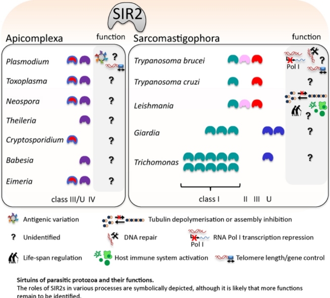
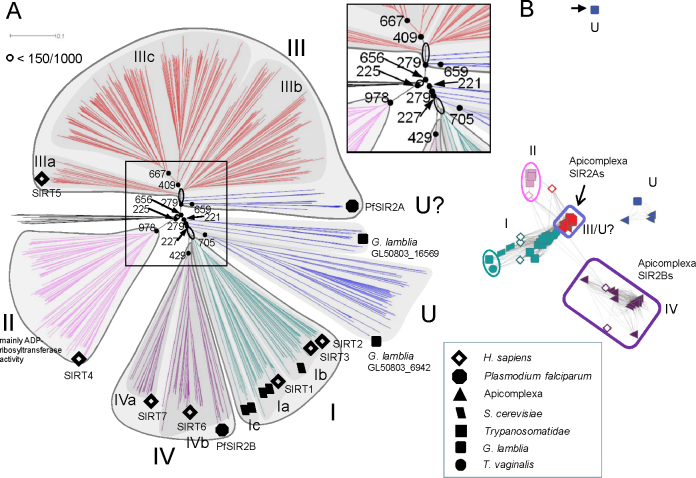
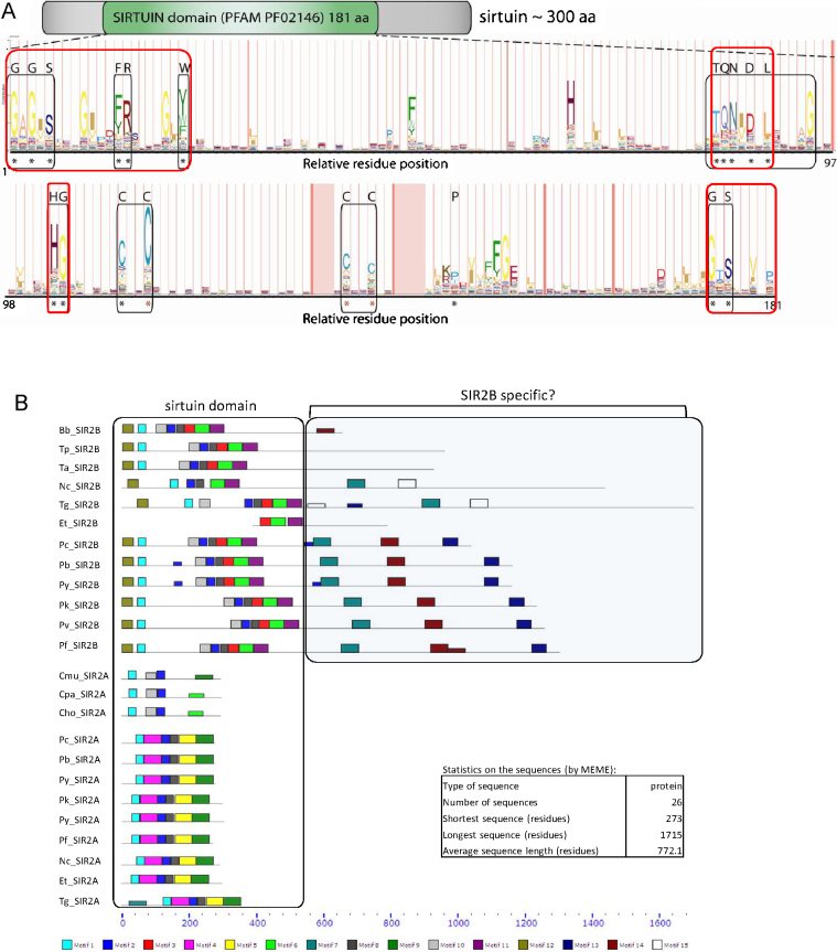
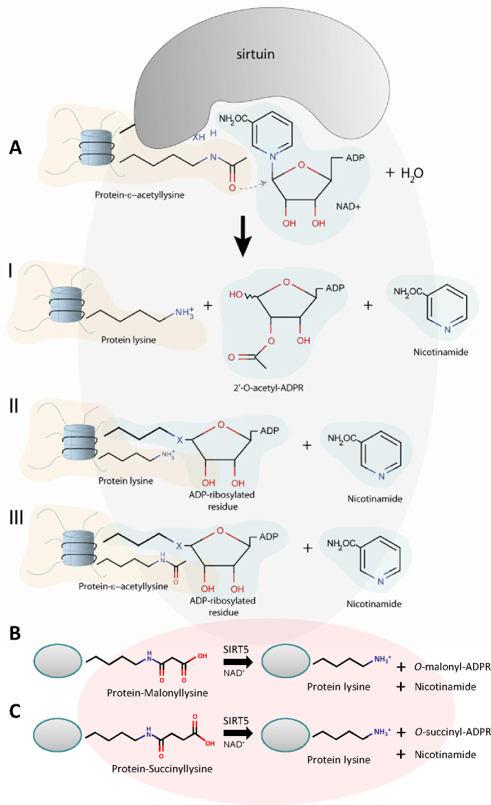
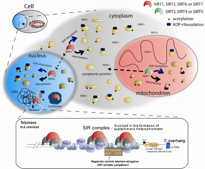
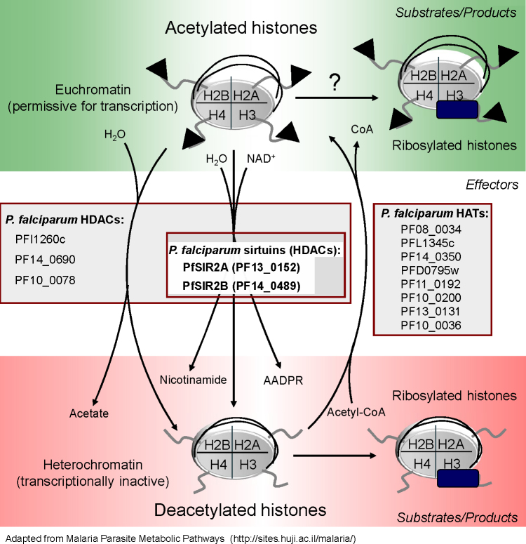
The SIR2 family of NAD(+)-dependent protein deacetylases, collectively called sirtuins, has been of central interest due to their proposed roles in life-span regulation and ageing. Sirtuins are one group of environment sensors of a cell interpreting external information and orchestrating internal responses at the sub-cellular level, through participation in gene regulation mechanisms. Remarkably conserved across all kingdoms of life SIR2 proteins in several protozoan parasites appear to have both conserved and intriguing unique functions. This review summarises our current knowledge of the members of the sirtuin families in Apicomplexa, including Plasmodium, and other protozoan parasites such as Trypanosoma and Leishmania. The wide diversity of processes regulated by SIR2 proteins makes them targets worthy of exploitation in anti-parasitic therapies.


Figures

None
Graphical abstract
Fig. 1
Fig. 1
Sirtuin phylogeny with focus on parasitic protozoa. (A) A phylogenetic tree of 778 annotated sirtuins from different organisms (sources NCBI, EuPathDB, GeneDB) produced in ClustalX2 using Neighbour-Joining (NJ) method, with a bootstrap value of 1000. The phylogenetic tree branches were coloured according to the previous classification into 5 main clades: class I–IV (teal, pink, red and purple, respectively) and class U (blue) . Note that several blue branches including Plasmodium SIR2As are assigned to group III (red) (as previously described in e.g.[10,12,13]) though with weak support. Therefore these sirtuins could also be grouped with class U sirtuins. Human sirtuin family members are marked with full diamonds. Incomplete protein sequences were removed from the analysis to facilitate the phylogenetic tree construction (for a list of sirtuins see Suppl. Table 1). The inset shows an enlargement clarification of the bootstrap values supporting particular nodes. (B) A Sequence Similarity Network (SSN) for the parasitic protozoa extracted from a global SSN built on BLAST pairwise alignments of sirtuin sequences from (A), with each node representing a single sirtuin and BLAST e-values as edges (cut-off threshold e15). The resulting SSN network intuitively represents the sirtuins class divisions (same colours as in B). Sirtuins of the parasitic protozoa contribute to every class of sirtuins. Apicomplexa sirtuins (triangles) belong mainly to class III/U and IV sirtuins (see text for details). H. sapiens (diamonds) and S. cerevisiae (parallelograms) SIRs are shown, and cluster accordingly to the previous classification . (For interpretation of the references to color in this figure legend, the reader is referred to the web version of the article.)
Fig. 2
Fig. 2
Structural organisation of the sirtuin domains of parasitic protozoa. (A) The average sirtuin of approximately 300 amino acids contains the canonical sirtuin domain (PFAM model PF02146) of 181 aa with several conserved regions (lower panel). Asterisks indicate residues conserved among parasitic protozoa according to ClustalX alignments (black = perfectly conserved, red = highly conserved). (B) Protein alignment of all Apicomplexa sirtuins with indicated subdomains characterised in silico by MEME sequence analysis tool (http://meme.sdsc.edu). The search was limited to 15 motifs/subdomains of between 6 and 50 aa. Sirtuin domain alignment (ClustalX2) with highlighted subdomains identified by MEME shows several highly conserved regions, with perfectly conserved residues within motif 1 (G[AS]GXS, FR), motif 2 (TQN[IV]D[SGN]L) and motif 8 (HG,CXXC). Motifs 7, 13 and 14 are present in most Apicomplexa SIR2B-like sirtuins, including all Plasmodium SIR2Bs, B. bovis SIR2B (Bb_SIR2B), N. caninum Nc_SIR2B, T. gondii Tg_SIR2B. Interestingly Cryptosporidium spp. sirtuins posesess some similarity in motif composition to Apicomplexa SIR2Bs. Note that Eimeria tenella Et_SIR2B sequence appears incomplete. Bb = Babesia bovis, Et = Eimeria tenella, Cryptosporidium: Cmu = C. muris, Cpa = C. parvum, Cho = C. hominis, Nc = Neospora caninum, Plasmodium: Pc = P. chabaudii, Pb = P. berghei, Py = P. yoelii, Pk = P. knowlesi, Pv = P. vivax, Pf = P. falciparum, Ta = Theileria annulata, Tp = Theileria parva, Tg = Toxoplasma gondii. (C) Top: 3D structure alignment of hSIRT5 and PfSIR2A (PDB IDs: 2B4Y gold and 3JWP blue respectively) using PyMOL (DeLano Scientific, www.pymol.org), structural alignment root mean squared (RMS) value = 2.0. PfSIR2A: The structure is shown with adenosine monophosphate (AMP) present in the NAD+-binding pocket in the Rossman fold (coloured red). hSIRT5: The structure is shown with adenosine-5-diphosphoribose (ADP) present in the NAD+-binding pocket in the Rossman fold (coloured green). The co-ordinated zinc ion is shown as grey (PfSIR2A) and black (hSIRT5) spheres. Bottom: 3D structure alignment of a class U sirtuin from Thermotoga maritima and PfSIR2A (PDB IDs: 3JR3 purple and 3JWP teal respectively). RMS value = 1.7. PfSIR2A: The structure is shown with adenosine monophosphate (AMP) present in the NAD+-binding pocket in the Rossman fold (coloured red). TmSIR2: The structure does not contain molecules in the NAD+-binding pocket. The co-ordinated zinc ion is shown as grey (PfSIR2A) and black (TmSIR2) spheres. (For interpretation of the references to color in this figure legend, the reader is referred to the web version of the article.)
Fig. 3
Fig. 3
A simplified mechanism of sirtuin deacetylation reaction. (A) An acetylated lysine (K) residue on a histone protein (red background) is a sirtuin substrate. Nicotinamide adenine dinucleotide (NAD+; blue background) and H2O are necessary for the reaction. Several reactions are possible within a sirtuin active site, yielding: deacetylated K residue, nicotinamide and 2′-O-acetyl-ADP-ribose, a potential second messenger (products I), deacetylated K residue, an ADP-ribosylated residue and nicotinamide (products II), or acetylated K residue, an ADP-ribosylated residue and nicotinamide (products III). Reactions B and C are demalonylation and desuccinylation of K residues performed by SIRT5 and not for other mammalian sirtuins. Both B and C are NAD+-dependent and analogously to A produce nicotinamide and O-malonyl-ADP-ribose and O-succinyl-ADP-ribose, respectively. (For interpretation of the references to color in this figure legend, the reader is referred to the web version of the article.)
Fig. 4
Fig. 4
General function and localisation of sirtuins. Sirtuins function as NAD+-dependent deacetylases and/or ADP-ribosylases affecting a variety of cellular proteins and processes. Commonly in cells of higher mammals the seven sirtuins (SIRT1–SIRT7) localise to the nucleus and nucleolus (light blue and dark blue structures), cytoplasm (grey) and mitochondrion (red). A number of proteins (various yellow shapes), which can be acetylated (black triangle) are sirtuin targets of deacetylation or ADP-ribosylation (indicated by dark blue elipse). Depending on the sirtuin type, tissue/cell type and current cellular state different sirtuin family members localise to various cellular compartments, with possible shuttling e.g. between the nucleus and cytoplasm as indicated by the dashed double-headed arrow. SIRT3 as the only mitochondrial sirtuins has recently been reported localised in the cytoplasm and nucleus under stress conditions (dashed double-headed arrow) . The nuclear sirtuins are involved in heterochromatin formation at several genomic locations, including telomeres (representation in the black box). In S. cerevisiae SIR2, SIR3 and SIR4 form a silencing complex at subtelomeric region. Heterochromatin formation is initiated by other proteins such as dsDNA-binding protein Rap1 and Ku70/Ku80 anchoring complex. Rif1–Rif2 compete for binding with the SIR complex. One of many other proteins at telomeres is Cdc13, here depicted interacting with Stn1–Ten1 proteins. This complex, binding to the G-rich 3′ overhang through OB domains, serves a protective function but also negatively regulates telomere elongation. (For interpretation of the references to color in this figure legend, the reader is referred to the web version of the article.)
Fig. 5
Fig. 5
Acetylation/deacetylation pathway in P. falciparum. A vast number of proteins in a cell can undergo acetylation. Histones are one of the best studied targets of epigenetic modifications including acetylation. Acetylated (black triangles) histone tails (specifically Lys residues) are usually a part of transcriptionally-permissive (eu)chromatin (see top green panel). On these histones DNA is usually less “tightly” wrapped enhancing the accessibility of the transcription machinery. Histones can also be ribosylated. Collected data suggests that in Plasmodium ADP-ribosylation can be performed by sirtuins following deacetylation (see bottom red panel). However it has been recently shown for TbSIR2rp1 that ADP-ribosylation can occur independently of, though at a much slower rate than the deacetylation reaction. In addition to NAD+-dependent sirtuins (middle panel – Effectors) P. falciparum genome contains other histone deacetylases which are listed (HDACs). The product of deacetylation by any of the HDACs is a non-acetylated histone residue. Global deacetylation usually leads to chromatin condensation yielding transcriptionally inactive (hetero)chromatin. Additional deacetylation products are nicotinamide and 2′-O-acetyl-ADP ribose in case of sirtuins and acetate for the remaining HDACs. Histone acetylases (HATs) complete the cycle by performing histone acetylation using acetyl-CoA as a co-substrate. (For interpretation of the references to color in this figure legend, the reader is referred to the web version of the article.)
None

Similar articles

Cited by

References

    1. Klar A.J., Fogel S., Macleod K. MAR1 – a regulator of the HMa and HMalpha loci in Saccharomyces cerevisiae. Genetics. 1979;93:37–50. - PMC - PubMed
    1. Rine J., Strathern J.N., Hicks J.B., Herskowitz I. A suppressor of mating-type locus mutations in Saccharomyces cerevisiae: evidence for and identification of cryptic mating-type loci. Genetics. 1979;93:877–901. - PMC - PubMed
    1. Herranz D., Munoz-Martin M., Canamero M., Mulero F., Martinez-Pastor B., Fernandez-Capetillo O. Sirt1 improves healthy ageing and protects from metabolic syndrome-associated cancer. Nature Communications. 2010;1:3. - PMC - PubMed
    1. Haigis M.C., Sinclair D.A. Mammalian sirtuins: biological insights and disease relevance. Annual Review of Pathology. 2010;5:253–295. - PMC - PubMed
    1. Li X., Kazgan N. Mammalian sirtuins and energy metabolism. International Journal of Biological Sciences. 2011;7:575–587. - PMC - PubMed

Publication types

LinkOut - more resources