. 2012 Nov;53(5):626-38.
doi: 10.1016/j.yjmcc.2012.08.002. Epub 2012 Aug 11.

Collagen XIV is important for growth and structural integrity of the myocardium

Affiliations

Collagen XIV is important for growth and structural integrity of the myocardium

Ge Tao et al. J Mol Cell Cardiol. 2012 Nov.

Abstract

Collagen XIV is a fibril-associated collagen with an interrupted triple helix (FACIT). Previous studies have shown that this collagen type regulates early stages of fibrillogenesis in connective tissues of high mechanical demand. Mice null for Collagen XIV are viable, however formation of the interstitial collagen network is defective in tendons and skin leading to reduced biomechanical function. The assembly of a tightly regulated collagen network is also required in the heart, not only for structural support but also for controlling cellular processes. Collagen XIV is highly expressed in the embryonic heart, notably within the cardiac interstitium of the developing myocardium, however its role has not been elucidated. To test this, we examined cardiac phenotypes in embryonic and adult mice devoid of Collagen XIV. From as early as E11.5, Col14a1(-/-) mice exhibit significant perturbations in mRNA levels of many other collagen types and remodeling enzymes (MMPs, TIMPs) within the ventricular myocardium. By post natal stages, collagen fibril organization is in disarray and the adult heart displays defects in ventricular morphogenesis. In addition to the extracellular matrix, Col14a1(-/-) mice exhibit increased cardiomyocyte proliferation at post natal, but not E11.5 stages, leading to increased cell number, yet cell size is decreased by 3 months of age. In contrast to myocytes, the number of cardiac fibroblasts is reduced after birth associated with increased apoptosis. As a result of these molecular and cellular changes during embryonic development and post natal maturation, cardiac function is diminished in Col14a1(-/-) mice from 3 months of age; associated with dilation in the absence of hypertrophy, and reduced ejection fraction. Further, Col14a1 deficiency leads to a greater increase in left ventricular wall thickening in response to pathological pressure overload compared to wild type animals. Collectively, these studies identify a new role for type XIV collagen in the formation of the cardiac interstitium during embryonic development, and highlight the importance of the collagen network for myocardial cell survival, and function of the working myocardium after birth.


Conflict of interest statement

Conflict of Interest. The authors of this paper have no conflicts of interest.

Figures

Figure 1
Figure 1. Collagen XIV is expressed in embryonic and adult mouse hearts
(A) Immunofluorescence to show Collagen XIV expression at low levels within the cardiac interstitium of the developing murine myocardium at E14.5 (arrows), and subepicardial region (arrowhead). (B–E) Collagen XIV immunoreactivity is more highly expressed in the myocardium at post natal (PN) stages, with comparatively stronger immunoreactivity within the compact (C) versus trabeculae (T) layers, and continued high expression in the subepicardial region (arrowhead). (C) At PN stages, expression is also detected in the leaflets (arrow) and chordae tendineae (arrowhead) of the valves (mitral valve shown), as well as the intramyocardial coronary vessels (arrow, D). (E) Higher magnification of Collagen XIV immunoreactivity within the cardiac interstitium of the PN heart (arrows). Laminin α2 indicates cell membranes and DAPI, nuclei. (F) By 3 months of age, Collagen XIV is still detectable within the adult myocardium, although levels are reduced. LV, left ventricle; IVS, interventricular septum; MV, mitral valve, PN, post natal.
Figure 2
Figure 2. Col14a1−/− mice show gross histological changes in ventricular morphology at 3 months of age
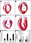
(A) Western blot of Collagen XIV expression in collagen extracts from post natal wild type (Col14a1+/+), and Col14a1−/− hearts. Note loss of protein in Col14a1−/− mice. Actin is used as a loading control. (B, C) Pentachrome staining to show cardiac morphology of 3 month old wild type (B) and Col14a1−/− (C) mouse hearts. Note the spherical appearance of the left ventricle of Col14a1−/− hearts. LV, left ventricle.
Figure 3
Figure 3. Echocardiography reveals differences in cardiac structure and function in adult Col14a1−/− male mice
M-mode Doppler analysis to show left ventricular end-diastolic volume (LV vol;d) (A), LV end-systolic volume during systole (LV vol;s) (B), LV posterior wall thickness (LVPW) during diastole (d) (C), LVPW during systole (s) (D), and ejection fraction (EF) in Col14a1−/− and Col14a1+/+ male mice at 3, 6, and 12 months of age. *, indicates statistical significance, p<0.05. (F) Stretch-force relationship assay to determine maximal stretch of skinned fibers from 3 month old Col14a1−/− and wild type mice.
Figure 4
Figure 4. Collagen homeostasis is disrupted in the myocardium of Col14a1−/− mice
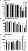
Taqman Low Density Array was used to examine changes in mRNA levels of fibril-forming (A), network and beaded-forming (B) and FACIT-forming (C) collagens in cDNA generated from E11.5, PND1 and 3 months Col14a1−/− and Col14a1+/+ (wild type) ventricles. *, indicate p<0.05 in samples from Col14a1−/− mice compared to wild types.
Figure 5
Figure 5. Collagen fiber organization and remodeling gene expression is altered in the myocardium of Col14a1−/− mice
(A) Western blot (left) and immunofluorescence (right) to show decreased Col1a1 expression in the ventricular myocardium of 3 month old Col14a1−/−, Col14a1+/− and wild type mice. (B) qPCR analysis to show fold changes in collagen processing enzymes in the ventricular myocardium of post natal Col14a1−/− compared to wild type controls. (C) TLDA analysis to show fold changes in expression of matrix remodeling genes in the ventricular myocardium of Col14a1−/− mice at E11.5, PN and 3 months of age. (D–F) Electron microscopy to examine collagen fiber organization in the ventricular myocardium of wild type (D) and Col14a1−/− mice (E–F) at PND1. Note lack of collagen fiber bundles in Col14a1−/− mice (E–F), compared to wild types (arrow, D).
Figure 6
Figure 6. Cardiomyocyte proliferation is prolonged in the myocardium of Col14a1−/− mice at post natal stages
(A–C) Immunostaining of phospho-Histone H3 (pHH3) and MF20 to examine proliferation of cardiomyocytes respectively in PND1 Col14a1−/− mice (B) and wild types (A). Quantitation of the number of cells double stained with MF20 and pHH3 at E11.5 and PND1. (D, E) Immunoreactivity of N-cadherin to detect intercalated discs of cardiomyocytes in tissue sections of ventricles from 3 month old Col14a1−/− (E) and wild type (D) mice. Wheat Germ Agglutinin (WGA) highlights the cell membrane, and DAPI indicates nuclei. (F) Quantitation of the average number of cells per microscopic field. (G, H) MF20 immunostaining of cultured ventricular cardiomyocytes isolated from 3 month old Col14a1−/− (H) and wild type (G) mice. (I) Quantitation to show fold change in cardiomyocyte cell area. *p<0.05, in Col14a1−/− mice compared to wild types.
Figure 7
Figure 7. Fibroblast cell number is decreased in the ventricular myocardium of Col14a1−/− mice at post natal stages
(A, B) Thy1 immunostaining to detect cardiac fibroblasts (arrows) within the ventricular myocardium of Col14a1−/− (B) and wild type (Col14a1−/−) mice (A) at PND1. (C) Percentage of Thy1 positive cells over the total number of nuclei. (D, E) Double immunostaining of Cleaved Caspase-3 and Thy1 within the ventricular myocardium of Col14a1−/− (E) and wild type (Col14a1−/−) mice (D) at the same time point. Arrowhead in D indicates apoptosis in a non-fibroblast cell, while arrows in E indicate double stained fibroblasts undergoing apoptosis. (F) Percentage of double stained (Cleaved Caspase-3 and Thy1) cells in Col14a1−/− and wild type mice over total number of Thy1-positive cells. (G, H) Double staining of phospho-Histone H3 (pHH3) and Thy1 within the ventricular myocardium of Col14a1−/− (H) and wild type (H) mice. (I) Percentage of double stained (pHH3 and Thy1) cells in Col14a1−/− and wild type mice over total number of Thy1-positive cells. *p<0.05, statistical significance in Col14a1−/−mice compared to wild types.[13]
Figure 8
Figure 8. Left ventricular wall thickness is further increased in Col14a1−/− mice following pressure overload
(A–H) Images from echocardiography B-mode short-axis view of the ventricular chambers of wild type (A–D) and Col14a1−/− (E–H) mice, prior to surgery (pre-surgery) (A, C, E, G) and two weeks post-surgery (B, D, F, H). Red lines indicate left ventricular wall thickness. White * indicate papillary muscles in the shortaxis view. (I) Quantitation of trichrome staining and (J) qPCR of fold changes in Col1a1 and Col3a1 gene expression in the ventricles of wild type and Col14a1−/− mice following surgeries. *, indicates significant differences in TAC-operated animals versus Sham mice of the same genotype. #, shows p<0.05 in differences between Sham and TAC in Col14a1−/− mice compared to differences observed between wild types.

References

    1. Bowers SL, Banerjee I, Baudino TA. The extracellular matrix: at the center of it all. J Mol Cell Cardiol. 2010;48:474–482. - PMC - PubMed
    1. Eghbali M, Weber KT. Collagen and the myocardium: fibrillar structure, biosynthesis and degradation in relation to hypertrophy and its regression. Molecular and cellular biochemistry. 1990;96:1–14. - PubMed
    1. Lockhart M, Wirrig E, Phelps A, Wessels A. Extracellular matrix and heart development. Birth Defects Res A Clin Mol Teratol. 2011;91:535–550. - PMC - PubMed
    1. Carver W, Terracio L, Borg TK. Expression and accumulation of interstitial collagen in the neonatal rat heart. Anat Rec. 1993;236:511–520. - PubMed
    1. Jourdan-Lesaux C, Zhang J, Lindsey ML. Extracellular matrix roles during cardiac repair. Life Sci. 2010;87:391–400. - PMC - PubMed

Publication types

MeSH terms