. 2012 Oct;92(10):1461-71.
doi: 10.1038/labinvest.2012.104. Epub 2012 Aug 20.

Liver inflammation in a mouse model of Th1 hepatitis despite the absence of invariant NKT cells or the Th1 chemokine receptors CXCR3 and CCR5

Affiliations

Liver inflammation in a mouse model of Th1 hepatitis despite the absence of invariant NKT cells or the Th1 chemokine receptors CXCR3 and CCR5

James G Cripps et al. Lab Invest. 2012 Oct.

Abstract

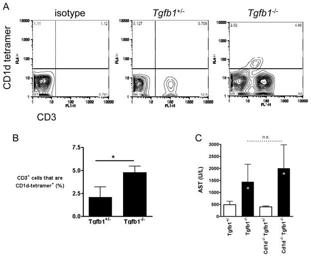
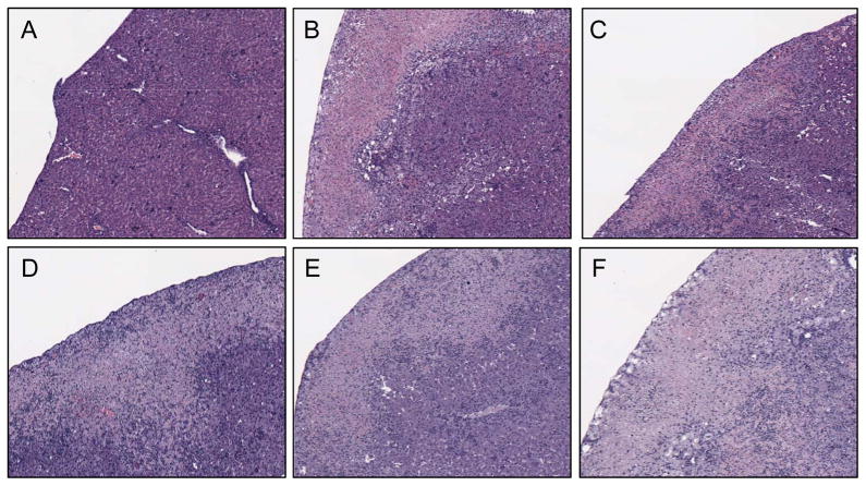
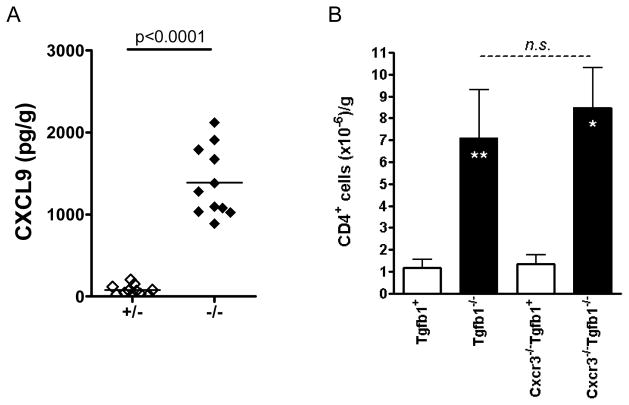
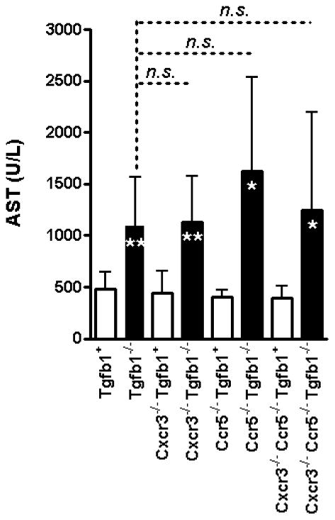
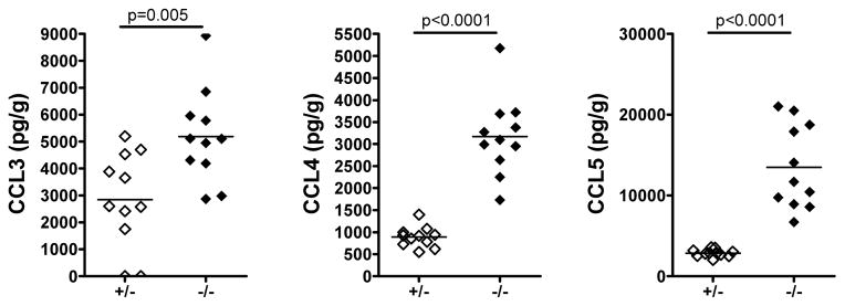
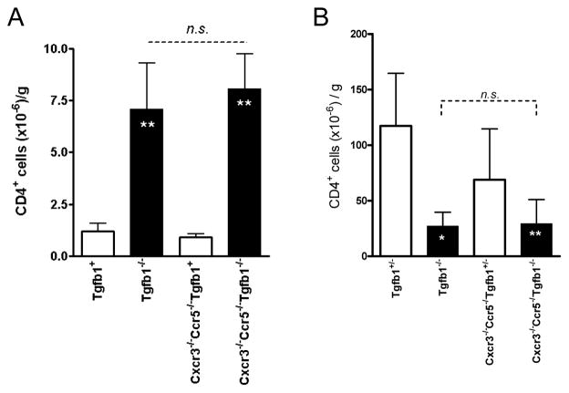
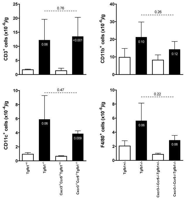
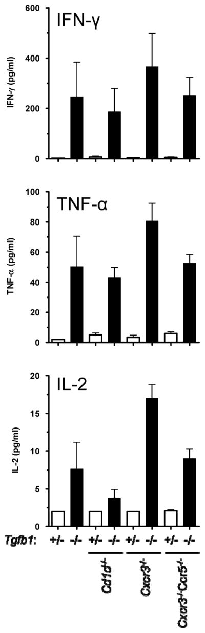
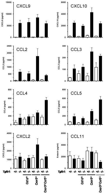
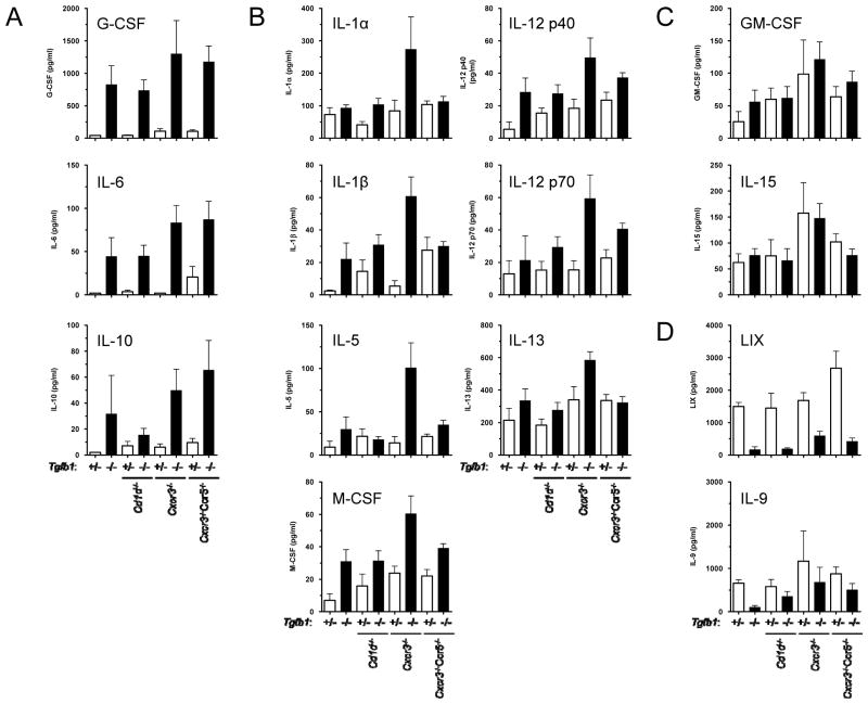
The specific mechanisms that mediate CD4(+) T-cell-mediated liver injury have not been fully elucidated. CD4(+) invariant natural killer T (iNKT) cells are required for liver damage in some mouse models of hepatitis, while the chemokine receptors CXCR3 and CCR5 are considered dominant Th1 chemokine receptors involved in Th1 trafficking in inflammatory conditions. BALB/c-Tgfb1(-/-) mice spontaneously develop Th1 hepatitis. Here, we directly test the hypotheses that iNKT cells or the Th1-cell chemokine receptors CXCR3 and CCR5 are required for development of liver disease in Tgfb1(-/-) mice. Tgfb1(-/-) mouse livers exhibited significant increases in iNKT cells and in ligands for CXCR3 or CCR5. Tgfb1(-/-) mice were rendered deficient in iNKT cells, CXCR3, CCR5, or both CXCR3 and CCR5, by cross-breeding with appropriate knockout mice. Tgfb1(-/-) mice developed severe liver injury, even in the absence of functional CD1d/iNKT cells, CXCR3, CCR5, or both CXCR3 and CCR5. Liver CD4(+) T cells accumulated to high numbers, and spleen CD4(+) T-cell numbers declined, regardless of the functionality of the CXCR3/CCR5 response pathways. Similarly, dendritic cells and macrophages accumulated in Tgfb1(-/-) livers even when CXCR3 and CCR5 were knocked out. Th1-associated cytokines (IFN-γ, TNF-α, IL-2) and chemokines (CXCL9, CXCL10) were strongly overexpressed in Tgfb1(-/-) mice despite knockouts in CD1d, CXCR3, or CCR5. These studies indicate that the cellular and biochemical basis for CD4(+) T-cell-mediated injury in liver can be complex, with myriad pathways potentially involved.


Figures

Figure 1
Figure 1. iNKT cell numbers are increased in liver in Tgfb1−/− mice but do not contribute to tissue damage
(A) Liver MNC from 11 day old mice were stained with anti-CD3 and CD1d-αgalcer tetramer and analyzed by flow cytometry. Representative flow plots are displayed. (B) Combined data showing the percentage of CD3+ cells that stain positive for the CD1d-α-galcer tetramer. n = 3 or more mice per genotype. *, p<0.05, Student’s t-test. Error bars indicate SD. (C) AST was measured from 11 day old mice of the indicated genotypes. White asterisks reflect statistical differences (* p<0.02) between Tgfb1−/− mice and their littermate control Tgfb1+/+ mice and Tgfb1+/− mice (aggregated as “Tgfb1+”). n = 6 mice per genotype, except for the Cd1d−/−Tgfb1+ genotype, for which n = 4. n.s indicates not significant. Mann-Whitney test. Error bars indicate SD.
Figure 2
Figure 2. Histologic liver damage in Tgfb1−/− mice is independent of CD1d/iNKT cells, CXCR3, and CCR5
Low power micrographs of H&E stained livers from mice of the following genotypes: (A) Tgfb1+; (B) Tgfb1−/−; (C) Cd1d−/−Tgfb1−/−; (D) Cxcr3−/−Tgfb1−/−; (E) Ccr5−/−Tgfb1−/−; (F) Cxcr3−/−Ccr5−/−Tgfb1−/−. Obvious necrosis of the liver is observed in Tgfb1−/− mice, independent of the presence or absence of CD1d/iNKT cells, CXCR3, CCR5, or CXCR3/CCR5.
Figure 3
Figure 3. CXCL9 is over-expressed in Tgfb1−/− mouse livers, but CD4+ T cell accumulation is independent of CXCR3
(A) The CXCR3 ligand CXCL9 was measured in liver lysates isolated from 11 day old Tgfb1+/− mice and Tgfb1−/− mice and expressed as pg/g wet weight (n=11 for each genotype). Chemokines were measured by Luminex bead assay. Horizontal lines indicate means. The p value from a statistical comparison of data (Student’s t test) is shown at the top of the graph. (B) Livers were dissected from 11 day old mice and weighed. Liver MNC were isolated, counted by hemacytometer, and stained for CD4+ T cells. CD4+ density (cells/g) was calculated. * p<0.01; ** p<0.001. n.s.: no significant difference. Mann-Whitney test. n = 4–6 mice per genotype. Error bars indicate SD.
Figure 4
Figure 4. AST elevation in Tgfb1−/− mice is independent of CXCR3 and CCR5
AST was measured from 11 day old mice of the indicated genotypes. White asterisks reflect statistical differences (*, p<0.01; **, p<0.001) between Tgfb1−/− mice and their littermate control Tgfb1+/+ mice and Tgfb1+/− mice (aggregated as “Tgfb1+”). n = 6 or more mice per genotype. For the comparisons indicated by the dashed lines, differences were not significant (n.s.). Mann-Whitney test. Error bars indicate SD.
Figure 5
Figure 5. Chemokine ligands for CCR5 are over-expressed in Tgfb1−/− mouse livers
Chemokine ligands that activate CCR5 were measured in liver lysates isolated from 11 day old Tgfb1+/− mice and Tgfb1−/− mice and expressed as pg/g wet weight. Each symbol represents one mouse of the indicated genotype. Chemokines were measured by Luminex bead assay. Horizontal lines indicate means. The p values from statistical comparisons of data (Student’s t test) are shown at the top of the graphs.
Figure 6
Figure 6. CD4+ T cell accumulation in Tgfb1−/− mouse liver and reduction in Tgfb1−/− mouse spleen are independent of both CXCR3 and CCR5
(A) Liver and (B) splenic MNC from 11 day old mice of the indicated genotypes were isolated, counted by hemacytometer, and stained for CD4+ T cells. CD4+ densities (cells/g) were calculated. * p<0.01; ** p<0.001; n.s.: no significant difference. Mann-Whitney test. n = 4–7 mice per genotype. Error bars indicate SD.
Figure 7
Figure 7. Accumulation of T cells, dendritic cells, and macrophages in Tgfb1−/− mouse liver is independent of both CXCR3 and CCR5
Liver MNC prepared as in the legend to figure 6 were stained for the markers CD3, CD11c, CD11b, and F4/80. p values for statistical comparisons are shown. Increases in some cell populations approached (p<0.10), but did not reach (p<0.05) statistical significance. Error bars indicate SD.
Figure 8
Figure 8. Overexpression of type 1 cytokines is independent of CD1d/iNKT cells, CXCR3, and CCR5
Plasma samples were isolated at day 11 for mice of the indicated genotypes. Cytokines were measured by Luminex bead assay and results shown as pg/ml. n = 3–10 mice per genotype, except for Tgfb1+ mice, for which n = 2. Error bars indicate SEM.
Figure 9
Figure 9. Overexpression of IFN-γ-induced chemokines is independent of CD1d/iNKT cells, CXCR3, and CCR5
Chemokines were analyzed in day 11 samples by Luminex, and results are displayed as in the legend to figure 8.
Figure 10
Figure 10. Expression levels of specific cytokines are differentially influenced by TGF-β1, CD1d/iNKT cells, CXCR3, and CCR5
Cytokines were analyzed in day 11 samples by Luminex, and results are displayed as in the legend to figure 8. (A) Some cytokines (G-CSF, IL-6, IL-10) are strongly over-expressed in plasma from Tgfb1−/− mice; expression levels are largely independent of functional Cd1d, Cxcr3, or Ccr5 genes. (B) These cytokines show highest expression when both Tgfb1 and Cxcr3 are knocked out, but levels revert when Ccr5 is additionally knocked out. (C) Expression levels of some cytokines (GM-CSF, IL-15) are unaffected in Tgfb1−/− mice. (D) Expression levels of some cytokines (LIX, IL-9) are reduced in Tgfb1−/− mice.

Similar articles

Cited by

References

    1. Senaldi G, et al. Immunohistochemical features of the portal tract mononuclear cell infiltrate in chronic aggressive hepatitis. Arch Dis Child. 1992;67(12):1447–53. - PMC - PubMed
    1. Rosen HR. Hepatitis C pathogenesis: mechanisms of viral clearance and liver injury. Liver Transpl. 2003;9(11):S35–S43. - PubMed
    1. Grakoui A, et al. HCV persistence and immune evasion in the absence of memory T cell help. Science. 2003;302(5645):659–62. - PubMed
    1. Schlaak JF, et al. Analysis of the in vitro cytokine production by liver-infiltrating T cells of patients with autoimmune hepatitis. Clin Exp Immunol. 1993;94(1):168–173. - PMC - PubMed
    1. Rymarchyk SL, et al. Widespread natural variation in murine natural killer T-cell number and function. Immunology. 2008;125(3):331–43. - PMC - PubMed

Publication types

MeSH terms