. 2012 Oct-Dec;3(4):219-24.
doi: 10.4161/sgtp.20755. Epub 2012 Aug 21.

R(h)oads to microvesicles

Affiliations

R(h)oads to microvesicles

Marc A Antonyak et al. Small GTPases. 2012 Oct-Dec.

Abstract

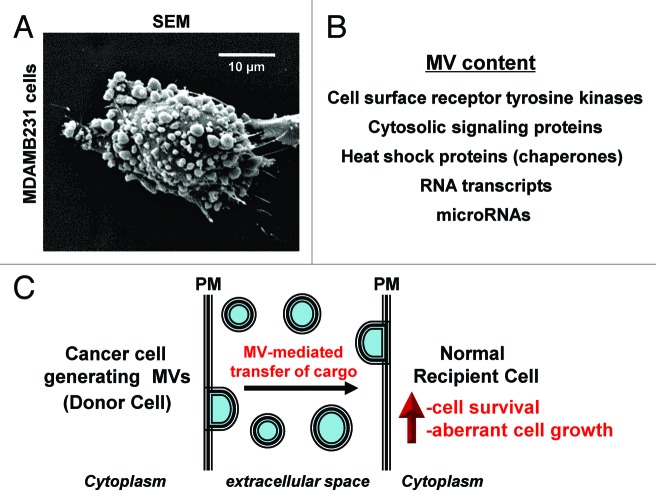
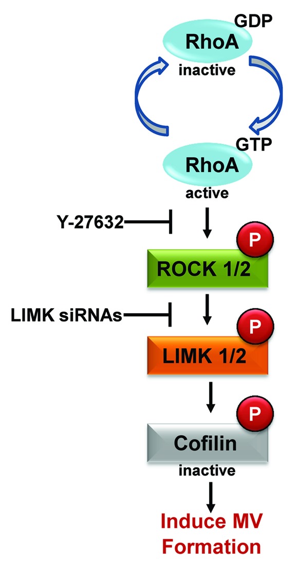
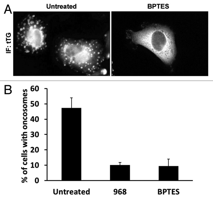
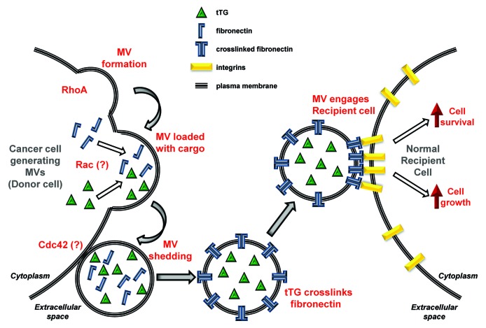
A novel form of cell-to-cell communication involving the formation and shedding of large vesicular structures, called microvesicles (MVs), from the surfaces of highly aggressive forms of human cancer cells has been attracting increasing amounts of attention. This is in large part due to the fact that MVs contain a variety of cargo that is not typically thought to be released from cells including cell-surface receptor tyrosine kinases, cytosolic and nuclear signaling proteins and RNA transcripts. MVs, by sharing their contents with other cells, can greatly impact cancer progression by increasing primary tumor growth, as well as by promoting the development of the pre-metastatic niche. We have recently shown that the small GTPase RhoA is critical for MV biogenesis in human cancer cells. Moreover, we have now obtained evidence that implicates the highly related small GTPases, Rac and Cdc42, in regulating the loading of specific cargo into MVs, as well as in the shedding of MVs from cancer cells. Thus, linking the Rho family of small GTPases to MV biogenesis has begun to shed some light on a new and unexpected way that these signaling proteins contribute to human cancer progression.


Figures

None
Figure 1. Highly aggressive forms of human cancer cells generate and shed MVs. (A) A scanning electron microscopy (SEM) image of a human MDAMB231 breast cancer cell covered with MVs. (B) A list highlighting some of the contents that have been identified in MVs. (C) Schematic showing that MVs generated and shed from a cancer cell (Donor Cell) can be transferred to a recipient normal cell, conferring upon the recipient cell the characteristics of a transformed cell (i.e., providing the cell with growth and survival advantages). PM stands for plasma membrane.
None
Figure 2. RhoA-dependent signaling induces MV biogenesis. Schematic showing the RhoA signaling pathway that is responsible for promoting MV formation in human cancer cells. The pathway culminates with the phosphorylation of cofilin, which inhibits its actin-severing activity. This results in increased actin polymerization and the generation of MVs. The ROCK inhibitor, Y-27632, and LIMK siRNAs (for knocking-down LIMK expression) are particularly useful reagents for blocking RhoA-induced MV formation in cancer cells.
None
Figure 3. The altered metabolism exhibited by human cancer cells is important for MV biogenesis. MDAMB231 breast cancer cells were left untreated or were treated with the glutaminase C inhibitors, 968 and BPTES, for 2 d prior to being fixed. The cells were subjected to immunofluorescence using a tTG antibody (IF: tTG) to label the MVs on the surfaces of the cells. (A) Representative images of MDAMB231 cells treated without (Untreated) or with BPTES. Note the lack of detectable MVs on the BPTES-treated cell. (B) Quantification of MV production by MDAMB231 cells treated without (Untreated) or with 968 or BPTES. The experiments were done three times and the results from each experiment were averaged together and graphed. The error bars indicate standard deviation.
None
Figure 4. Schematic depicting the lifecycle of a MV. Activation of RhoA in a cancer (Donor) cell initiates MV formation by inducing a “budding” event at a particular site along the plasma membrane. Our preliminary studies suggest that the loading of specific cargo into the MV involves Rac, whereas the release or shedding of the MV from the cancer cell is directed by Cdc42. The shed MV contains transforming cargo, including tTG and the extracellular matrix protein, fibronectin. tTG crosslinks fibronectin to itself within the shed MV, generating a fibronectin dimer that exhibits enhanced signaling capabilities when it engages integrins that are expressed on the surface of a normal recipient cell. The signaling induced by the crosslinked fibronectin-integrin complex aberrantly regulates signaling pathways that promote cell growth and survival, causing the recipient cell to acquire a transformed phenotype.

Comment on

  • Li B, Antonyak MA, Zhang J, Cerione RA. RhoA triggers a specific signaling pathway that generates transforming microvesicles in cancer cells. Oncogene. 2012 doi: 10.1038/onc.2011.636.

Similar articles

Cited by

References

    1. Al-Nedawi K, Meehan B, Micallef J, Lhotak V, May L, Guha A, et al. Intercellular transfer of the oncogenic receptor EGFRvIII by microvesicles derived from tumour cells. Nat Cell Biol. 2008;10:619–24. doi: 10.1038/ncb1725. - DOI - PubMed
    1. Antonyak MA, Li B, Boroughs LK, Johnson JL, Druso JE, Bryant KL, et al. Cancer cell-derived microvesicles induce transformation by transferring tissue transglutaminase and fibronectin to recipient cells. Proc Natl Acad Sci U S A. 2011;108:4852–7. doi: 10.1073/pnas.1017667108. - DOI - PMC - PubMed
    1. Skog J, Würdinger T, van Rijn S, Meijer DH, Gainche L, Sena-Esteves M, et al. Glioblastoma microvesicles transport RNA and proteins that promote tumour growth and provide diagnostic biomarkers. Nat Cell Biol. 2008;10:1470–6. doi: 10.1038/ncb1800. - DOI - PMC - PubMed
    1. Peinado H, Lavotshkin S, Lyden D. The secreted factors responsible for pre-metastatic niche formation: old sayings and new thoughts. Semin Cancer Biol. 2011;21:139–46. doi: 10.1016/j.semcancer.2011.01.002. - DOI - PubMed
    1. Schauer IG, Sood AK, Mok S, Liu J. Cancer-associated fibroblasts and their putative role in potentiating the initiation and development of epithelial ovarian cancer. Neoplasia. 2011;13:393–405. - PMC - PubMed

Publication types

Substances

LinkOut - more resources