

A human fallopian tube model for investigation of C. trachomatis infections
- PMID: 22907315
- PMCID: PMC3486760
- DOI: 10.3791/4036
A human fallopian tube model for investigation of C. trachomatis infections
Abstract
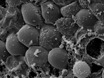
Genital tract infections with Chlamydia trachomatis (C. trachomatis) are the most frequent transmitted sexually disease in women worldwide. Inefficient clearance or persistence of the pathogens may lead to ascending infections of the upper genital tract and are supposed to cause chronic inflammatory damage to infected tissues (1,2). As a consequence, severe clinical sequelae like pelvic inflammatory disease (PID), tubal occlusion and infertility may occur (3,4). Most of the research with C. trachomatis has been conducted in epithelial cell lines (e.g. HEp-2 cells and HeLa-229) or in mice. However, as with cell- culture based models, they do neither reflect the physiology of native tissue nor the pathophysiology of C. trachomatis genital tract infections in vivo (5). Further limitations are given by the fact that central signaling cascades (e.g. IFN-γ mediated JAK/STAT signaling pathway) that control intracellular chlamydial growth fundamentally differ between mice and humans (6,7). We and others therefore established a whole organ fallopian tube model to investigate direct interactions between C. trachomatis and human fallopian tube cells ex vivo (8,9). For this purpose, human fallopian tubes from women undergoing hysterectomy were collected and infected with C. trachomatis serovar D. Within 24 h post infection, specimen where analyzed using scanning electron microscopy (SEM) and transmission electron microscopy (TEM) to detect Chlamydia trachomatis mediated epithelial damage as well as C. trachomatis inclusion formation in the fallopian tissue.
Similar articles
-
Pathogenesis of fallopian tube damage caused by Chlamydia trachomatis infections.Contraception. 2015 Aug;92(2):108-15. doi: 10.1016/j.contraception.2015.01.004. Epub 2015 Jan 13. Contraception. 2015. PMID: 25592078 Review.
-
Interleukin-1 is the initiator of Fallopian tube destruction during Chlamydia trachomatis infection.Cell Microbiol. 2007 Dec;9(12):2795-803. doi: 10.1111/j.1462-5822.2007.00996.x. Epub 2007 Jul 5. Cell Microbiol. 2007. PMID: 17614966
-
Recovery of Chlamydia trachomatis from endometrial and fallopian tube biopsies in women with infertility of tubal origin.Fertil Steril. 1989 Aug;52(2):232-8. doi: 10.1016/s0015-0282(16)60847-6. Fertil Steril. 1989. PMID: 2753172
-
Fallopian tubal infertility: the result of Chlamydia trachomatis-induced fallopian tubal fibrosis.Mol Cell Biochem. 2022 Jan;477(1):205-212. doi: 10.1007/s11010-021-04270-7. Epub 2021 Oct 15. Mol Cell Biochem. 2022. PMID: 34652537 Review.
-
Chlamydia trachomatis infection of human fallopian tube organ cultures.J Gen Microbiol. 1990 Jun;136(6):1109-15. doi: 10.1099/00221287-136-6-1109. J Gen Microbiol. 1990. PMID: 2384745
Cited by
-
Elaborations on Corallopyronin A as a Novel Treatment Strategy Against Genital Chlamydial Infections.Front Microbiol. 2019 May 7;10:943. doi: 10.3389/fmicb.2019.00943. eCollection 2019. Front Microbiol. 2019. PMID: 31134007 Free PMC article.
-
The prevalence and outcome of asymptomatic chlamydial infection screening among infertile women attending gynecological clinic in ibadan, South west Nigeria.Ann Med Health Sci Res. 2014 Mar;4(2):253-7. doi: 10.4103/2141-9248.129057. Ann Med Health Sci Res. 2014. PMID: 24761248 Free PMC article.
-
EphrinA2 receptor (EphA2) is an invasion and intracellular signaling receptor for Chlamydia trachomatis.PLoS Pathog. 2015 Apr 23;11(4):e1004846. doi: 10.1371/journal.ppat.1004846. eCollection 2015 Apr. PLoS Pathog. 2015. PMID: 25906164 Free PMC article.
-
Murine Endometrial Organoids to Model Chlamydia Infection.Front Cell Infect Microbiol. 2020 Aug 14;10:416. doi: 10.3389/fcimb.2020.00416. eCollection 2020. Front Cell Infect Microbiol. 2020. PMID: 32923409 Free PMC article.
-
Sorangicin A Is Active against Chlamydia in Cell Culture, Explanted Fallopian Tubes, and Topical In Vivo Treatment.Antibiotics (Basel). 2023 Apr 22;12(5):795. doi: 10.3390/antibiotics12050795. Antibiotics (Basel). 2023. PMID: 37237698 Free PMC article.
References
-
- Dean D, Suchland RJ, Stamm WE. Evidence for long-term cervical persistence of Chlamydia trachomatis by omp1 genotyping. J. Infect. Dis. 2000;182:909–916. - PubMed
-
- Campbell LA, Patton DL, Moore DE, Cappuccio AL, Mueller BA, Wang SP. Detection of Chlamydia trachomatis deoxyribonucleic acid in women with tubal infertility. Fertil. Steril. 1993;59:45–50. - PubMed
-
- Peipert JF. Clinical practice. Genital chlamydial infections. N. Engl. J. Med. 2003;349:2424–2430. - PubMed
-
- Mardh PA. Tubal factor infertility, with special regard to chlamydial salpingitis. Curr. Opin. Infect. Dis. 2004;17:49–52. - PubMed
Publication types
MeSH terms
LinkOut - more resources
Full Text Sources
Medical
Research Materials
