

Anterior delayed gadolinium-enhanced MRI of cartilage values predict joint failure after periacetabular osteotomy
- PMID: 22907475
- PMCID: PMC3492640
- DOI: 10.1007/s11999-012-2519-9
Anterior delayed gadolinium-enhanced MRI of cartilage values predict joint failure after periacetabular osteotomy
Abstract
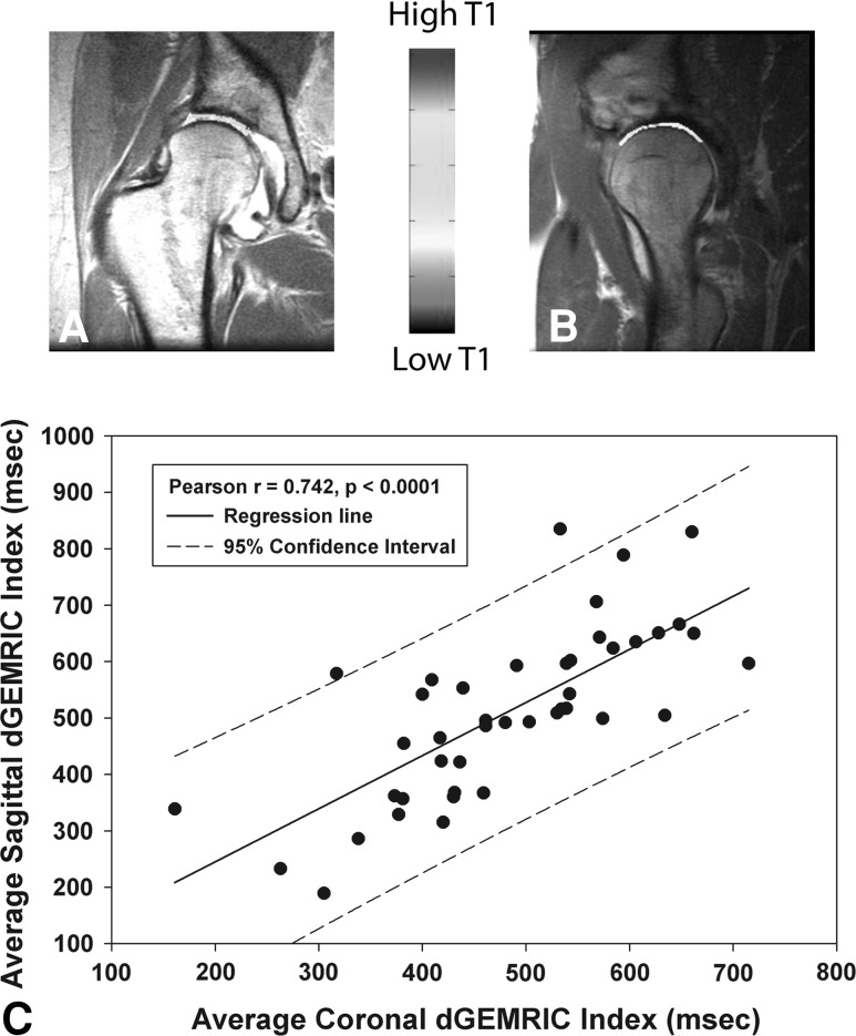
Background: Several available compositional MRIs seem to detect early osteoarthritis before radiographic appearance. Delayed gadolinium-enhanced MRI of cartilage (dGEMRIC) has been most frequently used in clinical studies and reportedly predicts premature joint failure in patients undergoing Bernese periacetabular osteotomies (PAOs).
Questions/purposes: We asked, given regional variations in biochemical composition in dysplastic hips, whether the dGEMRIC index of the anterior joint would better predict premature joint failure after PAOs than the coronal dGEMRIC index as previously reported.
Methods: We retrospectively reviewed 43 hips in 41 patients who underwent Bernese PAO for hip dysplasia. Thirty-seven hips had preserved joints after PAOs and six were deemed premature failures based on pain, joint space narrowing, or subsequent THA. We used dGEMRIC to determine regional variations in biochemical composition. Preoperative demographic and clinical outcome score, radiographic measures of osteoarthritis and severity of dysplasia, and dGEMRIC indexes from different hip regions were analyzed in a multivariable regression analysis to determine the best predictor of premature joint failure. Minimum followup was 24 months (mean, 32 months; range, 24-46 months).
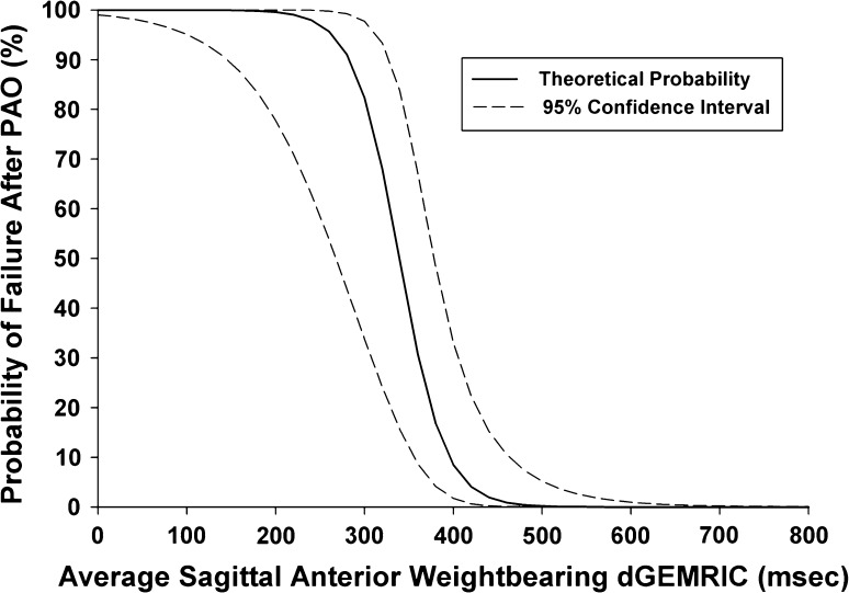
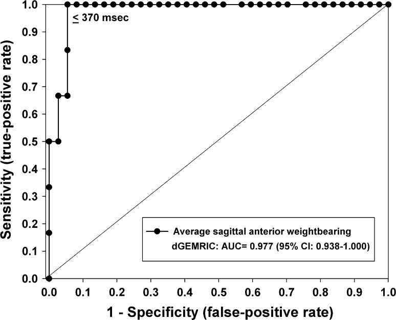
Results: The two cohorts were similar in age and sex distribution. Severity of dysplasia was similar as measured by lateral center-edge, anterior center-edge, and Tönnis angles. Preoperative pain, joint space width, Tönnis grade, and coronal and sagittal dGEMRIC indexes differed between groups. The dGEMRIC index in the anterior weightbearing region of the hip was lower in the prematurely failed group and was the best predictor.
Conclusions: Success of PAO depends on the amount of preoperative osteoarthritis. These degenerative changes are seen most commonly in the anterior joint. The dGEMRIC index of the anterior joint may better predict premature joint failure than radiographic measures of hip osteoarthritis and coronal dGEMRIC index.
Level of evidence: Level II, prognostic study. See Instructions for Authors for a complete description of levels of evidence.
Figures
Similar articles
-
One-third of Hips After Periacetabular Osteotomy Survive 30 Years With Good Clinical Results, No Progression of Arthritis, or Conversion to THA.Clin Orthop Relat Res. 2017 Apr;475(4):1154-1168. doi: 10.1007/s11999-016-5169-5. Clin Orthop Relat Res. 2017. PMID: 27905061 Free PMC article.
-
Does periacetabular osteotomy have depth-related effects on the articular cartilage of the hip?Clin Orthop Relat Res. 2015 Dec;473(12):3735-43. doi: 10.1007/s11999-015-4545-x. Clin Orthop Relat Res. 2015. PMID: 26329795 Free PMC article.
-
Delayed gadolinium-enhanced magnetic resonance imaging of cartilage to predict early failure of Bernese periacetabular osteotomy for hip dysplasia.J Bone Joint Surg Am. 2006 Jul;88(7):1540-8. doi: 10.2106/JBJS.E.00572. J Bone Joint Surg Am. 2006. PMID: 16818980
-
Patient selection criteria for periacetabular osteotomy or rotational acetabular osteotomy.Clin Orthop Relat Res. 2012 Dec;470(12):3342-54. doi: 10.1007/s11999-012-2516-z. Clin Orthop Relat Res. 2012. PMID: 22895690 Free PMC article.
-
Assessment of adult hip dysplasia and the outcome of surgical treatment.Dan Med J. 2012 Jun;59(6):B4450. Dan Med J. 2012. PMID: 22677250 Review.
Cited by
-
Periacetabular osteotomy and arthroscopic labral repair after failed hip arthroscopy due to iatrogenic aggravation of hip dysplasia.Knee Surg Sports Traumatol Arthrosc. 2014 Apr;22(4):911-4. doi: 10.1007/s00167-013-2540-x. Epub 2013 Jun 13. Knee Surg Sports Traumatol Arthrosc. 2014. PMID: 23760037
-
Biochemical MRI With dGEMRIC Corresponds to 3D-CT Based Impingement Location for Detection of Acetabular Cartilage Damage in FAI Patients.Orthop J Sports Med. 2021 Mar 19;9(3):2325967120988175. doi: 10.1177/2325967120988175. eCollection 2021 Mar. Orthop J Sports Med. 2021. PMID: 33816640 Free PMC article.
-
Arthroscopic Hip Joint Assessment can Impact the Indications for PAO Surgery.Iowa Orthop J. 2019;39(1):149-157. Iowa Orthop J. 2019. PMID: 31413688 Free PMC article.
-
Automated quantification of cartilage quality for hip treatment decision support.Int J Comput Assist Radiol Surg. 2022 Nov;17(11):2011-2021. doi: 10.1007/s11548-022-02714-z. Epub 2022 Aug 17. Int J Comput Assist Radiol Surg. 2022. PMID: 35976596 Free PMC article.
-
How Does the dGEMRIC Index Change After Surgical Treatment for FAI? A Prospective Controlled Study: Preliminary Results.Clin Orthop Relat Res. 2017 Apr;475(4):1080-1099. doi: 10.1007/s11999-016-5098-3. Clin Orthop Relat Res. 2017. PMID: 27709422 Free PMC article.
References
-
- Aronson J. Osteoarthritis of the young adult hip: etiology and treatment. Instr Course Lect. 1986;35:119–128. - PubMed
MeSH terms
Substances
LinkOut - more resources
Full Text Sources
Medical
Research Materials
