. 2012 Nov;32(21):4306-22.
doi: 10.1128/MCB.00382-12. Epub 2012 Aug 20.

The prolyl isomerase Pin1 targets stem-loop binding protein (SLBP) to dissociate the SLBP-histone mRNA complex linking histone mRNA decay with SLBP ubiquitination

Affiliations

The prolyl isomerase Pin1 targets stem-loop binding protein (SLBP) to dissociate the SLBP-histone mRNA complex linking histone mRNA decay with SLBP ubiquitination

Nithya Krishnan et al. Mol Cell Biol. 2012 Nov.

Abstract

Histone mRNAs are rapidly degraded at the end of S phase, and a 26-nucleotide stem-loop in the 3' untranslated region is a key determinant of histone mRNA stability. This sequence is the binding site for stem-loop binding protein (SLBP), which helps to recruit components of the RNA degradation machinery to the histone mRNA 3' end. SLBP is the only protein whose expression is cell cycle regulated during S phase and whose degradation is temporally correlated with histone mRNA degradation. Here we report that chemical inhibition of the prolyl isomerase Pin1 or downregulation of Pin1 by small interfering RNA (siRNA) increases the mRNA stability of all five core histone mRNAs and the stability of SLBP. Pin1 regulates SLBP polyubiquitination via the Ser20/Ser23 phosphodegron in the N terminus. siRNA knockdown of Pin1 results in accumulation of SLBP in the nucleus. We show that Pin1 can act along with protein phosphatase 2A (PP2A) in vitro to dephosphorylate a phosphothreonine in a conserved TPNK sequence in the SLBP RNA binding domain, thereby dissociating SLBP from the histone mRNA hairpin. Our data suggest that Pin1 and PP2A act to coordinate the degradation of SLBP by the ubiquitin proteasome system and the exosome-mediated degradation of the histone mRNA by regulating complex dissociation.


Figures

Fig 1
Fig 1
Phosphorylated SLBP forms a specific complex with the prolyl isomerase Pin1 in vitro. (A) Chemical shift mapping of uniformly 15N-labeled Pin1 was performed with the addition of increasing concentrations of unlabeled phosphorylated full-length SLBP. 15N-labeled Pin1 (0.25 mM) was titrated with increasing molar ratios of phosphorylated SLBP (phospho-SLBP) as indicated. The free (1H, 15N) HSQC Pin1 spectrum is shown in blue, and the (1H, 15N) HSQC Pin1 spectrum with SLBP is shown in red. All spectra were apodized identically and are plotted at the same contour level. At a substoichiometric ratio of 0.5:1 for phospho-SLBP to Pin1, several Pin1 amides in the WW domain and the catalytic domain that are known to be part of ligand binding sites disappear from the spectrum. These residues have been mapped onto the crystal structure of Pin1 (pdb code 1PIN in panel E). Only resonances from the disordered N terminus remain visible (residue numbers are indicated) in the spectrum in the presence of an equivalent amount of phospho-SLBP. For panel B, a similar titration was performed with baculovirus-expressed hSLBP RPD. The overlay of the free and RPD-bound Pin1 spectra is shown (C). No perturbations were observed in the Pin1 spectrum when dephosphorylated SLBP (baculovirus expressed and treated with calf intestinal phosphatase) was titrated into the spectrum. (D) 31P NMR spectra of phosphorylated human SLBP RPD are shown in the presence and absence of Pin1. Two resonances for a single phosphate on Thr171 are observed in the 1D 31P spectrum due to cis-trans isomerization about the Thr-Pro bond. Addition of Pin1 changes the populations of the two conformers. (E) The chemical shift perturbations from the NMR titration are depicted in red on the crystal structure of Pin1 (PDB code 1PIN).
Fig 2
Fig 2
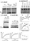
The SLBP-histone mRNA complex dissociates in the presence of Pin1 and PP2A. (A) Dissociation of the dSLBP-RNA complex monitored by EMSA. 32P-labeled histone stem-loop RNA was incubated with the baculovirus-expressed full-length (FL) dSLBP on ice either with or without the various enzymes as indicated. (Left) Addition of Pin1 alone does not dissociate the preformed dSLBP-RNA complex. SLBP was at 40 nmol in all lanes in a total reaction volume of 10 μl, whereas the Pin1 concentrations were 50 nmol (lane 1), 100 nmol (lane 2), 200 nmol (lane 3), and 400 nmol (lane 4). No Pin1 was added to the “0” lane. The 32P-labeled RNA was at 1 pmol in all lanes. (Middle) Addition of increasing concentration of Pin1 and a 10-fold excess of SL28 RNA (relative to the 32P-labeled RNA) results in accumulation of free [32P]ATP at the bottom of the gel. SLBP was at 40 nmol in all lanes in a total reaction volume of 10 μl, whereas the Pin1 concentrations were 50 nmol (lane 1), 100 nmol (lane 2), 200 nmol (lane 3), 400 nmol (lane 4), and 800 nmol (lane 5). No Pin1 was added to the “0” lane. (Right) The 32P-labeled histone stem-loop probe is released more efficiently when the complex is incubated with increasing concentrations of Pin1 and a fixed concentration (0.3 nmol) of PP2A. The concentrations of SLBP, RNA, and Pin1 are the same as those described for the middle panel. (B) The control panel shows that addition of neither cold RNA alone nor phosphatase alone results in dissociation. Dissociation is observed only when Pin1 is present in the presence of excess competitor RNA. (C) Binding of Pin1 to the full-length baculovirus-expressed phosphorylated human SLBP-RNA complex was monitored by EMSA in the presence and absence of the Pin1 inhibitor PiB, but without excess competitor RNA. No effect of Pin1 was apparent on the preformed complex in either reaction. (D) Binding of Pin1 of the baculovirus-expressed phosphorylated human SLBP-RPD RNA complex was monitored by EMSA in the presence and absence of the Pin1 inhibitor PiB and PP2A. Addition of Pin1 and PP2A to the human SLBP RPD-RNA complex (in the absence of competitor RNA) results in the complex running diffuse through the gel, indicating that some dissociation occurs even in the absence of cold RNA. The SLBP RBD was at 10 nmol, the 32P-labeled RNA was at 0.5 pmol, Pin1 was at 20 nmol (lane 1) and 40 nmol (lane 2), and PP2A was at 0.3 nmol. (D) Fluorescence anisotropy for the association of 3′ fluorescein-labeled histone stem-loop RNA with phosphorylated full-length human SLBP, Pin1, PP2A, and a 10-fold excess of unlabeled competitor RNA is shown. Addition of Pin1 to the SL28-SLBP binary complex results in an increase in fluorescence anisotropy. Addition of PP2A results in a small decrease in the anisotropy, and a complete decrease in anisotropy to baseline is observed when unlabeled SL28 RNA is added to the (SL28 plus hSLBP-Pin1 plus PP2A) mixture. (E) Binding isotherm for interaction of human SLBP with 3′ fluorescein-labeled histone stem-loop RNA (1), addition of Pin1 to the SLBP-RNA complex in the absence of competitor RNA (2), and addition of Pin1 and PP2A to the SLBP-RNA complex in the presence of competitor RNA (3) is shown. In panel F, the individual isotherms for these reactions are shown. The apparent dissociation constants derived from the observed change in anisotropy are indicated. The concentration of RNA for the fluorescence experiments was 50 nM, and 0.02 μg of a 0.1 mg/ml solution of PP2A (corresponding to 0.4 enzyme units) was added to the 500-μl reaction mixture. (G) The concentrations of hSLBP and Pin1 are indicated. All fluorescence experiments were performed at 25°C in PBS buffer (pH 7.4), and each data point was measured in triplicate.
Fig 3
Fig 3
Pin1 inhibition increases SLBP levels. (A) HeLa cells were treated with either S2 or C2 siRNA for up to 72 h using a standard two-hit method. The levels of Pin1, SLBP, and actin were probed by Western blotting (WB) as noted. (B) HEK293 cells were treated with either S2 or C2 Pin1 siRNA for 72 h, and the cells were probed for Pin1 and SLBP. (C) Pin1 activity was inhibited by the addition of 20 μM PiB inhibitor for 16 h, whereas control cells were mock treated with DMSO. The protein levels of SLBP, Pin1, and actin were determined by Western blotting. The SLBP mRNA levels were also quantified in the control and PiB-treated cells by RT-PCR. (D) The effect of the Pin1 siRNA on SLBP levels could be rescued by the transfection of exogenous WT Flag-tagged Pin1 for 24 h into HEK293 cells after Pin1 was knocked down by 50 nM siRNA for 48 h. (E) Pin1 knockdown results in increased accumulation of cells in G1 and reduced accumulation in S and G2 phases. Cell cycle profiles of HeLa cells treated with either 25 nM Pin1 siRNA (S2) or 25 nM control RNA (C2) for 72 h were pulse-labeled with propidium iodide; the DNA content was analyzed by flow cytometry, and the cell cycle distribution was determined using the ModFit software. (F) The Pin1 inhibitor PiB affects the stability of SLBP. The levels of SLBP were analyzed at different time points after treatment with cycloheximide in the presence and absence of the Pin1 inhibitor PiB. The protein levels were quantified using ImageQuant software. (G) Pin1 coimmunoprecipitates with HA-SLBP. HeLa cell lysates were prepared from a cell line that stably expressed HA-SLBP. The lysates were treated with Pin1 antibody as described in Materials and Methods. Anti-Myc antibody was used as the control. The immunoprecipitates were run on SDS-PAGE and probed for Pin1 and HA-SLBP. A total of 5% of the input was run in the first lane as a control. No SLBP is observed in the anti-Myc control lane (lane 2).
Fig 4
Fig 4
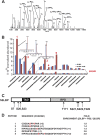
Identification of SLBP phosphorylation sites upregulated in response to PiB inhibitor by mass spectrometry. Several phosphorylation sites in human SLBP were mapped using high-resolution tandem mass spectrometry. The MS-MS spectrum in panel A illustrates a typical high-energy C-trap dissociation (HCD) fragmentation pattern obtained on an LTQ Orbitrap Elite MS instrument for an SLBP phosphorylated peptide (in this case, it is peptide 1 in panel D showing phosphorylation of S20 and S23). The bar graph in panel B compares the normalized abundance (based on a global scaling factor of all observable peptides in the LCMS experimental run) of SLBP phosphorylated peptides obtained from HEK293 cells treated without (blue) or with (red) 20 μM PiB inhibitor. Mass spectra were compared using Nonlinear Dynamics' Progenesis LCMS software (version 3.1.4003.30577) as described in Materials and Methods. The three inset chromatographs show the elution profiles of a phosphopeptide that was upregulated in the presence of PiB (top), a downregulated phosphopeptide (middle), and a phosphopeptide whose abundance was comparable (lower) in SLBP isolated in the presence or absence of PiB. The zoom in the inset illustrates additional phosphorylated SLPB peptides of lower abundance that show significant upregulation (labeled 1, 2, 3, 4, and 5) in the presence of PiB. (Note that the chromatographs corresponding to SLBP + PiB are offset.) (C) The domain organization of human SLBP is shown. TAD, translation activation domain; RPD, RNA binding and processing domain. Ser/Thr phosphorylation sites that show at least a 2-fold increase in abundance in the presence of PiB are shown. In panel D, the phosphorylated peptides identified are shown along with the corresponding fold enrichment in the presence of PiB. The numbering of the peptides corresponds to that depicted in panel B.
Fig 5
Fig 5
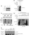
Pin1 regulates SLBP ubiquitination. (A) SLBP is degraded by the proteasome and Pin1. HEK293 cells were either treated with 20 μM PiB for 16 h or mock treated with DMSO to inhibit Pin1 as indicated, and/or treated with the proteasome inhibitor ALLN for 4 h before harvest. The SLBP and actin levels were probed in these cells by Western blotting. For panel B, HEK293 cells were treated with either control or Pin1 siRNA for 48 h, after which they were transiently cotransfected with (His)6-tagged ubiquitin and Flag-tagged WT SLBP followed by treatment with ALLN. The lysates were purified on a nickel column as described in Materials and Methods to capture ubiquitinated proteins and then probed for ubiquitinated SLBP using an anti-SLBP antibody. (C) Flag-tagged SLBP WT or T171A or T60A/T61A mutants were immunoprecipitated (IP) from HEK293 cells expressing these proteins along with HA-tagged ubiquitin in the presence or absence of PiB inhibitor. Anti-Myc antibody was used as a control. The ubiquitinated SLBP was probed using the anti-HA antibody. (D) Pin1 siRNA increases accumulation of ubiquitinated SLBP in all SLBP mutants except S20A/S23A SLBP. HEK293 cells were treated with control or Pin1 siRNA, followed by cotransfection of Flag-tagged WT or mutant SLBP forms with (His)6-tagged ubiquitin. The ubiquitinated proteins were purified over a nickel column as described in Materials and Methods. The lysates were probed for the presence of SLBP using the anti-SLBP antibody. A significant accumulation of ubiquitinated SLBP was observed when Pin1 was knocked down by siRNA, and no ubiquitination of SLBP was observed for the S20A/S23A mutant in either control or Pin1 siRNA-treated cells. (E) Flag-tagged SLBP WT and mutants were transiently transfected into HEK293 cells. The SLBP was immunoprecipitated using the Flag antibody and probed for Flag-tagged SLBP and Pin1. Only the T171A/T60A/T61A triple mutant significantly abrogated the SLBP-Pin1 interaction, indicating that multisite phosphorylation at these sites is required for interaction with Pin1.
Fig 6
Fig 6
Localization of SLBP T171 mutants and SLBP in Pin1 siRNA-treated cells. (A) The distribution of SLBP wild type and T171A and T171E mutants in HeLa cells was visualized in 0.5-μm optical sections following deconvolution. The SLBP is shown in red, the DAPI in blue, and the EdU in green. The first image in panel A shows a cell in early-mid-S phase with WT SLBP present predominantly in the nucleus, but some SLBP is also in the cytoplasm. The second panel shows two cells with T171A SLBP staining. One cell is in early S phase and shows nuclear and some cytoplasmic staining. The second cell is not in S phase and shows an abundance of T171A SLBP in the cytoplasm. The third and fourth panels show the localization of T171E and T171D SLBPs, which is predominantly nuclear in both S-phase and non-S-phase cells. The individual staining patterns are shown in Fig. S3 in the supplemental material. (B) Representative confocal microscopic section images of control HeLa cells (left) and Pin1 siRNA-treated HeLa cells (right) that were stained with DAPI for nuclear imaging (in blue). Immunofluorescence (IF) for Pin1, SLBP, CPSF160, and SC-35 is shown in red or green, and the merge panel is shown on the right of each data set. siRNA knockdown of Pin1 results in increased fluorescence intensity in the nucleus for SLBP, whereas CPSF160 partitions into the nucleoli. There is no change in speckle formation as determined by the localization of SC-35 foci. (C) HeLa cells stably expressing HA-SLBP were probed to confirm the increase in nuclear signal observed in Pin1 siRNA-treated cells. HA-SLBP is shown in red, and DAPI is shown in blue. Representative confocal sections are displayed. (D) ImageStream analysis of control and Pin1 siRNA-treated HeLa cells. Similarity score distributions as a measure of SLBP nuclear distribution for control HeLa cells (red) and Pin1 knockdown cells (black) are shown. Representative composite images of SLBP (green) and DRAQ5 (red) corresponding to similarity scores are shown below.
Fig 7
Fig 7
Effect of Pin1 inhibition on histone mRNA levels. (A) HeLa cells were treated with 50 nM Pin1 siRNA for 72 h. The change in histone mRNA abundance measured after Pin1 knockdown was validated by RT-PCR for five core histone genes, Pin1, and GAPDH. The average fold change determined from the CT values using the comparative CT method is depicted with the standard errors derived from three independent data sets. (B) The effect of chemical inhibition of Pin1 by the PiB inhibitor on histone gene abundance was determined in HEK293 cells by RT-PCR. The experiment was performed in triplicate. (C) HEK293 cells were treated with either control siRNA (designated with a “-C” extension) or Pin1 siRNA (designated with an “-R” extension). Forty-eight hours after the second hit, the cells either were left untransfected (UNTR) or were transfected with SLBP WT or the SLBP T171E or SLBP T171A mutant or Pin1 WT or the Pin1 W34A mutant as indicated. The cells were harvested after 24 h, and the histone mRNA abundance for HIST1 H4 was quantified by RT-PCR as described in Materials and Methods. (D) mRNA decay rates for four different histone mRNAs measured after the addition of hydroxyurea in the presence and absence of PiB in HEK293 cells. The experiments were done in triplicate. The mRNA was quantified for each time point using RT-PCR.
Fig 8
Fig 8
Model for the role of Pin1 in regulating histone mRNA decay. Our data suggest that T171 phosphorylation is required for efficient import of SLBP into the nucleus during S phase. During S phase and at the end of S phase, Pin1 acts with a phosphatase such as PP2A to dephosphorylate SLBP, thereby dissociating SLBP from the histone message in the cytoplasm. Pin1 may also regulate SLBP phosphorylation at the N terminus at Ser20 and Ser23, which we have identified as a phosphodegron that controls SLBP polyubiquitination. At the end of S phase, SLBP is also phosphorylated at Thr60 and Thr61, which is reported to trigger SLBP degradation (67). The histone mRNA is consequently degraded in the cytoplasm, whereas SLBP is degraded by the ubiquitin-proteasome pathway in the nucleus.

Similar articles

Cited by

References

    1. Albert A, Lavoie S, Vincent M. 1999. A hyperphosphorylated form of RNA polymerase II is the major interphase antigen of the phosphoprotein antibody MPM-2 and interacts with the peptidyl-prolyl isomerase Pin1. J. Cell Sci. 112(Part 15):2493–2500 - PubMed
    1. Alterman RB, et al. 1984. Cell cycle regulation of mouse H3 histone mRNA metabolism. Mol. Cell. Biol. 4:123–132 - PMC - PubMed
    1. Borchers CH, et al. 2006. Combined top-down and bottom-up proteomics identifies a phosphorylation site in stem-loop-binding proteins that contributes to high-affinity RNA binding. Proc. Natl. Acad. Sci. U. S. A. 103:3094–3099 - PMC - PubMed
    1. Crenshaw DG, Yang J, Means AR, Kornbluth S. 1998. The mitotic peptidyl-prolyl isomerase, Pin1, interacts with Cdc25 and Plx1. EMBO J. 17:1315–1327 - PMC - PubMed
    1. Dimitrova DS, Berezney R. 2002. The spatio-temporal organization of DNA replication sites is identical in primary, immortalized and transformed mammalian cells. J. Cell Sci. 115:4037–4051 - PubMed

Publication types

MeSH terms

LinkOut - more resources