. 2012 Sep;69(9):644-58.
doi: 10.1002/cm.21057. Epub 2012 Aug 20.

CdGAP regulates cell migration and adhesion dynamics in two-and three-dimensional matrix environments

Affiliations

CdGAP regulates cell migration and adhesion dynamics in two-and three-dimensional matrix environments

Duncan Wormer et al. Cytoskeleton (Hoboken). 2012 Sep.

Abstract

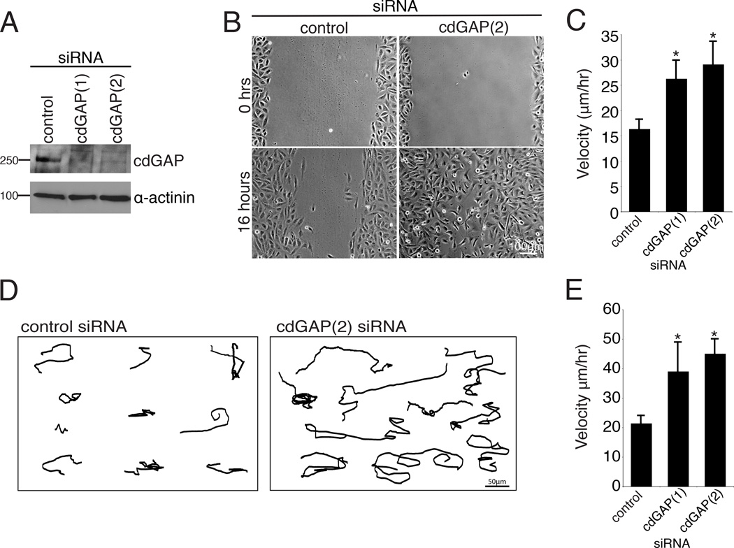
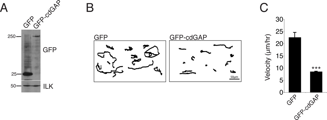
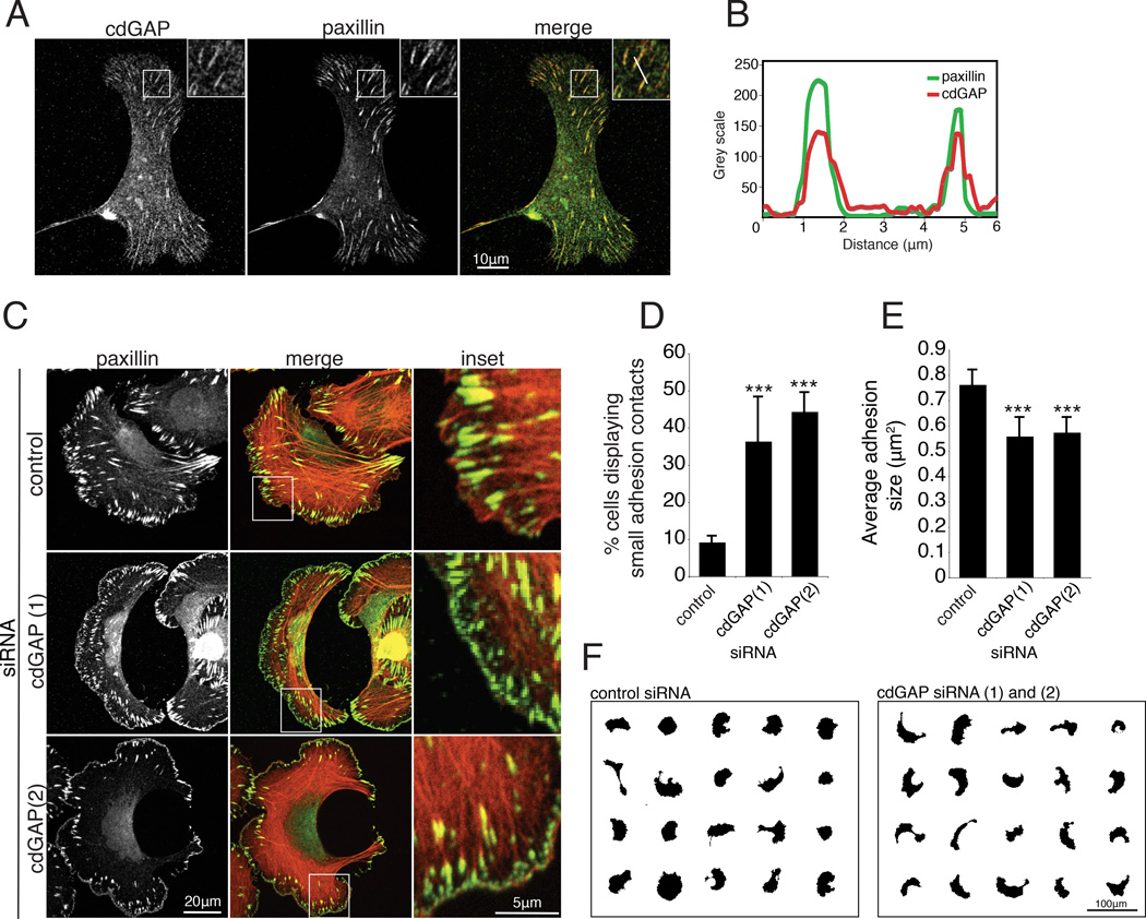
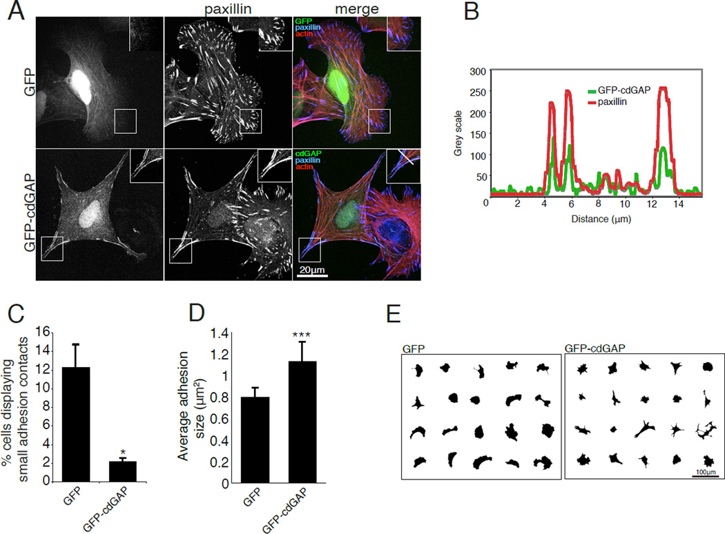
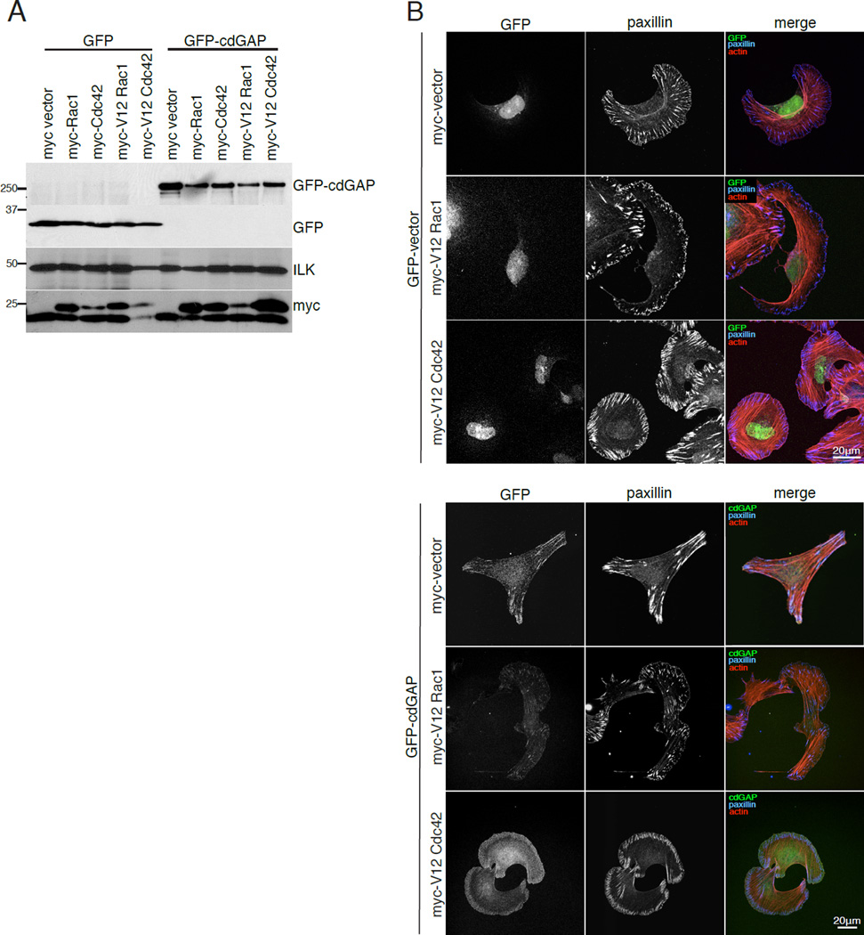
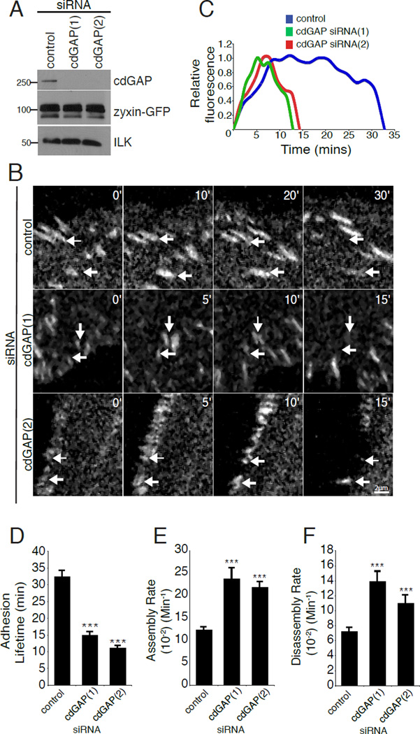
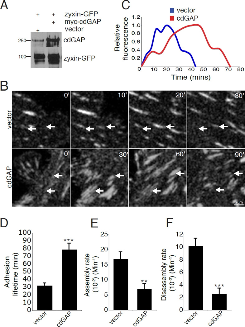
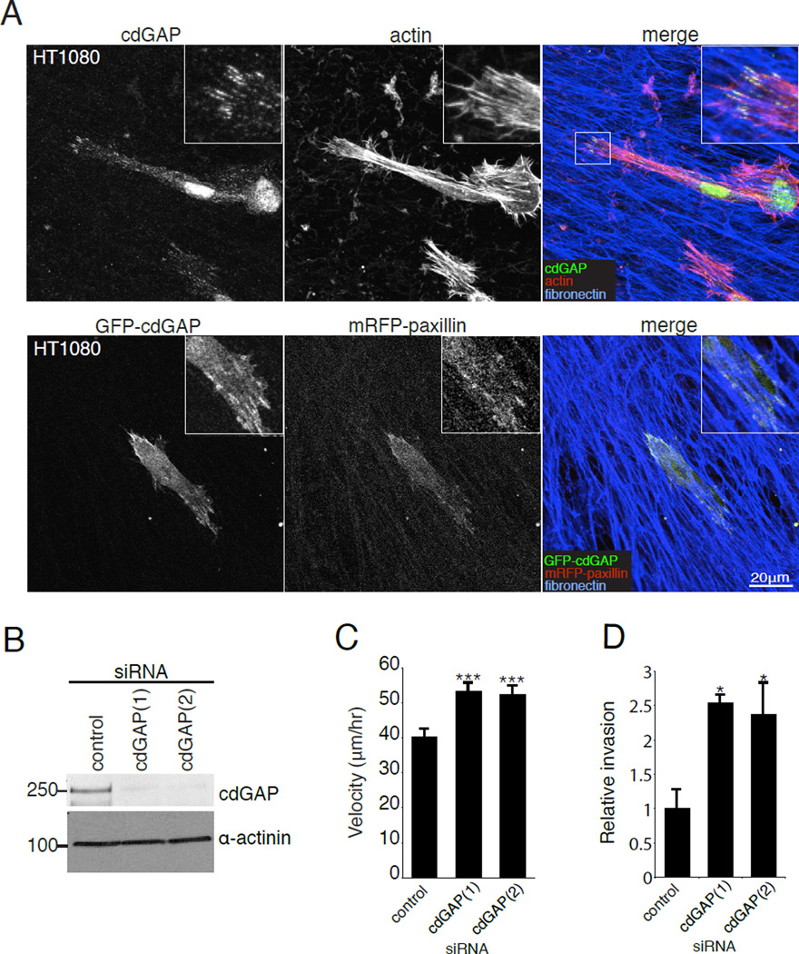
CdGAP is a Rac1/Cdc42 specific GTPase activating protein (GAP) that localizes to cell-matrix adhesions through an interaction with the adhesion scaffold α-parvin/actopaxin to regulate lamellipodia formation and cell spreading. Herein, we demonstrate, using a combination of siRNA-mediated silencing and overexpression, that cdGAP negatively regulates directed and random migration by controlling adhesion maturation and dynamics through the regulation of both adhesion assembly and disassembly. Interestingly, cdGAP was also localized to adhesions formed in three-dimensional (3D) matrix environments and cdGAP depletion promoted cancer cell migration and invasion through 3D matrices. These findings highlight the importance of GAP proteins in the regulation of Rho family GTPases and the coordination of the cell migration machinery..


Conflict of interest statement

Conflict of interest: The authors declare that they have no conflict of interest.

Figures

Figure 1
Figure 1. CdGAP silencing accelerates the speed of wound healing and random migration
(A) U2OS cells were transfected with two separate siRNAs targeted against human cdGAP and knockdown efficiency was assessed by western blotting using an antibody specific for human cdGAP. (B) CdGAP siRNA-treated cells closed a wound more quickly then control siRNA-treated cells. U2OS cells treated with control or cdGAP siRNA were plated at high density, scrape wounded, and imaged over 16 hours. (C) Tracking of individual cells at the wound edge confirmed that cells treated with cdGAP siRNA migrated at a higher velocity than control siRNA-treated cells. A minimum of 40 cells were tracked in each wound per siRNA condition from each of three independent experiments and compared using Student’s t-test, *, p < 0.05 for each cdGAP siRNA when compared to the control. (D) Representative tracks generated from control and cdGAP siRNA-treated U2OS cells plated at low density migrating randomly on 10µg/ml collagen for 16 hours demonstrating the longer distance traveled by cdGAP silenced cells. (E) cdGAP siRNA-treated cells migrate at a higher velocity than control siRNA-treated cells. For random migration velocity calculations, a minimum of 30 cells from each of three independent experiments were tracked. Velocity measurements from the control siRNA-treatment and each cdGAP siRNA-treatment were statistically compared using Student’s t-test, *, p < 0.05 for both siRNAs. For Figure 1, all graphs have error bars representing +/− the standard error of the mean.
Figure 2
Figure 2. CdGAP over expression slows random cell migration
(A) U2OS cells were transfected with GFP or GFP-cdGAP. (B) Representative tracks of transfected U2OS cells plated at low density migrating on 10µg/ml collagen for 16 hours, with GFP-cdGAP transfected cells migrating a shorter distance than GFP control cells. (C) GFP expressing cells migrate at a higher velocity than GFP-cdGAP expressing cells. A minimum of 30 cells from three independent experiments were analyzed per condition and GFP versus GFP-cdGAP expressing cells compared with Student’s t-test,***, p < 0.001. Velocity graph has error bars representing +/− the standard error of the mean.
Figure 3
Figure 3. CdGAP depletion promotes the formation of small adhesion contacts at the leading edge and decreases average adhesion area
(A) U2OS cells were spread, fixed and stained for endogenous cdGAP two hours post plating on 10µg/ml collagen. Endogenous cdGAP co-localizes to adhesions that are positive for paxillin staining. Inset shows a zoom of adhesions positive for cdGAP and paxillin staining used to generate the line profile in (B). (B) A line profile through adhesions demonstrates that cdGAP and paxillin fluorescence intensity peaks in adhesions and diminishes in the surrounding cytoplasm. (C) Control and cdGAP siRNA-treated U2OS cells were spread on 10µg/ml collagen coated coverslips and stained for actin and paxillin. Merged images are overlays of the paxillin and actin channels. Inset shows enlarged area from each white box; small adhesions are frequently observed in the leading edge of migrating U2OS cells treated with cdGAP siRNA. (D) CdGAP depletion increases the percentage of cells with small, leading edge or peripheral adhesions. Cells were scored as positive if > 20% of their leading edge or periphery contained small paxillin-positive adhesions. A minimum of 80 cells from three separate experiments were scored per condition and control versus siRNA-treated cells compared with Student’s t-test, ***, p < 0.001 for both siRNAs. (E) Average adhesion size decreases in cdGAP siRNA-treated cells, a minimum of 30 cells from three independent experiments were analyzed and control-treated cells compared to each siRNA-treatment with Student’s t-test, ***, p < 0.001 for both siRNAs. (F) Masks of cdGAP siRNA-treated cells were made to display representative cell morphology. CdGAP siRNA-treatment promoted the formation of a crescent morphology in U2OS cells. For Figure 3, all error bars represent +/− the standard error of the mean.
Figure 4
Figure 4. CdGAP over expression inhibits the formation of small adhesion contacts and increases adhesion area
(A) U2OS cells transfected with GFP or GFP-cdGAP were spread on 10µg/ml collagen coated coverslips and stained for paxillin and actin. Inset shows the localization of GFP-cdGAP to adhesions that are positive for paxillin staining. The white bar in the merged inset for the GFP-cdGAP expressing cell marks adhesions through which the line profile in (B) was drawn. (B) A line profile through adhesions demonstrates that GFP-cdGAP and paxillin fluorescence intensity peaks in adhesions and diminishes in the surrounding cytoplasm. (C) The percentage of cells with small leading edge or peripheral adhesions decreased in GFP-cdGAP expressing cells. The percentage of small adhesions was quantified from a minimum of 60 transfected cells from each of three independent experiments per condition and GFP versus GFP-cdGAP expressing cells compared with Student’s t-test, *, p < 0.05. (D) Average adhesion size was measured in U2OS cells expressing GFP and GFP-cdGAP constructs from a total of 30 transfected cells per condition from three independent experiments and compared using Student’s t-test, ***, p < 0.001. (E) Masks of cells over expressing GFP or GFP-cdGAP demonstrate that GFP-cdGAP over expressing cells have a smaller spread area and an angular morphology. For Figure 4, all error bars represent +/− the standard error of the mean.
Figure 5
Figure 5. Dominant active V12Rac1 and V12Cdc42 block the cdGAP over expression phenotype
(A) U2OS cells were co-transfected with either GFP or GFP-cdGAP and myc-tagged dominant active V12Rac1 or myc-tagged dominant active V12Cdc42 and the expression levels of each construct were evaluated by western blotting. (B) Cells expressing the indicated constructs were spread on 10µg/ml collagen coated coverslips and stained for paxillin and actin.
Figure 6
Figure 6. CdGAP silencing accelerates adhesion dynamics
(A) U2OS cells stably expressing zyxin-GFP were subjected to control or cdGAP siRNA-treatment and evaluated for cdGAP and zyxin-GFP expression by western blot. (B) Zyxin-GFP stable U2OS cells treated with control or cdGAP siRNA were imaged to quantify focal adhesion dynamics. White arrows indicate adhesions that are turning over during the time course and labels indicate the duration of each sequence in minutes. (C) The relative fluorescence intensity of representative individual adhesions with siRNA treatments plotted over time. (D) CdGAP siRNA shortens the average adhesion lifetime. (E) CdGAP siRNA accelerates adhesion assembly rates. (F) CdGAP siRNA enhances adhesion disassembly rates. Confocal image sequences were used to calculate the lifetime, adhesion assembly, and adhesion disassembly rates for each siRNA treatment. All lifetime and rate measurements were made from three independent experiments for a total of 50–70 adhesions, 4–7 cells per condition and compared with Student’s t-test, ***, p < 0.001 for assembly, disassembly, and lifetime calculations in comparison to control siRNA for both cdGAP siRNAs. For Figure 6 all error bars represent +/− the standard error of the mean.
Figure 7
Figure 7. CdGAP over expression slows adhesion dynamics
(A) U2OS cells were transiently co-transfected with zyxin-GFP and either vector or myc-cdGAP and evaluated for cdGAP and zyxin-GFP expression by western blot. (B) Zyxin-GFP positive U2OS cells expressing myc-control vector or myc-cdGAP were imaged to quantify focal adhesion dynamics. White arrows indicate adhesions that are turning over during the time course and labels indicate the duration of each sequence in minutes. (C) The relative fluorescence intensity of representative individual adhesions plotted over time. (D) Myc-cdGAP over expression lengthens adhesion lifetime. (E) Over expression of myc-cdGAP inhibits adhesion assembly rates. (F) Over expression of myccdGAP inhibits adhesion disassembly rates. Confocal image sequences were used to calculate the lifetime, adhesion assembly, and adhesion disassembly rates for each condition. All lifetime and rate measurements were made from four independent experiments for a minimum of 50 adhesions from 6–10 cells per condition. Statistical comparisons were made between control and myc-cdGAP over expressing cells for the lifetime and adhesion dynamics measurements with a Student’s t-test,**, p < 0.005 for adhesion assembly rate, *** , p < 0.001 for lifetime and disassembly rate comparisons. For Figure 7, all error bars represent +/− the standard error of the mean.
Figure 8
Figure 8. CdGAP localizes to 3D matrix adhesions and cdGAP siRNA enhances Matrigel invasion and migration velocity in 3D CDMs
(A) Endogenous cdGAP and GFP-cdGAP both localize to 3D matrix adhesions. HT1080 cells were plated onto coverslips coated with 3D CDMs before being fixed and stained for endogenous cdGAP, actin and fibronectin. HT1080 cells co-transfected with GFP-cdGAP and mRFP-paxillin were fixed and stained for fibronectin. (B) HT1080 cells were treated with cdGAP siRNAs and knockdown efficiency evaluated by western blotting. (C) CdGAP siRNA-treatment enhanced cell migration velocity in 3D CDMs. Migration velocity was calculated for individual cells moving within the 3D matrix from three independent experiments with a minimum of 30 cells per experiment and siRNA treatment, with ***, p < 0.001 for comparisons between control and cdGAP silenced cells. (D) HT1080 cells treated with cdGAP siRNA invade more efficiently through Matrigel than control siRNA-treated cells. Relative invasion was calculated from three independent experiments and compared using Student’s t-test. *, p < 0.05 for comparisons between control and each cdGAP siRNA-treatment. For Figure 8, all error bars represent +/− the standard error of the mean.

Similar articles

Cited by

References

    1. Abercrombie M, Dunn GA. Adhesions of fibroblasts to substratum during contact inhibition observed by interference reflection microscopy. Exp. Cell Res. 1975;92:57–62. - PubMed
    1. Allen WE, Zicha D, Ridley AJ, Jones GE. A role for Cdc42 in macrophage chemotaxis. J. Cell Biol. 1998;141:1147–1157. - PMC - PubMed
    1. Aoki K, Nakamura T, Matsuda M. Spatio-temporal Regulation of Rac1 and Cdc42 activity during nerve growth factor-induced neurite outgrowth in PC12 cells. J. Biol. Chem. 2004;279:713–719. - PubMed
    1. Arthur WT, Noren NK, Burridge K. Regulation of rho family GTPases by cell-cell and cell-matrix adhesion. Biol. Res. 2002;35:239–246. - PubMed
    1. Bacac M, Stavenkovic I. Metastatic Cancer Cell. Annu. Rev. Path. Mech. Dis. 2008;3:221–247. - PubMed

Publication types