

Cutaneous hybrid cyst in a sprague-dawley rat
- PMID: 22907984
- PMCID: PMC3392906
- DOI: 10.1293/tox.25.175
Cutaneous hybrid cyst in a sprague-dawley rat
Abstract
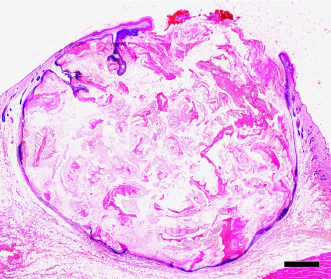
This report describes a spontaneous hybrid cyst in a Sprague-Dawley (SD) rat. A 52-week-old, male SD rat had a cutaneous cyst on the left mystacial pad. Histologically, the cyst wall showed infundibular differentiation with keratohyalin granules in the granular layer and matrical differentiation comprising basaloid epithelial cells with trichohyalin granules. The cyst cavity was filled with lamellar, flaky keratin and aggregates of shadow cells. Immunohistochemically, the infundibular-type epithelium was positive for cytokeratin (CK) AE1/AE3, CK KL1 and CK14 but negative for CK15, whereas the matrical-type epithelium was negative for all four CK isoforms examined. These immunohistochemical properties of the infundibular- and matrical-type epithelia were similar to those of the infundibulum and inferior segment of normal hair follicles, respectively. Based on these findings, the cyst was diagnosed as a hybrid cyst, comprising more than one type of cyst arising from various parts of the pilosebaceous unit.
Keywords: Sprague-Dawley rat; cytokeratin; epidermal inclusion cyst; hybrid cyst; kin.
Figures


References
-
- Stephen G, Lake LH-E, Robert EM, Barry PS. Nonneoplaspic lesions. In: Integument and Mammary Glands (Monographs on Pathology of Laboratory Animals), Springer-Verlag, Heidelberg, Germany. 130–157. 1989
-
- Goldschmidt MH, Dunstan RW, Stannard AA. Cyst. In: Histological Classifcation of Epithelial and Melanocytic Tumors of the Skin of Domestic Animals, Armed Forces Institute of Pathology, Washington DC, USA. 33–35. 1998
-
- Gross TL, Ihrke PJ, Walder EJ, Affolter VK. Follicular Tumors. In: Skin Diseases of the Dog and Cat, 2nd ed. TL Gorss, PJ Ihrke, EJ Walder, and VK Affolter (eds). Blackwell Publishing, Oxford, UK. 604–640. 2005
-
- Brownstein MH. Hybrid cyst: a combined epidermoid and trichilemmal cyst. J Am Acad Dermatol. 9: 872–875 1983. - PubMed
-
- McGavran MH, Binnington B. Keratinous cysts of the skin. Identification and differentiation of pilar cysts from epidermal cysts. Arch Dermatol. 94: 499–508 1966. - PubMed
LinkOut - more resources
Full Text Sources
Research Materials
Miscellaneous