. 2012 May 23;22(10):2027-2039.
doi: 10.1002/adfm.201101662. Epub 2012 Feb 21.

Functional Human Vascular Network Generated in Photocrosslinkable Gelatin Methacrylate Hydrogels

Affiliations

Functional Human Vascular Network Generated in Photocrosslinkable Gelatin Methacrylate Hydrogels

Ying-Chieh Chen et al. Adv Funct Mater. .

Abstract

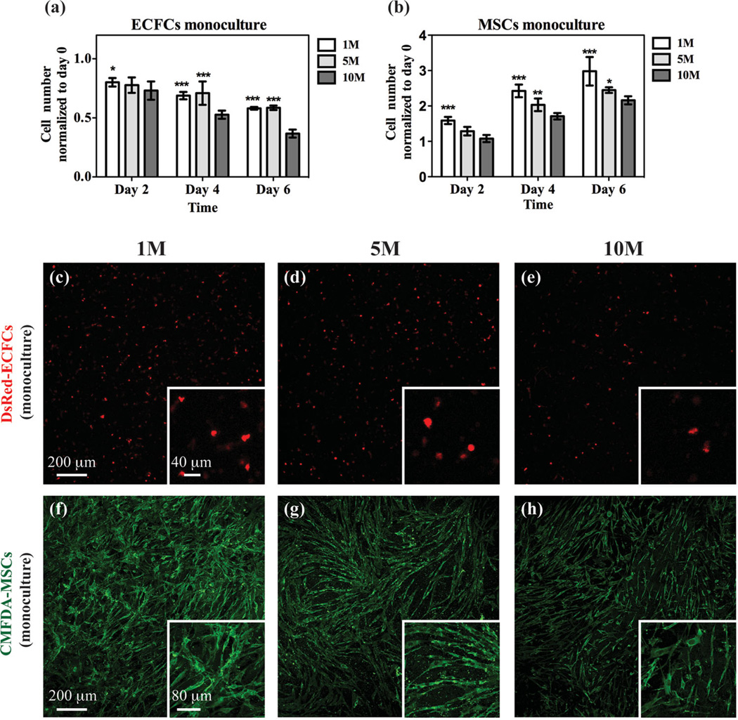
The generation of functional, 3D vascular networks is a fundamental prerequisite for the development of many future tissue engineering-based therapies. Current approaches in vascular network bioengineering are largely carried out using natural hydrogels as embedding scaffolds. However, most natural hydrogels present a poor mechanical stability and a suboptimal durability, which are critical limitations that hamper their widespread applicability. The search for improved hydrogels has become a priority in tissue engineering research. Here, the suitability of a photopolymerizable gelatin methacrylate (GelMA) hydrogel to support human progenitor cell-based formation of vascular networks is demonstrated. Using GelMA as the embedding scaffold, it is shown that 3D constructs containing human blood-derived endothelial colony-forming cells (ECFCs) and bone marrow-derived mesenchymal stem cells (MSCs) generate extensive capillary-like networks in vitro. These vascular structures contain distinct lumens that are formed by the fusion of ECFC intracellular vacuoles in a process of vascular morphogenesis. The process of vascular network formation is dependent on the presence of MSCs, which differentiate into perivascular cells occupying abluminal positions within the network. Importantly, it is shown that implantation of cell-laden GelMA hydrogels into immunodeficient mice results in a rapid formation of functional anastomoses between the bioengineered human vascular network and the mouse vasculature. Furthermore, it is shown that the degree of methacrylation of the GelMA can be used to modulate the cellular behavior and the extent of vascular network formation both in vitro and in vivo. These data suggest that GelMA hydrogels can be used for biomedical applications that require the formation of microvascular networks, including the development of complex engineered tissues.


Figures

Figure 1
Figure 1
a–f) Characterization of GelMA hydrogels with different methacrylation degrees: compressive modulus (a); degradation profiles upon incubation with collagenase (b); average pore size (c); SEM images (d–f). The data are presented as the mean ± the standard deviation (SD). **: p < 0.01, ***: p < 0.001, compared with the 10M GelMA.
Figure 2
Figure 2
Monocultures of vascular cells embedded in GelMA. a,b) Monocultures containing either ECFCs (a) or MSCs (b) were evaluated for cell number at 2, 4 and 6 d using GelMA hydrogels with different methacrylation degrees. c–h) Representative confocal microscopy images of embedded DsRed-ECFCs (c–e) and CMFDA-labeled MSCs (f–h) after day 6 in monoculture for each methacrylation degree. The insets show images taken at higher magnification. The data are presented as the mean ± SD. *: p < 0.1, **: p < 0.01, ***: p < 0.001, compared with the 10M GelMA.
Figure 3
Figure 3
Co-culture of vascular cells in GelMA. Constructs containing both DsRed-ECFCs and CMFDA-labeled MSCs were cultured for 2, 4 and 6 d using GelMA hydrogels with different methacrylation degrees. a,b) Numbers of DsRed-ECFCs (a) and CMFDA-MSCs (b) were separately counted at each time point under a fluorescence microscope. c–h) Representative confocal microscopy images of DsRed-ECFCs (c–e) and CMFDA-MSCs (f–h) after day 6 in co-culture for each methacrylation degree. The data are presented as the mean ± SD. *: p < 0.1, **: p < 0.01, ***: p < 0.001, compared with the 10M GelMA.
Figure 4
Figure 4
Extent of capillary-like network formation. Constructs containing both DsRed-ECFCs and CMFDA-labeled MSCs were cultured in the GelMA hydrogels with different methacrylation degrees. The DsRed-ECFCs were imaged in whole-mount constructs using confocal microscopy at day 6. a–c) Representative 2D projections (xy plane) of images collected along the z -axis of the constructs. d–f) Representative 3D reconstruction of constructs from confocal microscopy images: z -plane cross-sections covering a thickness of 400 µm are in the direction of the white arrows. A representative video of a rotating 3D reconstruction of the confocal images showing interconnected vascular networks is available (Video 1, Supporting Information) g–j) Quantitative analysis of the extent of capillary-like network formation after 2, 4 and 6 d in vitro was carried out by measuring total capillary-like length per unit of area (g), the average length of capillaries (h), the number of branch points per unit of area (i) and the number of branches per unit of area (j). The data are presented as the mean ± SD. *: p < 0.1, **: p < 0.01, ***: p < 0.001, compared with the 10M GelMA.
Figure 5
Figure 5
Stabilization of the ECFC-lined capillaries by perivascular cells. Constructs containing both DsRed-ECFCs and MSCs were cultured for 7 d in GelMA hydrogels with different methacrylation degrees. The ability of the MSCs to differentiate into perivascular cells was analyzed by confocal microscopy after immunoflourescence staining with antibodies against smooth muscle markers. a,c) Representative confocal images showing the spatial distribution of the DsRed-ECFC-lined capillaries surrounded by αSMA-expressing MSCs. Higher magnification images depicting details of a capillary (top) and a cross-section image taken in the direction of the white arrows (bottom) are shown to the right of the these images in panels (a) and (c). b,d) Representative confocal images showing the spatial distribution of both DsRed-ECFC-lined capillaries surrounded by sm-MHC-expressing MSCs. A representative video of a rotating 3D reconstruction of confocal images showing MSC-wrapped capillaries is available (Video 2, Supporting Information).
Figure 6
Figure 6
Formation of ECFC-lined lumens by fusion of intracellular vacuoles. Constructs containing both DsRed-ECFCs and MSCs were cultured for 7 d using 1M GelMA hydrogels. The presence of DsRed-ECFC-lined lumens was investigated by a confocal microscopy examination of whole-mounted constructs. Analyses of the constructs at 3, 6 and 10 d of culture reveal a progressive process of lumen morphogenesis. a) Initial accumulation of ECFC-specific intracellular vacuoles (asterisks) at day 3; the vacuoles were intracellularly arranged into rows and were present in the majority of the ECFC-lined capillary structures. The surface of these vacuoles were stained positive for human CD31. b) By day 6, larger vacuoles coalesced forming large, intracellular luminal structures. c) At day 10, hollow ECFC-lined lumens were identified unequivocally and uniformly distributed in discrete locations within the capillary-like structures. d) Lumens were lined exclusively by ECFCs and were surrounded by αSMA-expressing MSCs (yellow arrowhead). e) Schematic diagram depicting the steps of endothelial lumen formation in GelMA.
Figure 7
Figure 7
Evaluation of GelMA hydrogels in vivo. GelMA hydrogels with 1M and 10M methacrylation degrees and containing no cells were surgically implanted into the subcutaneous space on the backs of 6-week old nude mice. Rat-tail type-1-collagen gel served as a control. a) Macroscopic views of explanted gels after 7 and 28 d. b,c) In vivo gel degradation profiles based on volume (b) and weight (c) loss: n = 3 per gel type and time point. The data are presented as the mean ± SD. *: p < 0.1, **: p < 0.01, ***: p < 0.001, compared with collagen. d–l) Representative images of hematoxylin/eosin (H&E)-stained sections from collagen and the 1M and 10M GelMA implants, harvested at day 7 (d–i) and day 28 (j–l). The insets show representative photographs taken at the time of harvesting and depict the integration of implants with murine subcutaneous tissue at the site of implantation. The dashed lines delineate the implant border at day 28. “†” indicates adipocytes.
Figure 8
Figure 8
In vivo formation of functional vascular networks. a) Schematic diagram: the 1M and 10M GelMA hydrogels containing both ECFCs and MSCs were incubated in vitro for 24 h and surgically implanted into the subcutaneous space on the backs of 6-week old nude mice (n = 7 per gel type). Rat-tail type-1-collagen gel served as a control. The implants were retrieved after 7 d in vivo. b–d) Representative images of H&E-stained sections from collagen and the 1M and 10M GelMA explants, revealing the presence of numerous blood vessels containing murine erythrocytes (yellow arrowheads). Macroscopic views of the explants are depicted in the insets. e–g) Immunohistochemistry showed that the engineered microvessels were stained positive for human CD31 (red arrowheads), while the murine capillaries (green arrowheads) were not. h–j) Images at higher magnification showing a single human-CD31-expressing vessel (asterisks) carrying murine erythrocytes. k–m) Representative images of fluorescently stained sections using rhodamine-conjugated UEA-1 lectin (to mark human-ECFC-lined vessels) and fluorescein isothiocyanate (FITC)-conjugated anti-αSMA antibodies (to mark perivascular cells; red arrowheads). The UEA-1 lectin did not bind to the murine vessels (green arrowheads). The nuclei were stained with 4’,6-diamidino-2-phenylindole (DAPI). n–p) The extent of the vascular-network formation was quantified by measuring the microvessel density (n), the percentage of human-CD31-expressing blood vessels (o), and the size distribution of the luminal cross-sectional area (p). The data are presented as the mean ± SD. **: p < 0.01, ***: p < 0.001, compared with the collagen implants.

References

    1. Levenberg S, Rouwkema J, Macdonald M, Garfein ES, Kohane DS, Darland DC, Marini R, van Blitterswijk CA, Mulligan RC, D’Amore PA, Langer R. Nat. Biotechnol. 2005;23:879. - PubMed
    1. Langer R, Vacanti JP. Science. 1993;260:920. - PubMed
    1. Jain RK, Au P, Tam J, Duda DG, Fukumura D. Nat. Biotechnol. 2005;23:821. - PubMed
    1. Isner JM, Asahara T. J. Clinical Investigation. 1999;103:1231. - PMC - PubMed
    1. Isner JM, Pieczek A, Schainfeld R, Blair R, Haley L, Asahara T, Rosenfield K, Razvi S, Walsh K, Symes JF. Lancet. 1996;348:370. - PubMed

LinkOut - more resources