

Lipocalin 2 is present in the EAE brain and is modulated by natalizumab
- PMID: 22907989
- PMCID: PMC3414908
- DOI: 10.3389/fncel.2012.00033
Lipocalin 2 is present in the EAE brain and is modulated by natalizumab
Abstract
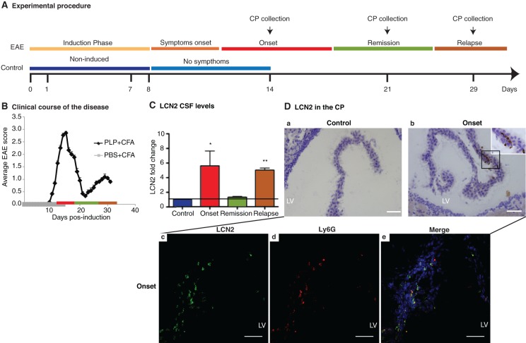
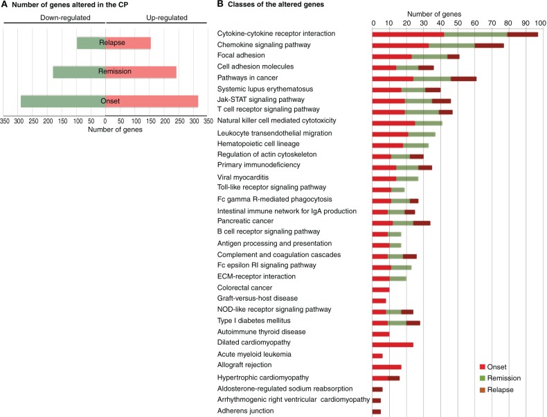
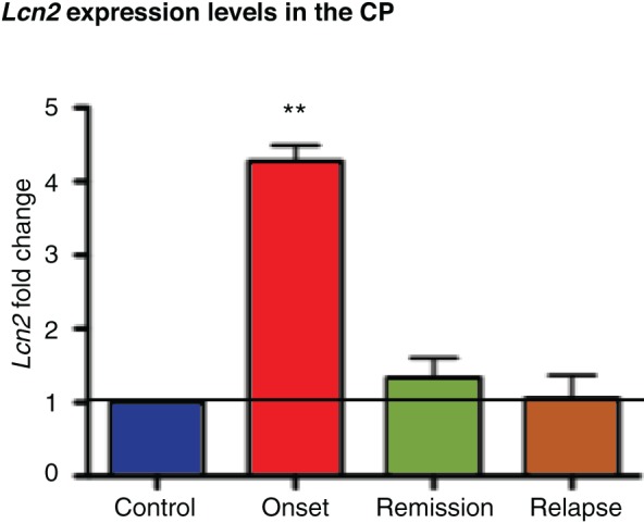
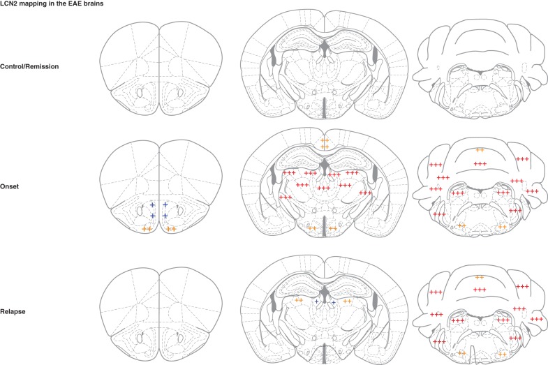
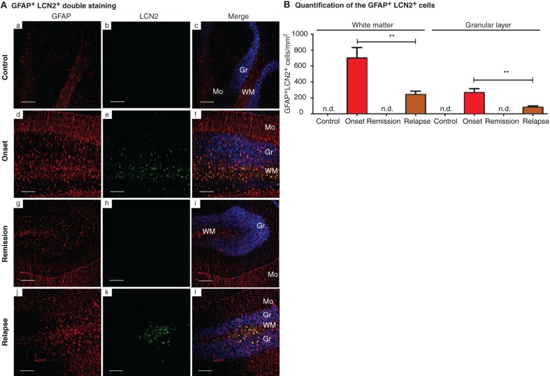
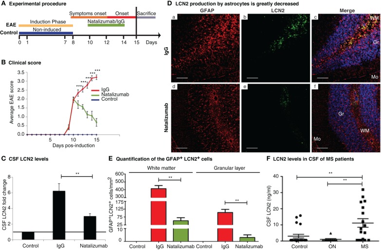
Multiple sclerosis (MS) is a demyelinating disease that causes major neurological disability in young adults. A definitive diagnosis at the time of the first episode is still lacking, but since early treatment leads to better prognosis, the search for early biomarkers is needed. Here we characterized the transcriptome of the choroid plexus (CP), which is part of the blood-brain barriers (BBBs) and the major site of cerebrospinal fluid production, in the experimental autoimmune encephalomyelitis (EAE) mouse model of MS. In addition, cerebrospinal fluid samples from two cohorts of patients with MS and with optic neuritis (ON) were analyzed to confirm the clinical relevance of the findings. Genes encoding for adhesion molecules, chemokines and cytokines displayed the most altered expression, supporting the role of CP as a site of immune-brain interaction in MS. The gene encoding for lipocalin 2 was the most up-regulated; notably, the cerebrospinal fluid lipocalin 2 levels coincided with the active phases of the disease. Immunostaining revealed that neutrophils infiltrating the CP were the source of the increased lipocalin 2 expression in this structure. However, within the brain, lipocalin 2 was also detected in astrocytes, particularly in regions typically affected in patients with MS. The increase of lipocalin 2 in the cerebrospinal fluid and in astrocytes was reverted by natalizumab treatment. Most importantly, the results obtained in the murine model were translatable into humans since patients from two different cohorts presented increased cerebrospinal fluid lipocalin 2 levels. The findings support lipocalin 2 as a valuable molecule for the diagnostic/monitoring panel of MS.
Keywords: astrocytes; cerebrospinal fluid; experimental autoimmune encephalomyelitis; lipocalin 2; multiple sclerosis; natalizumab.
Figures
Similar articles
-
Lipocalin 2 is a novel immune mediator of experimental autoimmune encephalomyelitis pathogenesis and is modulated in multiple sclerosis.Glia. 2012 Jul;60(7):1145-59. doi: 10.1002/glia.22342. Epub 2012 Apr 12. Glia. 2012. PMID: 22499213
-
Active induction of experimental autoimmune encephalomyelitis by MOG35-55 peptide immunization is associated with differential responses in separate compartments of the choroid plexus.Fluids Barriers CNS. 2012 Aug 7;9(1):15. doi: 10.1186/2045-8118-9-15. Fluids Barriers CNS. 2012. PMID: 22870943 Free PMC article.
-
The choroid plexus is modulated by various peripheral stimuli: implications to diseases of the central nervous system.Front Cell Neurosci. 2015 Apr 13;9:136. doi: 10.3389/fncel.2015.00136. eCollection 2015. Front Cell Neurosci. 2015. PMID: 26236190 Free PMC article. Review.
-
Cerebrospinal fluid fetuin-A is a biomarker of active multiple sclerosis.Mult Scler. 2013 Oct;19(11):1462-72. doi: 10.1177/1352458513477923. Epub 2013 Feb 25. Mult Scler. 2013. PMID: 23439582
-
Involvement of the choroid plexus in central nervous system inflammation.Microsc Res Tech. 2001 Jan 1;52(1):112-29. doi: 10.1002/1097-0029(20010101)52:1<112::AID-JEMT13>3.0.CO;2-5. Microsc Res Tech. 2001. PMID: 11135454 Review.
Cited by
-
Associations between Microglia and Astrocytic Proteins and Tau Biomarkers across the Continuum of Alzheimer's Disease.Int J Mol Sci. 2024 Jul 9;25(14):7543. doi: 10.3390/ijms25147543. Int J Mol Sci. 2024. PMID: 39062786 Free PMC article.
-
Uncovering mechanisms of brain inflammation in Alzheimer's disease with APOE4: Application of single cell-type lipidomics.Ann N Y Acad Sci. 2022 Dec;1518(1):84-105. doi: 10.1111/nyas.14907. Epub 2022 Oct 6. Ann N Y Acad Sci. 2022. PMID: 36200578 Free PMC article. Review.
-
Immune Thymic Profile of the MOG-Induced Experimental Autoimmune Encephalomyelitis Mouse Model.Front Immunol. 2018 Oct 11;9:2335. doi: 10.3389/fimmu.2018.02335. eCollection 2018. Front Immunol. 2018. PMID: 30369926 Free PMC article.
-
Different Kinetics of Serum ADAMTS13, GDF-15, and Neutrophil Gelatinase-Associated Lipocalin in the Early Phase of Aneurysmal Subarachnoid Hemorrhage.Int J Mol Sci. 2023 Jul 2;24(13):11005. doi: 10.3390/ijms241311005. Int J Mol Sci. 2023. PMID: 37446186 Free PMC article.
-
Hormones in experimental autoimmune encephalomyelitis (EAE) animal models.Transl Neurosci. 2021 May 6;12(1):164-189. doi: 10.1515/tnsci-2020-0169. eCollection 2021 Jan 1. Transl Neurosci. 2021. PMID: 34046214 Free PMC article. Review.
References
-
- Calabrese M., Mattisi I., Rinaldi F., Favaretto A., Atzori M., Bernardi V., Barachino L., Romualdi C., Rinaldi L., Perini P., Gallo P. (2010). Magnetic resonance evidence of cerebellar cortical pathology in multiple sclerosis. J. Neurol. Neurosurg. Psychiatry 81, 401–404 10.1136/jnnp.2009.177733 - DOI - PubMed
-
- Columba-Cabezas S., Griguoli M., Rosicarelli B., Magliozzi R., Ria F., Serafini B., Aloisi F. (2006). Suppression of established experimental autoimmune encephalomyelitis and formation of meningeal lymphoid follicles by lymphotoxin beta receptor-Ig fusion protein. J. Neuroimmunol. 179, 76–86 10.1016/j.jneuroim.2006.06.015 - DOI - PubMed
-
- Comi G., Colombo B., Martinelli V. (2000). Prognosis-modifying therapy in multiple sclerosis. Neurol. Sci. 21, S893–899 - PubMed
LinkOut - more resources
Full Text Sources
Other Literature Sources
Molecular Biology Databases
Miscellaneous