. 2012 Aug 10:6:50.
doi: 10.3389/fncir.2012.00050. eCollection 2012.

Activation of cortical 5-HT(3) receptor-expressing interneurons induces NO mediated vasodilatations and NPY mediated vasoconstrictions

Affiliations

Activation of cortical 5-HT(3) receptor-expressing interneurons induces NO mediated vasodilatations and NPY mediated vasoconstrictions

Quentin Perrenoud et al. Front Neural Circuits. .

Abstract

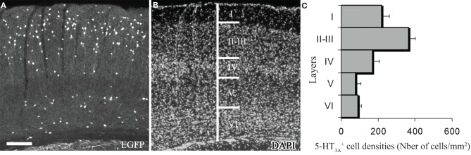
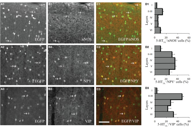
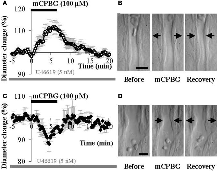
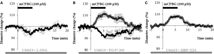
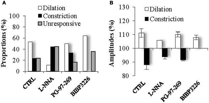
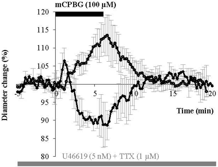
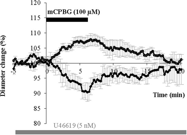
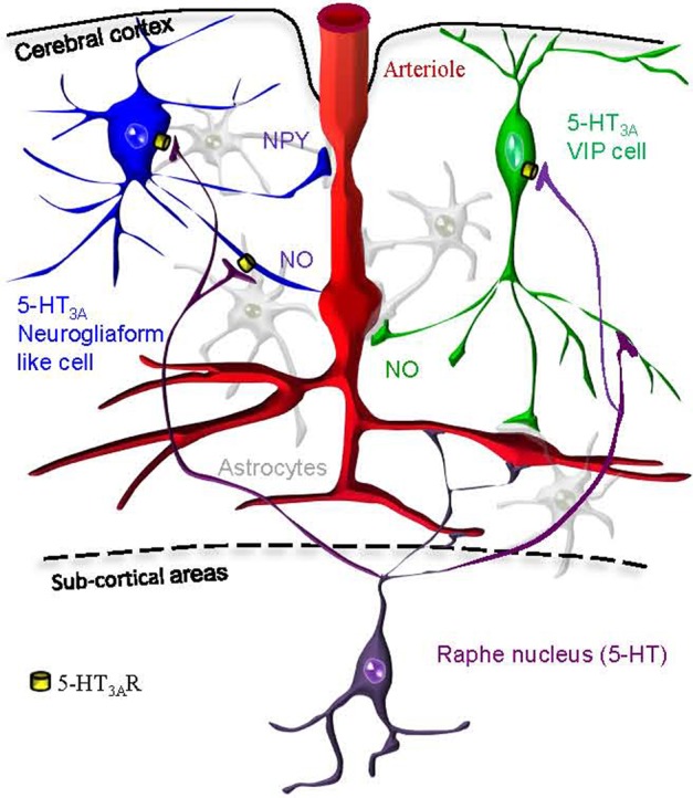
GABAergic interneurons are local integrators of cortical activity that have been reported to be involved in the control of cerebral blood flow (CBF) through their ability to produce vasoactive molecules and their rich innervation of neighboring blood vessels. They form a highly diverse population among which the serotonin 5-hydroxytryptamine 3A receptor (5-HT(3A))-expressing interneurons share a common developmental origin, in addition to the responsiveness to serotonergic ascending pathway. We have recently shown that these neurons regroup two distinct subpopulations within the somatosensory cortex: Neuropeptide Y (NPY)-expressing interneurons, displaying morphological properties similar to those of neurogliaform cells and Vasoactive Intestinal Peptide (VIP)-expressing bipolar/bitufted interneurons. The aim of the present study was to determine the role of these neuronal populations in the control of vascular tone by monitoring blood vessels diameter changes, using infrared videomicroscopy in mouse neocortical slices. Bath applications of 1-(3-Chlorophenyl)biguanide hydrochloride (mCPBG), a 5-HT(3)R agonist, induced both constrictions (30%) and dilations (70%) of penetrating arterioles within supragranular layers. All vasoconstrictions were abolished in the presence of the NPY receptor antagonist (BIBP 3226), suggesting that they were elicited by NPY release. Vasodilations persisted in the presence of the VIP receptor antagonist VPAC1 (PG-97-269), whereas they were blocked in the presence of the neuronal Nitric Oxide (NO) Synthase (nNOS) inhibitor, L-NNA. Altogether, these results strongly suggest that activation of neocortical 5-HT(3A)-expressing interneurons by serotoninergic input could induces NO mediated vasodilatations and NPY mediated vasoconstrictions.

Keywords: Pet1 knock-out mouse; U46619; brain slices; mCPBG; neurogliaform cells; neurovascular coupling; serotonin; vasoactive intestinal peptide.


Figures

Figure 1
Figure 1
Expression of 5-HT3A within the somatosensory cortex. (A,B) Coronal section of a 5-HT3A:GFP mouse counterstained with DAPI showing the preferential location of 5-HT3A-expressing cells in supragranular layers. (C) Density of 5-HT3A-expressing cells from transgenic 5-HT3A:GFP mice in the different layers of the primary somatosensory cortex. Scale bar: 250 μm.
Figure 2
Figure 2
Expression of vasoactive molecules in 5-HT3A-expressing interneurons. (A1–3) Immunodetection of GFP-expressing neurons within the primary somatosensory cortex of 5-HT3A:GFP+ mice. (B1–3) Immunodetection of nNOS (B1), NPY (B2) and VIP (B3) within the same areas as in (A). (C1–3) Overlays showing the colocalization of GFP with nNOS (C1), NPY (C2), or VIP (C3). (D1–3), Density of 5-HT3A-expressing cells colocalized with nNOS (D1), NPY (D2), or VIP (D3) in the different layers of the somatosensory cortex. Examples of co-labeled neurons are pointed by arrows. Scale bar: 65 μm.
Figure 3
Figure 3
5-HT3A:GFP+ neurons in relation with the closest large penetrating blood vessel. (A) In layer I, 5-HT3A:GFP+ neurons do not show specific preferences to be located close to large penetrating blood vessels. (B,C) By contrast in layer II multipolar and bipolar/bitufted 5-HT3A:GFP+ neurons are preferentially positioned in the vicinity of penetrating blood vessels. (D) Confocal picture of a coronal section taken at the level of the supragranular layers of the somatosensory cortex showing the distribution of 5-HT3A:GFP+ neurons (green) in relation with blood vessels stained by collagen IV (red). Large penetrating blood vessel is indicated by a star. (E) Higher power view of the caption shown in (D). The arrow points to a multipolar 5-HT3A:GFP+ neurons displaying a large soma. The filled arrowhead points to a bipolar 5-HT3A:GFP+ neurons displaying a fusiform shape. Scale bars: (D) 130 μm; (E) 35 μm.
Figure 4
Figure 4
mCPBG induces both vasodilations and vasoconstrictions in somatosensory cortical slices. (A) Mean vascular dilation (n = 9) induced by mCPBG (100 μM). (B) Infrared images of a penetrating blood vessel that reversibly dilated in response to bath application of mCPBG (100 μM). Arrows indicate region of high vascular reactivity. (C) Mean vascular constriction (n = 4) induced by mCPBG (100 μM). (D) Infrared images of a penetrating blood vessel that reversibly constricted in response to bath application of mCPBG (100 μM). Scale bar: 10 μm.
Figure 5
Figure 5
mCPBG induced vasodilations are mediated by NO while constrictions are mediated by NPY. (A) Mean vasoconstriction (n = 4) induced by mCPBG (100 μM) in the presence of nNOS inhibitor L-NNA (100 μM) and the preconstricting agent U46619 (5 nM). (B) Mean vascular dilation (n = 4; white circle) and mean vascular constriction (n = 2; black diamond) induced by mCPBG (100 μM) in the presence of the VIP receptor VPAC1 antagonist PG-97-269 (100 nM) and U46619 (5 nM). (C) Vasodilation (n = 9) induced by mCPBG (100 μM) in the presence of the NPY Y1 receptor antagonist BIBP 3226 (1 μM) and U46619 (5 nM).
Figure 6
Figure 6
Proportions (A) and amplitudes (B) of dilating or constricting blood vessels in control condition (CTRL) and in the presence of the pharmacological blockers used in Figure 5. Note that in presence of the nNOS inhibitor, L-NNA, as in the presence the Y1 NPY receptor antagonist, BIBP 3226, the occurrence of dilations versus contractions was reduced or favored respectively, without impacting their amplitudes. On the opposite, in the presence of the VPAC1 receptor antagonist of VIP, PG-97-269, no changes were observed compared to control conditions.
Figure 7
Figure 7
mCPBG induces TTX-insensitive vasodilations and vasoconstrictions. Mean vascular dilation (n = 5) and constriction (n = 2) induced by mCPBG (100 μM) in the presence of TTX (1 μM) and U46619 (5 nM).
Figure 8
Figure 8
mCPBG induces vasodilations and vasoconstrictions in Pet1−/− mice. Mean vasodilation (n = 6) and vasoconstriction (n = 3) induced by mCPBG (100 μM) in Pet1−/− mice.
Figure 9
Figure 9
Hypothetical schematic representation of the 5-HT3AR induction of neurovascular coupling within the somatosensory cortex. We find that pharmacological stimulation of 5-HT3A-expressing neurogliaform like cells (in blue) or VIP cells (in green) could either lead to vasodilation via NO release or vasoconstriction via NPY release. Cortical penetrating arterioles (in red) are also directly innervated by serotoninergic fibers originating from raphe nuclei (in purple). Astrocytes are shown in gray.

Similar articles

Cited by

References

    1. Abounader R., Elhusseiny A., Cohen Z., Olivier A., Stanimirovic D., Quirion R., Hamel E. (1999). Expression of neuropeptide Y receptors mRNA and protein in human brain vessels and cerebromicrovascular cells in culture. J. Cereb. Blood Flow Metab. 19, 155–163 10.1097/00004647-199902000-00007 - DOI - PubMed
    1. Abounader R., Villemure J. G., Hamel E. (1995). Characterization of neuropeptide Y (NPY) receptors in human cerebral arteries with selective agonists and the new Y1 antagonist BIBP 3226. Br. J. Pharmacol. 116, 2245–2250 - PMC - PubMed
    1. Andrade R. (2011). Serotonergic regulation of neuronal excitability in the prefrontal cortex. Neuropharmacology 61, 382–386 10.1016/j.neuropharm.2011.01.015 - DOI - PMC - PubMed
    1. Ascoli G. A., Alonso-Nanclares L., Anderson S. A., Barrionuevo G., Benavides-Piccione R., Burkhalter A., Buzàki G., Cauli B., Defelipe J., Faién A., Feldmeyer D., Fishell G., Fregnac Y., Freund T. F., Gardner E. P., Goldberg J. H., Helmstaedter M., Hestrin S., Karube F., Kisvàrday Z. F., Lambolez B., Lewis D. A., Marin O., Markram H., Munoz A., Packer A., Petersen C. C., Rockland K. S., Rossier J., Rudy B., Somogy P., Staiger J. F., Tamas G., Thomson A. M., Toledo-Rodriguez M., Wang Y., West D. C., Yuste R. (2008). Petilla terminology: nomenclature of features of GABAergic interneurons of the cerebral cortex. Nat. Rev. Neurosci. 9, 557–568 10.1038/nrn2402 - DOI - PMC - PubMed
    1. Baraban S. C., Tallent M. K. (2004). Interneuron diversity series: interneuronal neuropeptides– endogenous regulators of neuronal excitability. Trends Neurosci. 27, 135–142 10.1016/j.tins.2004.01.008 - DOI - PubMed

LinkOut - more resources