. 2012 Nov;56(11):5603-11.
doi: 10.1128/AAC.00612-12. Epub 2012 Aug 20.

Toxicity mechanisms of amphotericin B and its neutralization by conjugation with arabinogalactan

Affiliations

Toxicity mechanisms of amphotericin B and its neutralization by conjugation with arabinogalactan

Sarah Kagan et al. Antimicrob Agents Chemother. 2012 Nov.

Abstract

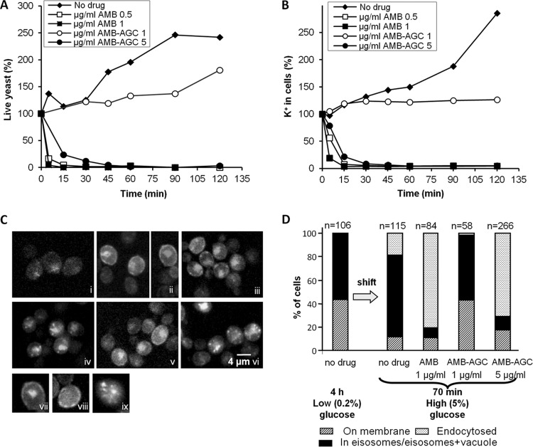
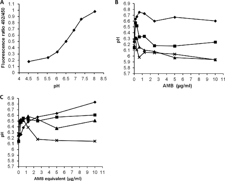
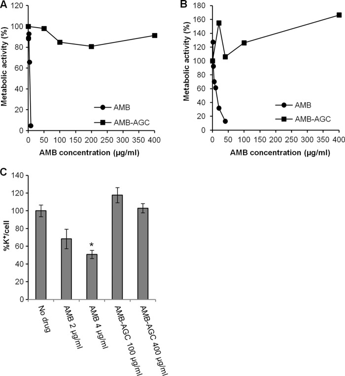
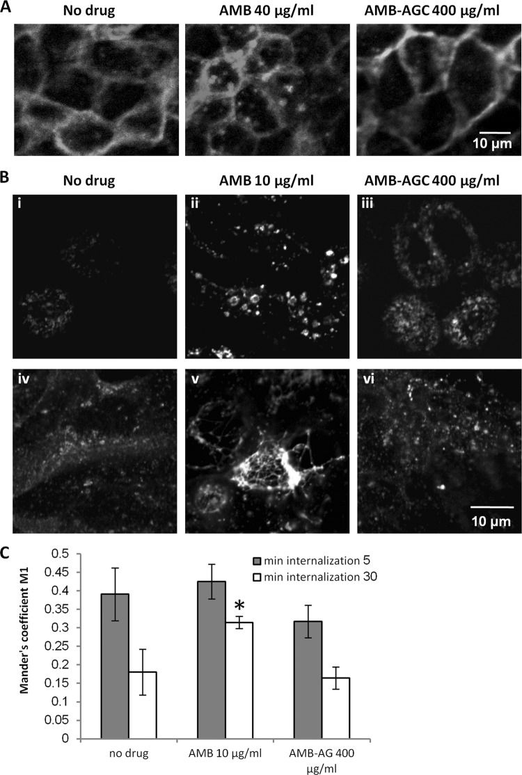
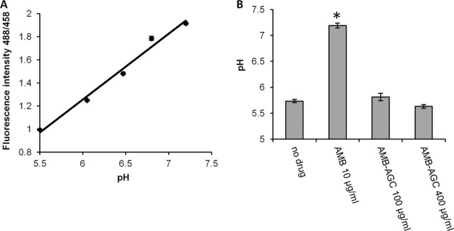
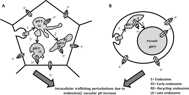
Amphotericin B (AMB) is an effective antifungal agent. However, its therapeutic use is hampered by its toxicity, mainly due to channel formation across kidney cell membranes and the disruption of postendocytic trafficking. We previously described a safe injectable AMB-arabinogalactan (AG) conjugate with neutralized toxicity. Here we studied the mechanism of the toxicity of free AMB and its neutralization by conjugation with AG. AMB treatment of a kidney cell line modulated the trafficking of three receptors (C-X-C chemokine receptor type 4 [CXCR4], M1 receptor, and human transferrin receptor [hTfnR]) due to an increase in endosomal pH. Similar data were also obtained in yeast but with an increase in vacuolar pH and the perturbation of Hxt2-green fluorescent protein (GFP) trafficking. The conjugation of AMB with AG neutralized all elements of the toxic activity of AMB in mammalian but not in fungal cells. Based on these results, we provide an explanation of how the conjugation of AMB with AG neutralizes its toxicity in mammalian cells and add to the knowledge of the mechanism of action of free AMB in both fungal and mammalian cells.


Figures

Fig 1
Fig 1
AMB and AMB-AGC killing kinetics, induction of K+ leakage, and internalization of Hxt2-GFP from the PM to endosomal compartments in yeast. (A) C. albicans yeast cells were treated with AMB or AMB-AGC in YPD medium. Aliquots were collected at the indicated time points, and survival rates were determined (%CFU). Similar results were obtained when PBS was used instead of the YPD medium. (B) C. albicans yeast cells were treated with AMB or AMB-AGC. Aliquots were collected at the indicated time points and washed, and the internal K+ concentration was determined by atomic absorption spectroscopy. (C) S. cerevisiae expressing the Hxt2-GFP construct were incubated in 5% glucose for 18 h (i) and then washed and transferred to 0.2% glucose for 4 h (ii). The yeast cells were then transferred again to 5% glucose for 70 min of no drug (control) (iii), 1 μg/ml AMB (iv), 1 μg/ml AMB-AGC (v), or 5 μg/ml AMB-AGC (vi). The yeast suspension was fixed with 4% paraformaldehyde (PFA) and visualized by fluorescence microscopy. Also shown are a cell with Hxt2-GFP in its PM and vacuole (vii), with Hxt2-GFP in the eisosomes (viii), and with Hxt2-GFP in the endosomal compartment (ix). (D) Quantification of the experiment shown in panel C. The cells were classified according to the localization of Hxt2-GFP (n = number of cells).
Fig 2
Fig 2
Increased vacuolar pH in C. albicans following treatment with AMB or AMB-AGC. (A) BCECF calibration curve. (B) BCECF-AM-labeled C. albicans treated with AMB. (C) BCECF-AM-labeled C. albicans treated with AMB-AGC. Fluorescence intensities were measured in panels B and C at different time points: ⧫, 9 min; ■, 20 min; ▲, 35 min; x, 60 min. The pHs were calculated according to the calibration curve. For each examined concentration, statistical significance was found at maximal pH increase (P < 0.05).
Fig 3
Fig 3
AMB-induced toxicity and K+ leakage in kidney cell lines are abolished by the conjugation of AMB with AG. Vero cells were treated for 1 h (A) and MDCK-PTR9 cells for 19 h (B) with the specified concentrations of AMB or AMB-AGC, and the metabolic activity was measured by an XTT assay. (C) Vero cells were treated for 1 h with the indicated concentrations of AMB or AMB-AGC. The internal K+ concentration was measured by atomic absorption spectrometry and normalized to the number of cells. The error bars show concentrations ± the SDs. One asterisk represents a P value of 0.0032 compared to the value for the no-drug control.
Fig 4
Fig 4
AMB, but not AMB-AGC, modulates the distribution of receptors in MDCK cells. (A) MDCK cells stably expressing M1-GFP were grown on glass coverslips and treated for 1 h with no drug (control), 40 μg/ml AMB, or 400 μg/ml AMB-AGC. The cells were visualized by fluorescence microscopy after fixation. (B) MDCK-PTR9 cells grown on glass coverslips in MEM/BSA medium were treated for 19 h (i to iii) or 24 h (iv to vi) with AMB (10 μg/ml), AMB-AGC (400 μg/ml), or no drug (control) and then incubated with 60 mg/ml of FITC-hTfn for 60 min on ice to induce binding. After intensive washing, the cells were transferred to fresh medium and incubated at 37°C for 30 min to enable endocytosis. The cells were then washed with acidic buffer to strip membrane-attached FITC-hTfn from the membrane, fixed, and visualized by confocal microscopy. (C) After FITC-hTfn internalization and fixation (19 h treatment), the cells were labeled with mouse anti-EEA1 and then with Cy5-conjugated goat anti-mouse IgG. The samples were documented by confocal microscopy, and quantification of FITC-hTfn colocalization with EEA1 was performed. The Manders' M1 coefficients are presented. Colocalization analyses were performed for at least 20 cells in at least four different microscopic fields. The error bars represent Manders' M1 coefficients ± the SDs. One asterisk represents a P value of 0.00197 compared to the value for the no-drug control. Gray and white columns represent internalization for 5 and 30 min, respectively.
Fig 5
Fig 5
The AMB-induced increase in endosomal pH in kidney cells is abolished by conjugation with AG. (A) Calibration curve of FITC-hTfn fluorescence intensity in MDCK-PTR9 cell endosomes as a function of pH. (B) MDCK-PTR9 cells were treated with AMB or AMB-AGC for 18 h, followed by 60 min of incubation on ice with 60 μg/ml FITC-hTfn to induce binding. After intensive washing, the cells were transferred to fresh medium and incubated at 37°C for 30 min to enable endocytosis. The samples were documented by confocal microscopy, and the pH was calculated as described in Materials and Methods. The columns show the average pHs (± the standard errors of the mean) of 145 to 170 endosomes in at least 16 different cells. One asterisk represents a P value of <0.0001 compared to the value for the no-drug control.
Fig 6
Fig 6
A proposed model for AMB activity in mammalian and fungal cells. AMB forms polar channels in mammalian PM (A) and fungal PM (B), which results in ion and metabolite leakage out of the cells. In addition, the AMB channels endocytose into endosomes (steps 1 and 4). In fungal cells, they arrive at the vacuole by postendocytic trafficking (step 5), while in mammalian cells they are transferred from early endosomes to the recycling endosomes and late endosomes (steps 2 and 3). The AMB channels in endosomes and vacuoles allow H+ leakage to the cytosol, leading to an endosomal/vacuolar pH increase, which in turn perturbs the trafficking steps.

References

    1. Ali R, Brett CL, Mukherjee S, Rao R. 2004. Inhibition of sodium/proton exchange by a Rab-GTPase-activating protein regulates endosomal traffic in yeast. J. Biol. Chem. 279:4498–4506 - PubMed
    1. Altschuler Y, et al. 1999. ADP-ribosylation factor 6 and endocytosis at the apical surface of Madin-Darby canine kidney cells. J. Cell Biol. 147:7–12 - PMC - PubMed
    1. Arnold TM, Dotson E, Sarosi GA, Hage CA. 2010. Traditional and emerging antifungal therapies. Proc. Am. Thorac. Soc. 7:222–228 - PubMed
    1. Baginski M, Resat H, Borowski E. 2002. Comparative molecular dynamics simulations of amphotericin B-cholesterol/ergosterol membrane channels. Biochim. Biophys. Acta 1567:63–78 - PubMed
    1. Bolard J. 1986. How do the polyene macrolide antibiotics affect the cellular membrane properties? Biochim. Biophys. Acta 864:257–304 - PubMed

MeSH terms

LinkOut - more resources