Review
. 2012 Aug 1;2(8):a007658.
doi: 10.1101/cshperspect.a007658.

Islet autoantigens: structure, function, localization, and regulation

Affiliations
Review

Islet autoantigens: structure, function, localization, and regulation

Peter Arvan et al. Cold Spring Harb Perspect Med. .

Abstract

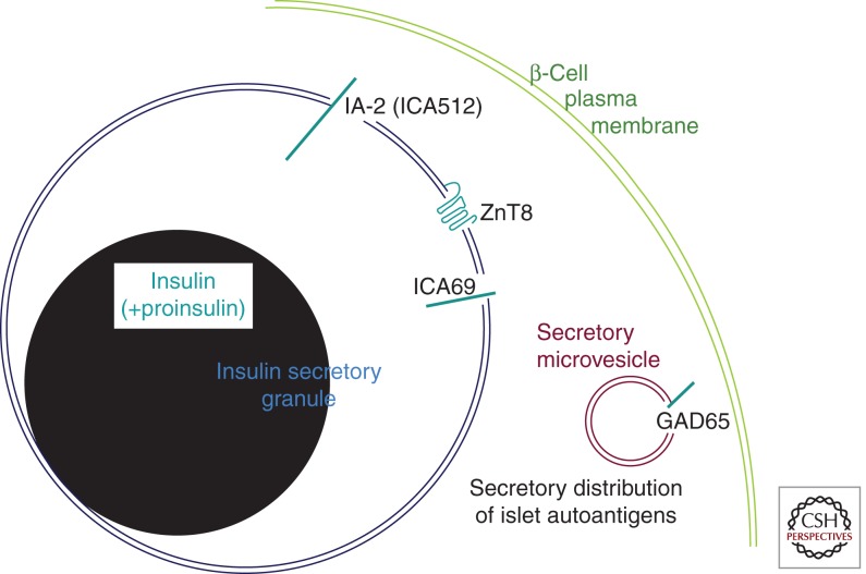
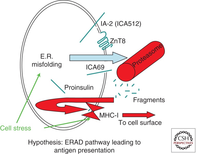
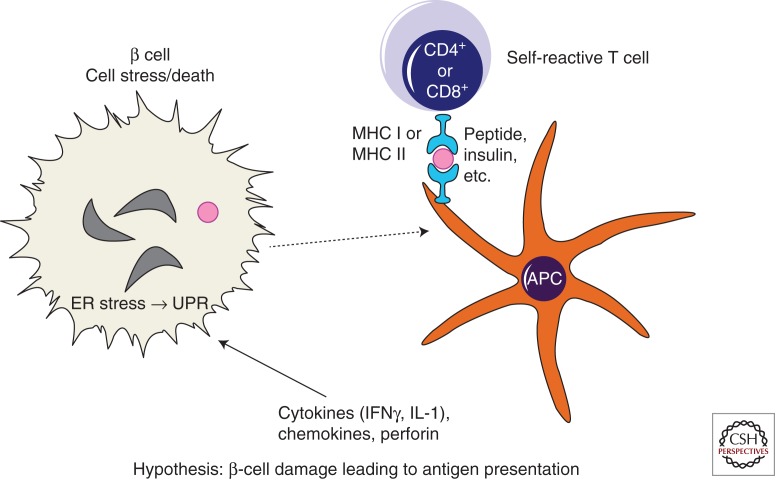
Islet autoantigens associated with autoimmune type 1 diabetes (T1D) are expressed in pancreatic β cells, although many show wider patterns of expression in the neuroendocrine system. Within pancreatic β cells, every T1D autoantigen is in one way or another linked to the secretory pathway. Together, these autoantigens play diverse roles in glucose regulation, metabolism of biogenic amines, as well as the regulation, formation, and packaging of secretory granules. The mechanism(s) by which immune tolerance to islet-cell antigens is lost during the development of T1D, remains unclear. Antigenic peptide creation for immune presentation may potentially link to the secretory biology of β cells in a number of ways, including proteasomal digestion of misfolded products, exocytosis and endocytosis of cell-surface products, or antigen release from dying β cells during normal or pathological turnover. In this context, we evaluate the biochemical nature and immunogenicity of the major autoantigens in T1D including (pro)insulin, GAD65, ZnT8, IA2, and ICA69.


Figures

Figure 1.
Figure 1.
Insulin residues B9–B23 contribute to the dimerization interface between insulin monomers. (A) The crystal structure (Protein Data Bank code 2R34) of two insulin monomers is displayed. Atoms underlying the molecular surface are colored blue for nitrogen, red for oxygen, and green for carbon for monomer 1 A chain, cyan for carbon for monomer 1 B chain, yellow for carbon for monomer 2 B chain, and magenta for carbon for monomer 2 A chain. A chloride ion is depicted as a green sphere. A manganese (II) ion is depicted as a purple sphere. (B) The relative contribution of insulin B-chain residues from each monomer that contributes to the dimerization interface is shown. PROTORP was used to analyze the interfaces between insulin chains, which shows that B9–B23 residues participate in the dimerization interface. B16 tyrosine is buried at the interface contributing more (% interface surface accessible area) to the dimerization interface (21%) than other residues.
Figure 2.
Figure 2.
Predominant intracellular distribution of major T1D autoantigens in pancreatic β cells. Two organelles of the β-cell secretory pathway are shown bearing autoantigens (in blue-green). The secretory granule contains primarily insulin (shown in black, similar to the appearance of the insulin crystal by transmission electron microscopy). A small fraction of unconverted proinsulin is also contained within secretory granules. The “clear space” surrounding the insulin granule core is thought to be enriched in the soluble C-peptide, which is not specifically denoted in the figure. The secretory granule membrane is the primary site of distribution of three additional β-cell autoantigens: ZnT8 is a polytopic membrane protein, IA-2 is a single-spanning transmembrane protein with both extensive luminal and cytosolic domains, and ICA69 is a type 1 transmembrane protein predominantly residing on the lumenal side of the membrane. As noted in the text, GAD65 localizes away from these other autoantigens, residing primarily on the cytosolic side of the membrane of secretory microvesicles, also known as “synaptic-like vesicles” (SNLVs).
Figure 3.
Figure 3.
Hypothesis: ERAD of misfolded secretory pathway proteins triggers MHC class I loading and presentation of autoantigens. In the case of misfolded secretory pathway proteins, retrotranslocation from the ER to the cytosol triggers degradation via the ubiquitin-proteasome system. The generation of small cleavage fragments and their transport back into the ER lumen allows for peptide loading of MHC class I (via the TAP/tapasin complex). Cell stress can promote ER misfolding of secretory and membrane proteins (Kuznetsov and Nigam 1998) and also may promote expression of major histocompatibility complex class I-related genes (Groh et al. 1996). Thus, it is a plausible hypothesis that the net result of these two effects is enhanced β-cell autoantigen presentation.
Figure 4.
Figure 4.
Simplified model of β-cell damage leading to antigen presentation. T cells can directly kill β cells through a cytotoxic process, but they can also influence β-cell destruction via release of mediators such as cytokines, chemokines, or perforin. Cytokine activation of inducible nitric oxide synthase can activate ER stress response signaling (Oyadomari et al. 2001)—pathways collectively known as the unfolded protein response (UPR). It is therefore conceivable that cell stress including UPR may be a potential contributor to β-cell toxicity in T1D. Processing of autoantigens within β cells generates peptides that are then taken up by antigen-presenting cells (APCs), either as whole dead β cells or β-cell fragments, for eventual further processing/presentation of these islet peptides to self-reactive T cells.

References

    1. Achenbach P, Koczwara K, Knopff A, Naserke H, Ziegler A-G, Bonifacio E 2004. Mature high-affinity immune responses to (pro)insulin anticipate the autoimmune cascade that leads to type 1 diabetes. J Clin Invest 589–597 - PMC - PubMed
    1. Achenbach P, Lampasona V, Landherr U, Koczwara K, Krause S, Grallert H, Winkler C, Pfluger M, Illig T, Bonifacio E, et al. 2009. Autoantibodies to zinc transporter 8 and SLC30A8 genotype stratify type 1 diabetes risk. Diabetologia 52: 1881–1888 - PubMed
    1. Akirav E, Kushner JA, Herold KC 2008. β-cell mass and type 1 diabetes: Going, going, gone? Diabetes 57: 2883–2888 - PMC - PubMed
    1. Alarcón C, Lincoln B, Rhodes CJ 1993. The biosynthesis of the subtilisin-related proprotein covertase PC3, but not that of the PC2 convertase, is regulated by glucose in parallel to proinsulin biosynthesis in rat pancreatic islets. J Biol Chem 268: 4276–4280 - PubMed
    1. Allen JS, Pang K, Skowera A, Ellis R, Rackham C, Lozanoska-Ochser B, Tree T, Leslie RD, Tremble JM, Dayan CM, et al. 2009. Plasmacytoid dendritic cells are proportionally expanded at diagnosis of type 1 diabetes and enhance islet autoantigen presentation to T-cells through immune complex capture. Diabetes 58: 138–145 - PMC - PubMed

Publication types

MeSH terms