

Review
doi: 10.1101/cshperspect.a011056.
PlGF: a multitasking cytokine with disease-restricted activity
Affiliations
- PMID: 22908198
- PMCID: PMC3405829
- DOI: 10.1101/cshperspect.a011056
Item in Clipboard
Review
PlGF: a multitasking cytokine with disease-restricted activity
Cold Spring Harb Perspect Med.
.
Abstract
Placental growth factor (PlGF) is a member of the vascular endothelial growth factor (VEGF) family that also comprises VEGF-A (VEGF), VEGF-B, VEGF-C, and VEGF-D. Unlike VEGF, PlGF is dispensable for development and health but has diverse nonredundant roles in tissue ischemia, malignancy, inflammation, and multiple other diseases. Genetic and pharmacological gain-of-function and loss-of-function studies have identified molecular mechanisms of this multitasking cytokine and characterized the therapeutic potential of delivering or blocking PlGF for various disorders.
Figures
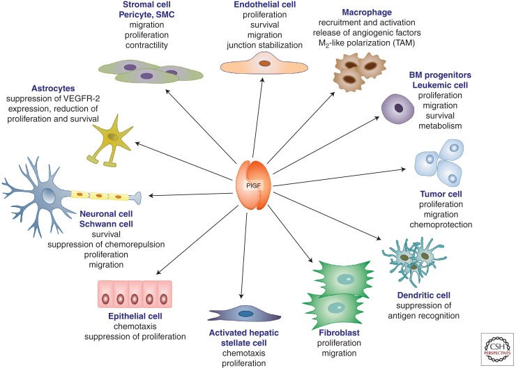
PlGF is a multitasking cytokine affecting various cellular activities. Scheme illustrating the pleiotropic actions of PlGF, including effects on survival, migration, proliferation, metabolism, and activation effects on vascular (endothelial cells, pericytes/smooth muscle cells) as well as nonvascular cells (macrophages, bone marrow–derived progenitors, tumor cells, dendritic cells, fibroblasts, hepatic stellate cells, epithelial cells, neurons, Shwann cells, astrocytes). BM, Bone marrow; SMC, smooth muscle cell; TAM, tumor-associated macrophage.
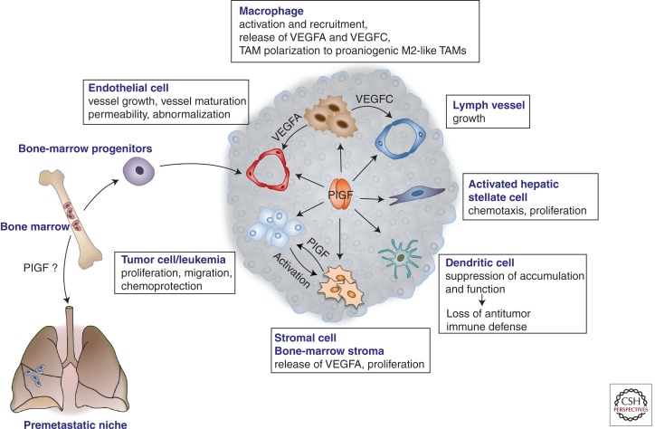
Roles of PlGF in cancer and metastasis. PlGF affects various cellular components and processes in the tumor. It affects angiogenesis by promoting proliferation and migration of endothelial cells, maturation of the vessels by recruiting smooth muscle cells, mobilization of vasculogenic bone-marrow progenitors, and recruitment of macrophages, which produce additional angiogenic and lymphangiogenic factors. PlGF also enhances the “disorganization” of tumor vessels, characterized by an irregular appearance and discontinuous endothelial lining, including “sinusoidal capillarization” in HCC (see also Fig. 3). PlGF also promotes the proliferation and migration of activated hepatic stellate cells in HCC and liver fibrosis. PlGF reduces dendritic cell accumulation and function, thereby suppressing antitumor immune defense responses. PlGF directly stimulates proliferation of tumor cells, which cross talk to and activate stromal cells to produce PlGF (see also Fig. 4). PlGF was also implicated in the mobilization of bone marrow–derived progenitor cells to the “premetastic niche,” although conflicting data have been reported. TAM, Tumor-associated macrophage; VEGFA, vascular endothelial growth factor A; VEGFC, vascular endothelial growth factor C.
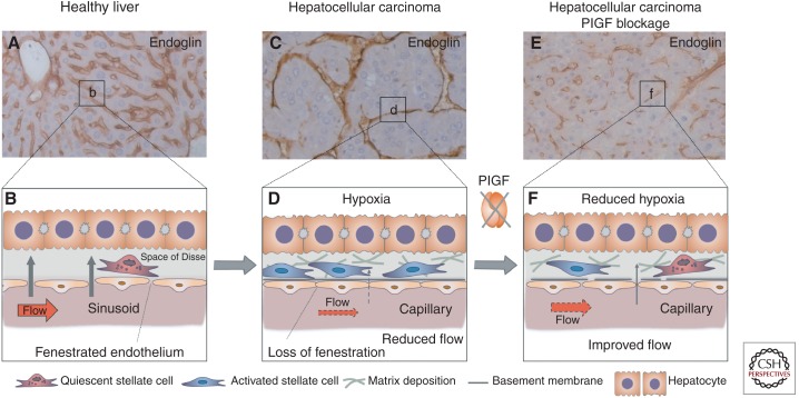
Role of PlGF in sinusoidal disorganization in HCC. (A,B) In healthy liver, the hepatic microvascular units, the sinusoids (visualized by endoglin immunostaining in A), are characterized by a porous fenestrated endothelial lining and absence of a basement membrane, facilitating oxygen and metabolic exchange between the bloodstream and the hepatocytes; the latter are separated from the sinusoids by the perisinusoidal space (space of Disse), where quiescent hepatic stellate cells reside (B). (C,D) In hepatocellular carcinoma, sinusoids undergo “capillarization,” characterized by loss of fenestration, increased numbers of activated stellate cells that deposit matrix and release angiogenic factors, change of shape and size of the capillarized sinusoids to more tortuous vessels, and lumen loss in a fraction of the capillaries. These changes result in impaired blood flow (red arrow in D) along with reduced oxygen transfer from the blood to the liver parenchyma (gray arrow in D), resulting in increased hypoxia, which further promotes tumor cell proliferation resulting in enlarged intercapillary distances (C). (E,F) PlGF blockage by gene silencing or by inhibition with neutralizing antibody partially prevented this disorganization of hepatic sinusoids. The abnormal tortuous appearance, abnormal size, and loss of lumenization of the sinusoids were attenuated (E), and sinusoidal “capillarization” was reduced (F). This partial “normalization” by PlGF blockage functionally improved tissue oxygenation and reduced proliferation of HCC cells (Van de Veire et al. 2010; Rolny et al. 2011).
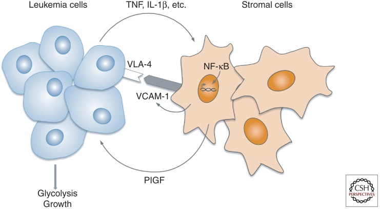
Bidirectional cross talk between leukemia and bone marrow stromal cells. In chronic myeloid leukemia (CML), tumor cells “educate” bone marrow stromal cells to produce PlGF. This induction involves NF-κB activation in the stromal cells and requires leukemia cell/stromal cell contact, which is in part mediated by VLA-4/VCAM-1 interaction. VLA-4/VCAM-1 interaction is reported to activate NF-κB (Zohlnhofer et al. 2000), and NF-κB, in turn, up-regulates VCAM-1 (Rajan et al. 2008), suggesting a positive feedback loop reinforcing VLA4+ CML cell binding to the VCAM-1-expressing stromal cells, thereby ensuring PlGF production. Stromal cell-derived PlGF then promotes proliferation and metabolism of the CML cells. Overall, PlGF creates a fertile microenvironmental soil for the seeding tumor cells to foster cancer cell survival and expansive growth (Schmidt et al. 2011).
Similar articles
-
Synergism between vascular endothelial growth factor and placental growth factor contributes to angiogenesis and plasma extravasation in pathological conditions.Nat Med. 2001 May;7(5):575-83. doi: 10.1038/87904. Nat Med. 2001. PMID: 11329059
-
Structure and function of placental growth factor.Trends Cardiovasc Med. 2002 Aug;12(6):241-6. doi: 10.1016/s1050-1738(02)00168-8. Trends Cardiovasc Med. 2002. PMID: 12242046 Review.
-
Prevention of elastase-induced emphysema in placenta growth factor knock-out mice.Respir Res. 2009 Nov 23;10(1):115. doi: 10.1186/1465-9921-10-115. Respir Res. 2009. PMID: 19930612 Free PMC article.
-
Placental growth factor and its receptor, vascular endothelial growth factor receptor-1: novel targets for stimulation of ischemic tissue revascularization and inhibition of angiogenic and inflammatory disorders.J Thromb Haemost. 2003 Jul;1(7):1356-70. doi: 10.1046/j.1538-7836.2003.00263.x. J Thromb Haemost. 2003. PMID: 12871269 Review.
-
Loss of placental growth factor protects mice against vascular permeability in pathological conditions.Biochem Biophys Res Commun. 2002 Jul 12;295(2):428-34. doi: 10.1016/s0006-291x(02)00677-0. Biochem Biophys Res Commun. 2002. PMID: 12150967
Cited by
-
PlGF and VEGF-A Regulate Growth of High-Risk MYCN-Single Copy Neuroblastoma Xenografts via Different Mechanisms.Int J Mol Sci. 2016 Sep 23;17(10):1613. doi: 10.3390/ijms17101613. Int J Mol Sci. 2016. PMID: 27669225 Free PMC article.
-
Recent molecular discoveries in angiogenesis and antiangiogenic therapies in cancer.J Clin Invest. 2013 Aug;123(8):3190-200. doi: 10.1172/JCI70212. Epub 2013 Aug 1. J Clin Invest. 2013. PMID: 23908119 Free PMC article. Review.
-
Interdependence of Angiogenesis and Arteriogenesis in Development and Disease.Int J Mol Sci. 2022 Mar 31;23(7):3879. doi: 10.3390/ijms23073879. Int J Mol Sci. 2022. PMID: 35409246 Free PMC article. Review.
-
Epigenetic control of hypoxia inducible factor-1α-dependent expression of placental growth factor in hypoxic conditions.Epigenetics. 2014 Apr;9(4):600-10. doi: 10.4161/epi.27835. Epub 2014 Feb 6. Epigenetics. 2014. PMID: 24504136 Free PMC article.
-
Elastase induced lung epithelial cell apoptosis and emphysema through placenta growth factor.Cell Death Dis. 2013 Sep 5;4(9):e793. doi: 10.1038/cddis.2013.329. Cell Death Dis. 2013. PMID: 24008737 Free PMC article.
References
-
- Adini A, Kornaga T, Firoozbakht F, Benjamin LE 2002. Placental growth factor is a survival factor for tumor endothelial cells and macrophages. Cancer Res 62: 2749–2752 - PubMed
-
- Akrami H, Soheili ZS, Sadeghizadeh M, Ahmadieh H, Rezaeikanavi M, Samiei S, Khalooghi K 2011. PlGF gene knockdown in human retinal pigment epithelial cells. Graefes Arch Clin Exp Ophthalmol 249: 537–546 - PubMed
-
- Apple FS, Pearce LA, Chung A, Ler R, Murakami MM 2007. Multiple biomarker use for detection of adverse events in patients presenting with symptoms suggestive of acute coronary syndrome. Clin Chem 53: 874–881 - PubMed
-
- Autiero M, Waltenberger J, Communi D, Kranz A, Moons L, Lambrechts D, Kroll J, Plaisance S, De Mol M, Bono F, et al. 2003. Role of PlGF in the intra- and intermolecular cross talk between the VEGF receptors Flt1 and Flk1. Nat Med 9: 936–943 - PubMed
-
- Babiak A, Schumm AM, Wangler C, Loukas M, Wu J, Dombrowski S, Matuschek C, Kotzerke J, Dehio C, Waltenberger J 2004. Coordinated activation of VEGFR-1 and VEGFR-2 is a potent arteriogenic stimulus leading to enhancement of regional perfusion. Cardiovasc Res 61: 789–795 - PubMed
Publication types
MeSH terms
Substances
LinkOut - more resources
Full Text Sources