. 2012 Oct 19;287(43):36208-21.
doi: 10.1074/jbc.M112.400705. Epub 2012 Aug 20.

Crystal structure of reaction intermediates in pyruvate class II aldolase: substrate cleavage, enolate stabilization, and substrate specificity

Affiliations

Crystal structure of reaction intermediates in pyruvate class II aldolase: substrate cleavage, enolate stabilization, and substrate specificity

Mathieu Coincon et al. J Biol Chem. .

Abstract

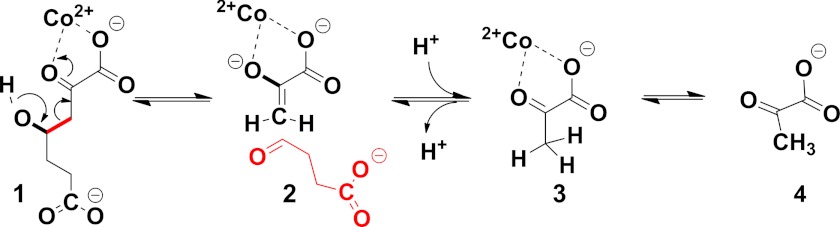
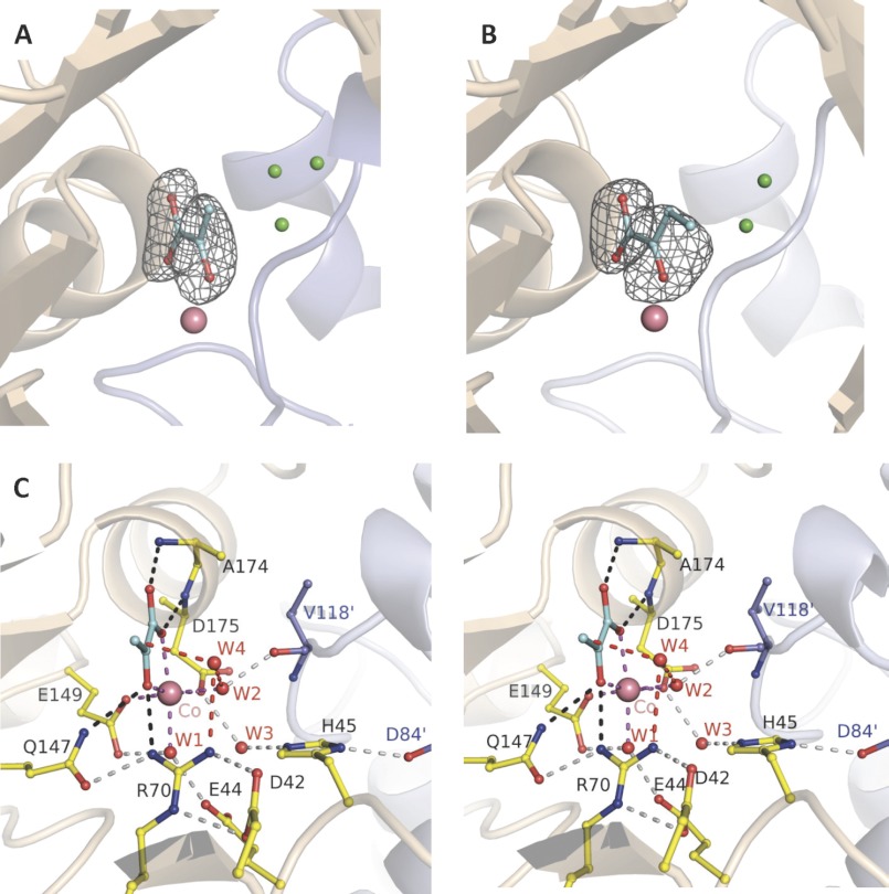
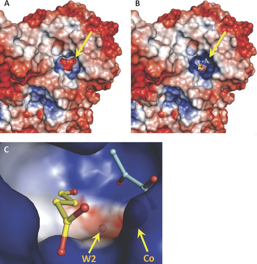
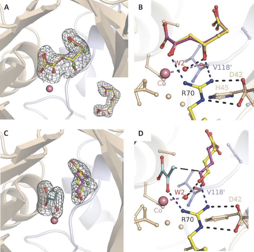
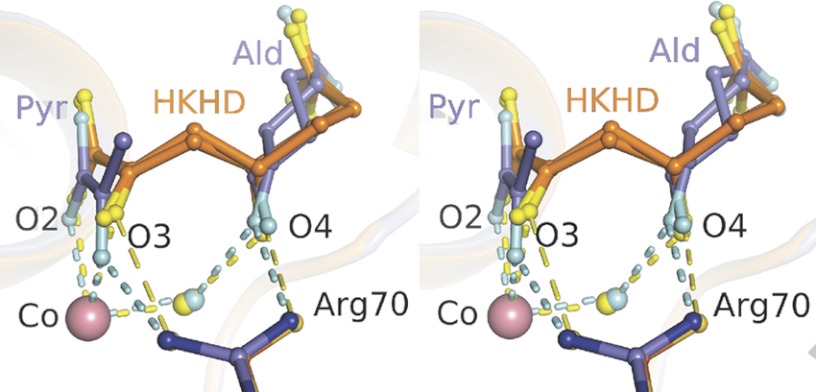
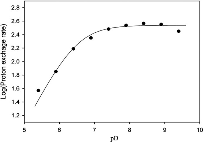
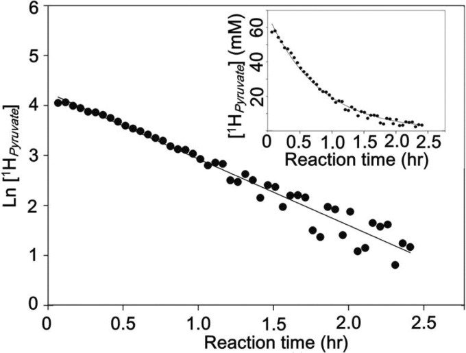
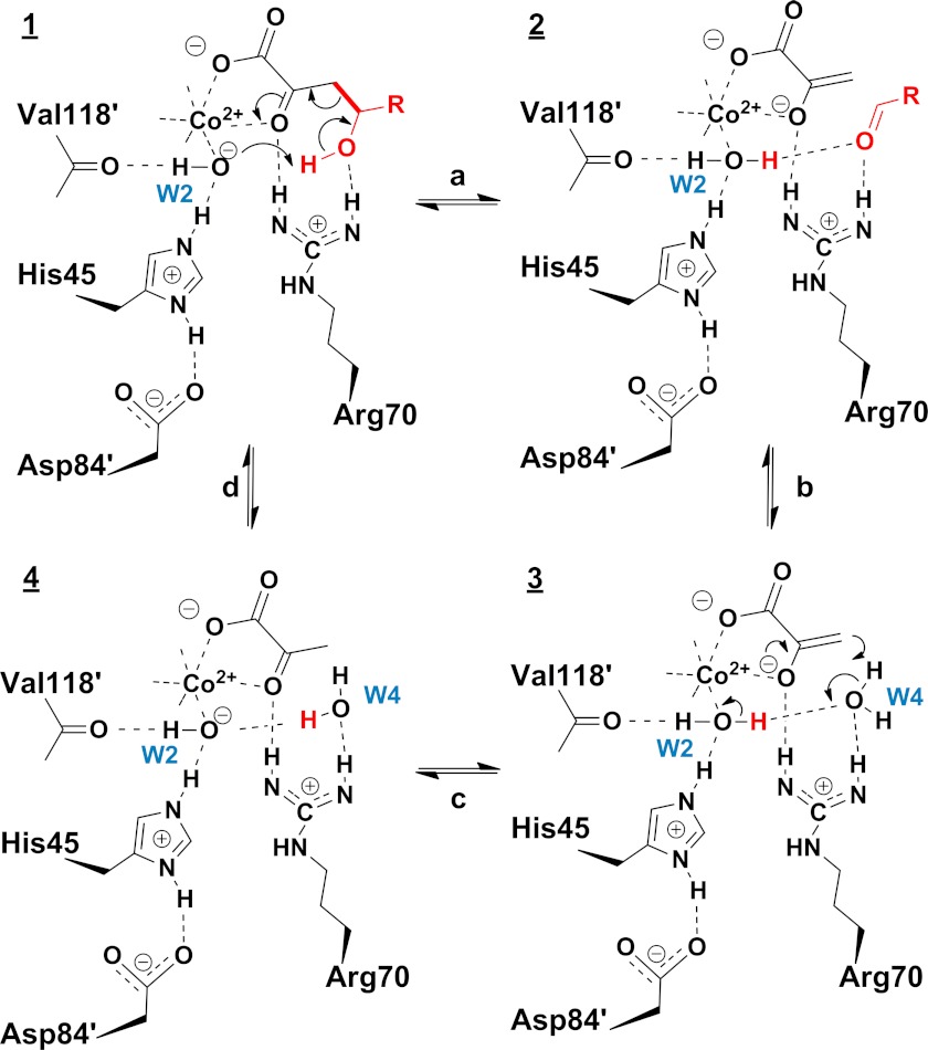
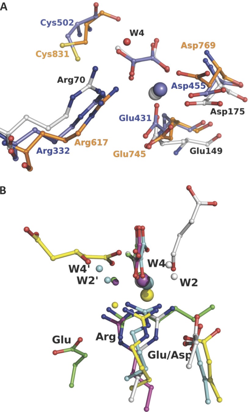
Crystal structures of divalent metal-dependent pyruvate aldolase, HpaI, in complex with substrate and cleavage products were determined to 1.8-2.0 Å resolution. The enzyme·substrate complex with 4-hydroxy-2-ketoheptane-1,7-dioate indicates that water molecule W2 bound to the divalent metal ion initiates C3-C4 bond cleavage. The binding mode of the aldehyde donor delineated a solvent-filled capacious binding locus lined with predominantly hydrophobic residues. The absence of direct interactions with the aldehyde aliphatic carbons accounts for the broad specificity and lack of stereospecific control by the enzyme. Enzymatic complex structures formed with keto acceptors, pyruvate, and 2-ketobutyrate revealed bidentate interaction with the divalent metal ion by C1-carboxyl and C2-carbonyl oxygens and water molecule W4 that is within close contact of the C3 carbon. Arg(70) assumes a multivalent role through its guanidinium moiety interacting with all active site enzymatic species: C2 oxygen in substrate, pyruvate, and ketobutyrate; substrate C4 hydroxyl; aldehyde C1 oxygen; and W4. The multiple interactions made by Arg(70) stabilize the negatively charged C4 oxygen following proton abstraction, the aldehyde alignment in aldol condensation, and the pyruvate enolate upon aldol cleavage as well as support proton exchange at C3. This role is corroborated by loss of aldol cleavage ability and pyruvate C3 proton exchange activity and by a 730-fold increase in the dissociation constant toward the pyruvate enolate analog oxalate in the R70A mutant. Based on the crystal structures, a mechanism is proposed involving the two enzyme-bound water molecules, W2 and W4, in acid/base catalysis that facilitates reversible aldol cleavage. The same reaction mechanism promotes decarboxylation of oxaloacetate.


Figures

SCHEME 1.
SCHEME 1.
Intermediates (1–4) of the catalytic mechanism in pyruvate class II aldolases.
FIGURE 1.
FIGURE 1.
Trapped 2-keto acids in active site of HpaI pyruvate aldolase. A, electron density of pyruvate bound in the active site of the native enzyme. B, electron density of ketobutyrate trapped in the active site of the native enzyme. C, stereoview showing active site interactions when pyruvate binds to HpaI aldolase. In A and B, water molecules occupying the aldehyde binding site (presented in Fig. 3) are shown in green. Magenta dashes depict coordination of Co2+ made by the two conserved water molecules (W1 and W2), pyruvate (blue), and residues Glu149 and Asp175. The backbone nitrogens of Ala174 and Glu175 direct the carboxylate oxygens of the pyruvate molecule, and Gln147 and Arg70 interact with the keto oxygen; these interactions are shown by black dashes. These interactions are used to specifically orient pyruvate in the active site. Hydrogen bonding interactions not directly contacting the metal ion or pyruvate (gray dots) organize remaining active site residues and water molecules. Interactions from Val118′ and Asp84′ of an adjacent contacting subunit (blue) complete the active site binding locus. The consistency in hydrogen bonding interactions made with W3 implies that Glu44 and Glu172 are hydrogen bond acceptors, whereas His45 is a hydrogen bond donor. Electron density encompassing the ligands was calculated from a simulated annealing FoFc omit map and contoured at 3.5 σ.
FIGURE 2.
FIGURE 2.
The electrostatic surface of an HpaI pyruvate aldolase evaluated by adaptive Poisson-Boltzmann solver. Views of the pyruvate aldolase active site (shown by yellow arrow) and of pyruvate aldolase in complex with substrates, pyruvate (cyan) and succinic semialdehyde (yellow) are shown. The color code has blue as positive charged and red as negative charged. A, electrostatic surface of apoenzyme where divalent cobalt ion and ligands were not included in electrostatic surface calculations. B, the positive charge in the active site in native holoenzyme with cobalt facilitates the binding of pyruvate and the aldehyde moiety of succinic semialdehyde, shown as stick models. Crystallographic symmetry operations were used to generate a complete active site. C, close-up of the active site cavity depicting bound pyruvate and succinic semialdehyde as stick models and showing the large neutral cavity (white) adjacent to the aldehyde ligand. W2, which hydrogen bonds the aldehyde carbonyl oxygen, and the divalent cobalt are also shown as semitransparent spheres. The electrostatic surface at W2 has a slight negative charge.
FIGURE 3.
FIGURE 3.
Substrate and cleavage products trapped in HpaI pyruvate aldolase active site. A, electron density of substrate HKHD trapped in the native enzyme. HKHD orientation corresponding to R chirality at C4 is shown in yellow, and that of S chirality is shown in red. The inset shows the same electron density looking down and corresponds to a counterclockwise rotation in the vertical plane of ∼90°. B, active site interactions implicating the metal cofactor, HKHD, Arg70, and W2. C, electron density of cleavage products pyruvate and succinic semialdehyde (re face orientation in yellow; si face in pink). D, interactions highlighting succinic semialdehyde binding and recognition. Hydrogen bonds are shown as short dashes, whereas long dashes connect atoms that are chemically bound upon aldol condensation. Water molecules in alternate conformation with the aldehyde binding site are shown in green. Upon pyruvate binding, Arg70 and the conserved water W2 (red) orient the carbonyl moiety of the aldehyde substrate. In B and D, the angle subtended by either C4 hydroxyl or aldehyde oxygen, W2, and Val118′ corresponds to tetrahedral hybridization geometry. Electron density encompassing the ligands was calculated from a simulated annealing FoFc omit map and contoured at 3.5 σ. The divalent cobalt ion is depicted by a salmon sphere.
FIGURE 4.
FIGURE 4.
Superposition of the active site binding geometries of HKHD and resultant cleavage products trapped in the active site of HpaI pyruvate aldolase. Substrate HKHD (orange) is depicted for both R and S conformations at its C4 hydroxyl, whereas the aldehyde (dark gray) is shown in re and si face orientations. Hydrogen bonds are shown as short dashes. Close van der Waals contacts made by Arg70 with HKHD O3 and O4 atoms are shown by long dashes (yellow). The divalent cobalt ion (Co) is depicted by a salmon sphere. The composite image corresponds to superposition of one HpaI subunit bound with HKHD and the equivalent subunit in complex with its cleavage products. The interactions by HKHD with Co(II) and Arg70 are weaker, indicated by longer interatomic distances, compared with the equivalent interactions made with Co(II) and Arg70 by the oxygen atoms of the 2-keto moiety, O2 and O3, as well as aldehyde C1 oxygen in the structure binding the cleavage products. Ald, aldehyde; Pyr, pyruvate.
FIGURE 5.
FIGURE 5.
pD profile of pyruvate C3 proton exchange reaction in HpaI aldolase. Assays were performed in deuterated constant ionic strength buffer containing 100 mm Tris, 50 mm acetic acid, 50 mm MES, and 1.0 mm CoCl2 with pD ranging from 5.4 to 9.4. The data were fitted to Equation 1 by non-linear regression using Leonora and showed a pK1 of 6.5 ± 0.1.
FIGURE 6.
FIGURE 6.
α-Proton exchange of 2-ketobutyrate catalyzed by Co2+-HpaI aldolase. The proton exchange was monitored in 20 mm deuterated MOPS buffer, pD 8.0 by 1H NMR. The loss of proton signal at the C3 carbon of 2-ketobutyrate followed a single exponential decay model.
SCHEME 2.
SCHEME 2.
Proposed catalytic mechanism (reactions a–d and intermediates 14) of C–C bond cleavage in pyruvate aldolase. R = CH2CH2COOH.
FIGURE 7.
FIGURE 7.
Active site architecture comparison of evolutionary related enzymes and non-homologous pyruvate lyases. A shows superposition of the HpaI·pyruvate complex structure with the P-enolpyruvate binding domain of enzyme I for bacterial P-enolpyruvate-dependent carbohydrate:phosphotransferase systems (Protein Data Bank code 2BG5, residues 261–573) and pyruvate, phosphate dikinase (Protein Data Bank code 1KC7, residues 534–874). B shows superposition of HpaI pyruvate aldolase (white), human HMG-CoA lyase (Protein Data Bank code 2CW6) (yellow), DmpG aldolase (Protein Data Bank code 1NVM) (cyan), bacterial oxaloacetate decarboxylase (Protein Data Bank code 3B8I) (magenta), and HMG/CHA aldolase (Protein Data Bank code 3NOJ) (green). Structures are presented from the same perspective, taking the pyruvate analog and the metal ion (large sphere) as reference except for human HMG-CoA lyase as noted under “Material and Methods.” Succinic semialdehyde in HpaI aldolase is shown in white, and 3-hydroxyglutarate, a cleavage product of HMG-CoA lyase, is shown in yellow. For HpaI, the condensing aldehyde will approach from the distal side of the pyruvate, whereas for DmpG aldolase, HMG-CoA lyase, HMG/CHA aldolase, and oxaloacetate decarboxylase it will approach from the proximal side. Water molecules identified as W2′ and W4′, equivalent to W2 and W4, which are described as part of the catalytic machinery in HpaI aldolase, also show the same spatial disposition as would their respective condensing aldehydes. The guanidinium moieties of adjacent arginine residues cluster tightly (identified as Arg) and interact with their respective metal-binding ligand and water molecule identified as W4/W4′ where present. Water molecules within hydrogen bonding distance of W4/W4′ and bound to the metal ion are identified as W2/W2′. Note that the Arg70-Asp42 pair has a direct analog in HMG-CoA lyase (Arg41-Glu72), HMG/CHA aldolase (Arg123-Glu36), and DmpG aldolase (Arg17-Glu48), whereas only in oxaloacetate decarboxylase is the equivalent arginine residue, Arg159, not ion-paired with a carboxylate group of an amino acid. The carboxylate groups of the interacting aspartate or glutamate are identified (Asp or Glu).

Similar articles

Cited by

References

    1. Takayama S., McGarvey G. J., Wong C. H. (1997) Microbial aldolases and transketolases: new biocatalytic approaches to simple and complex sugars. Annu. Rev. Microbiol. 51, 285–310 - PubMed
    1. Samland A. K., Sprenger G. A. (2006) Microbial aldolases as C–C bonding enzymes—unknown treasures and new developments. Appl. Microbiol. Biotechnol. 71, 253–264 - PubMed
    1. M. J., Floriano B., J. J., Santero E. (2002) Identification of a hydratase and a class II aldolase involved in biodegradation of the organic solvent tetralin. Appl. Environ. Microbiol. 68, 4841–4846 - PMC - PubMed
    1. Joerger A. C., Gosse C., Fessner W. D., Schulz G. E. (2000) Catalytic action of fuculose 1-phosphate aldolase (class II) as, derived from structure-directed mutagenesis. Biochemistry 39, 6033–6041 - PubMed
    1. Kroemer M., Merkel I., Schulz G. E. (2003) Structure and catalytic mechanism of l-rhamnulose-1-phosphate aldolase. Biochemistry 42, 10560–10568 - PubMed

Publication types

LinkOut - more resources