. 2012 Nov 23;287(48):40140-9.
doi: 10.1074/jbc.M112.348763. Epub 2012 Aug 20.

Estrogen represses hepatocellular carcinoma (HCC) growth via inhibiting alternative activation of tumor-associated macrophages (TAMs)

Affiliations

Estrogen represses hepatocellular carcinoma (HCC) growth via inhibiting alternative activation of tumor-associated macrophages (TAMs)

Weiwei Yang et al. J Biol Chem. .

Abstract

Background: Hepatocarcinoma cancer (HCC) occurs more often in men than in women, and little is known about its underlying molecular mechanisms.

Results: We identify that 17β-estradiol (E2) could suppress tumor growth via regulating the polarization of macrophages.

Conclusion: Estrogen functions as a suppressor for macrophage alternative activation.

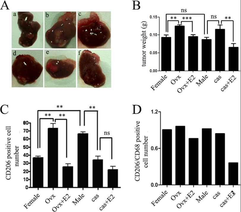
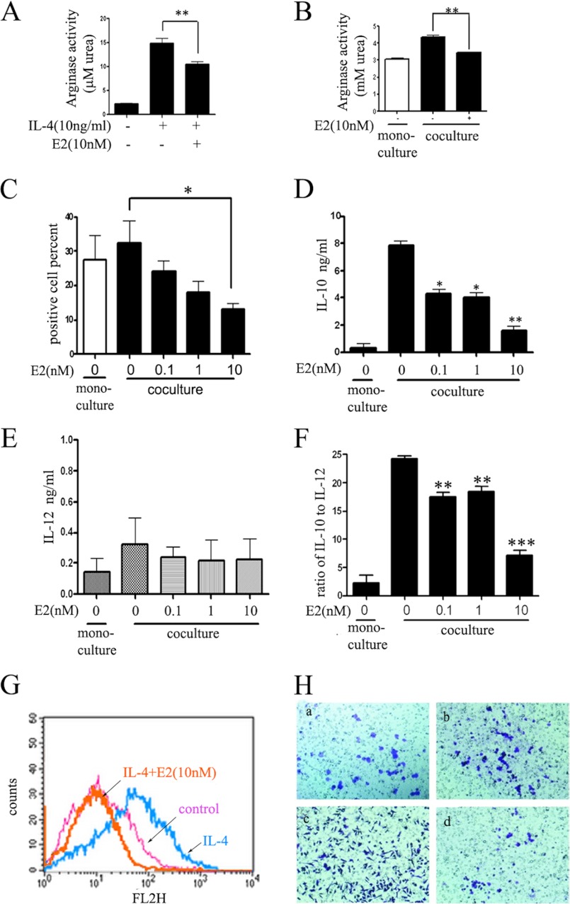
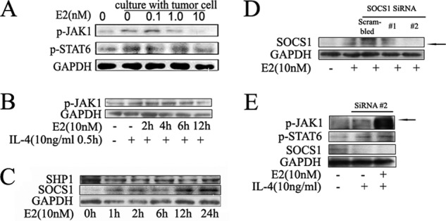
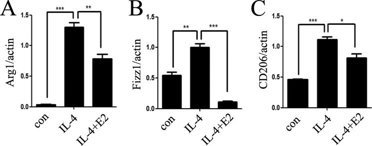
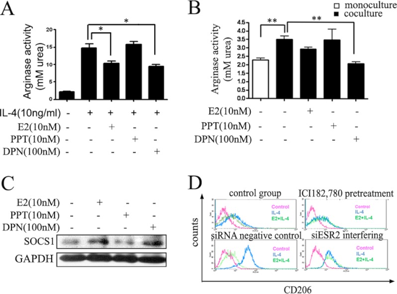
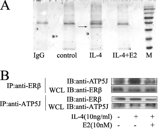
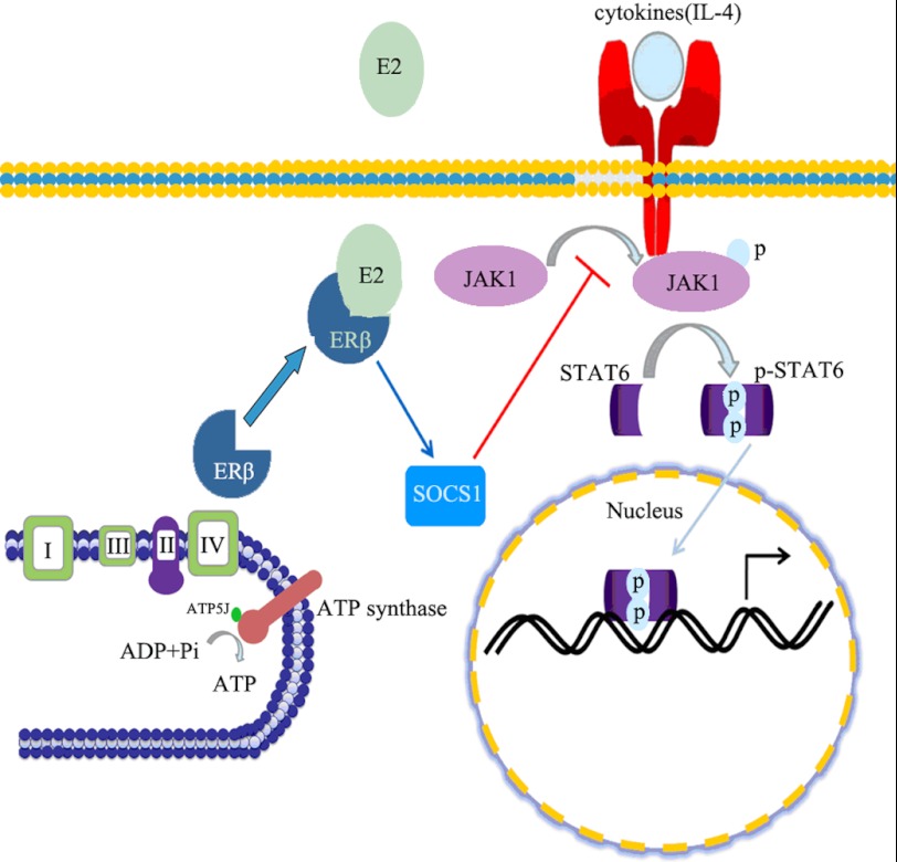
Significance: These studies introduce a novel mechanism for suppressing male-predominant HCC. Hepatocarcinoma cancer (HCC), one of the most malignant cancers, occurs significantly more often in men than in women; however, little is known about its underlying molecular mechanisms. Here we identified that 17β-estradiol (E2) could suppress tumor growth via regulating the polarization of macrophages. We showed that E2 re-administration reduced tumor growth in orthotopic and ectopic mice HCC models. E2 functioned as a suppressor for macrophage alternative activation and tumor progression by keeping estrogen receptor β (ERβ) away from interacting with ATP5J (also known as ATPase-coupling factor 6), a part of ATPase, thus inhibiting the JAK1-STAT6 signaling pathway. These studies introduce a novel mechanism for suppressing male-predominant HCC.


Figures

FIGURE 1.
FIGURE 1.
Estrogen inhibits macrophage alternative activation in orthotopic mouse liver tumor models. A, tumors developed in the left hepatic lobe of BALB/c mice 6 weeks after intrahepatic inoculation of 106 Heps cells in each group. a–c, females. a, female control; b, females that underwent ovariotomy; c, E2 (50 μg/kg) administration after ovariotomy. d–f, males. d, male control; e, males that underwent castration; f, E2 (50 μg/kg) administration after castration (n = 8–9). In the OVX female mice, intrahepatic metastasis of liver cancer existed. B, average weights of tumors from each group. Tumors were stripped and weighed. The data are presented as mean ± S.E. (n = 8–9). In OVX mice, the liver tumor size was larger than in the intact mice. After administration of E2, tumor size for OVX mice was small compared with growth in OVX mice without E2 treatment. In male mice, we also found that estrogen administration after castration can result in relatively smaller liver tumors than in the castrated group. C, the macrophage cell numbers in tumor tissue were enumerated directly. Columns show the mean of three different experiments. Error bars, S.E. *, p < 0.05; **, p < 0.01. D, the ratio of CD206 and CD68 was used to demonstrate the relative cell numbers of CD206+ macrophage. E2 supplement decreased the CD206+ macrophage proportion compared with the surgical groups.
FIGURE 2.
FIGURE 2.
Estrogen inhibits alternative activation of tumor-associated macrophage in vitro. A, E2 inhibits IL-4-induced arginase activity of ANA-1 cells. Values are expressed as means ± S.E. (n = 3 from three separate experiments; **, p < 0.01). B, E2 inhibits cocultured macrophage arginase activity. ANA-1 cells were pretreated with E2 for 2 days before coculture with Hepa1-6 cells. C, E2 inhibits cocultured macrophage mannose receptor expression in a dose-dependent manner. ANA-1 cells were pretreated with E2 for 2 days before coculture with Hepa1-6 cells. Anti-mouse CD206 (PE-conjugated; red) was used to label alternatively activated macrophage, and the percentage was calculated by flow cytometry analysis. Values are expressed as means ± S.E. (n = 3 from three separate experiments; *, p < 0.05). D–F, inflammatory cytokine protein secreted into culture medium. Values were expressed as means ± S.E. (n = 3 from three separate experiments; *, p < 0.05; **, p < 0.01; ***, p < 0.001). D, IL-10; E, IL-12p70; F, ratio of IL-10 to IL-12. G, E2 treatment could significantly inhibit IL-4-induced CD206 expression, as measured by flow cytometry analysis. H, Hepa1-6 cancer cell mobility is inhibited by E2-treated alternative macrophage. Mobility assays were carried out in 24-well Transwell units (cells per treatment condition in triplicate). Hepa1-6 cells were cocultured with no cells (a), control ANA-1 (b), alternatively activated ANA-1 (c), and E2-pretreated alternatively activated ANA-1 (d). After a 6-h incubation period, the moved cells that had passed through the membrane were stained and photographed (magnification, ×100). Error bars, S.E.; * and **, statistically significant p values <0.05 and <0.01, respectively.
FIGURE 3.
FIGURE 3.
Estrogen inhibits the alternative activation of macrophage through the Jak1-Stat6 pathway. A, estrogen inhibits phosphorylation of Jak1-Stat6 in a dose-dependent manner in macrophages. ANA-1 macrophage cell lines were cocultured with murine hepatocarcinoma cell line Hepa1-6, and different estrogen concentrations (0.1, 1.0, 5.0, and 10 nm) were used to treat cells. Blots were probed with antibodies against p-Jak1 and p-Stat6. Blots were then stripped and reprobed with antibody against Gapdh as an internal control for equal loading. B, estrogen inhibits phosphorylation of Jak1 in a time-dependent manner in macrophages by IL-4 treatment. The cytoplasm protein was extracted, and the protein level was detected by Western blot. Results shown are representative of three independent experiments. C, Western blot analysis of the protein level of Jak1-Stat6 signaling pathway negative regulators after estrogen administration. Results shown are representative of three independent experiments. D and E, ANA-1 cells were transfected with scrambled siRNA (20 pm) and Socs1 siRNA (20 pm), and then treated with E2 (10 nm). The black arrow points to the position of the Socs1 band. D, Western blot analysis of total extracts (E2 36-h treatment after transfection). E, p-Jak1, p-Stat6, and Socs1 from extracts (E2 36-h treatment after transfection and then IL-4 24-h treatment) were analyzed by Western blot. The black arrow points to the position of the p-Jak1 band.
FIGURE 4.
FIGURE 4.
Estrogen inhibits the downstream genes of the Jak1-Stat6 pathway. Total mRNA was extracted from ANA-1 cells treated with IL-4 and/or E2, and downstream genes Arg1 (A), Fizz1 (B), and CD206 (C) were analyzed by quantitative PCR. Error bars, S.E.
FIGURE 5.
FIGURE 5.
Estrogen exerts its inhibitory effects via ERβ but not ERα. The effects of estrogen receptor agonists on macrophage arginase activity were analyzed. Cells were precultured with E2 (nonspecific ER agonist), PPT (ERα-specific agonist) and DPN (ERβ-specific agonist) at the indicated concentration for 48 h and stimulated with IL-4 for another 24 h (A) or cocultured with Hepa1-6 (B). Values were expressed as means ± S.E. (error bars) (n = 6 from three separate experiments; *, p < 0.05 versus IL-4 treated group). C, Western blot analysis of total extracts from macrophages that were exposed to E2 (10 nm), ERα-specific agonist PPT (10 nm), and ERβ-specific agonist DPN (100 nm) for 24 h. Socs1 level was analyzed by immunoblotting after treatment with E2, DPN, and PPT. D, extracellular surface receptor CD206 of ANA-1 macrophage was detected by flow cytometry. Macrophages were pretreated with Estrogen receptor antagonist (ICI182,780) or siRNA to knock down ERβ (siESR2). The treatment in each group was independent from the others. siESR2 could abolish the inhibitory effects of estrogen.
FIGURE 6.
FIGURE 6.
Estrogen damages the interaction of ERβ and ATPase in IL-4-stimulated cells. A, whole cell lysates (WCL) were prepared for immunoprecipitation with anti-ERβ. ANA-1 macrophages were pretreated with E2 for 48 h and stimulated by IL-14 for another 12 h. After SDS-PAGE, Coomassie G250/silver staining was carried out. A band (black arrow) was found with IL-4 treatment, but it disappeared when pretreated with E2. B, coimmunoprecipitation (IP) of ATP5J and ERβ. IB, immunoblot.
FIGURE 7.
FIGURE 7.
The speculative model of macrophage alternative activation regulation by estrogen. When estrogen interacts with ERβ, it can keep ERβ away from ATPase. Consequently, this process may enhance Socs1 expression, a negative regulator of the Jak1-Stat6 signaling pathway, to suppress the phosphorylation of Stat6 and inhibit the macrophage alternative phenotype.

References

    1. Thomas M. B., Jaffe D., Choti M. M., Belghiti J., Curley S., Fong Y., Gores G., Kerlan R., Merle P., O'Neil B., Poon R., Schwartz L., Tepper J., Yao F., Haller D., Mooney M., Venook A. (2010) Hepatocellular carcinoma. Consensus recommendations of the National Cancer Institute Clinical Trials Planning Meeting. J. Clin. Oncol. 28, 3994–4005 - PMC - PubMed
    1. Rogers A. B., Theve E. J., Feng Y., Fry R. C., Taghizadeh K., Clapp K. M., Boussahmain C., Cormier K. S., Fox J. G. (2007) Hepatocellular carcinoma associated with liver-gender disruption in male mice. Cancer Res. 67, 11536–11546 - PubMed
    1. Ghebranious N., Sell S. (1998) Hepatitis B injury, male gender, aflatoxin, and p53 expression each contribute to hepatocarcinogenesis in transgenic mice. Hepatology 27, 383–391 - PubMed
    1. Maeda S., Kamata H., Luo J. L., Leffert H., Karin M. (2005) IKKβ couples hepatocyte death to cytokine-driven compensatory proliferation that promotes chemical hepatocarcinogenesis. Cell 121, 977–990 - PubMed
    1. Bosch F. X., Ribes J., Díaz M., Cléries R. (2004) Primary liver cancer. Worldwide incidence and trends. Gastroenterology 127, S5–S16 - PubMed

Publication types

MeSH terms