Breathing fluctuations in position-specific DNA base pairs are involved in regulating helicase movement into the replication fork
- PMID: 22908246
- PMCID: PMC3437866
- DOI: 10.1073/pnas.1212929109
Breathing fluctuations in position-specific DNA base pairs are involved in regulating helicase movement into the replication fork
Abstract
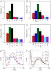
We previously used changes in the near-UV circular dichroism and fluorescence spectra of DNA base analogue probes placed site specifically to show that the first three base pairs at the fork junction in model replication fork constructs are significantly opened by "breathing" fluctuations under physiological conditions. Here, we use these probes to provide mechanistic snapshots of the initial interactions of the DNA fork with a tight-binding replication helicase in solution. The primosome helicase of bacteriophage T4 was assembled from six (gp41) helicase subunits, one (gp61) primase subunit, and nonhydrolyzable GTPγS. When bound to a DNA replication fork construct this complex advances one base pair into the duplex portion of the fork and forms a stably bound helicase "initiation complex." Replacement of GTPγS with GTP permits the completion of the helicase-driven unwinding process. Our spectroscopic probes show that the primosome in this stable helicase initiation complex binds the DNA of the fork primarily via backbone contacts and holds the first complementary base pair of the fork in an open conformation, whereas the second, third, and fourth base pairs of the duplex show essentially the breathing behavior that previously characterized the first three base pairs of the free fork. These spectral changes, together with dynamic fluorescence quenching results, are consistent with a primosome-binding model in which the lagging DNA strand passes through the central hole of the hexagonal helicase, the leading strand binds to the "outside" surfaces of subunits of the helicase hexamer, and the single primase subunit interacts with both strands.
Conflict of interest statement
The authors declare no conflict of interest.
Figures


Similar articles
-
A single-molecule view of the assembly pathway, subunit stoichiometry, and unwinding activity of the bacteriophage T4 primosome (helicase-primase) complex.Biochemistry. 2013 May 7;52(18):3157-70. doi: 10.1021/bi400231s. Epub 2013 Apr 24. Biochemistry. 2013. PMID: 23578280 Free PMC article.
-
Assembly and subunit stoichiometry of the functional helicase-primase (primosome) complex of bacteriophage T4.Proc Natl Acad Sci U S A. 2012 Aug 21;109(34):13596-601. doi: 10.1073/pnas.1210040109. Epub 2012 Aug 6. Proc Natl Acad Sci U S A. 2012. PMID: 22869700 Free PMC article.
-
Interactions of bacteriophage T4-coded primase (gp61) with the T4 replication helicase (gp41) and DNA in primosome formation.J Biol Chem. 1999 Sep 17;274(38):27287-98. doi: 10.1074/jbc.274.38.27287. J Biol Chem. 1999. PMID: 10480949
-
Coordinated DNA Replication by the Bacteriophage T4 Replisome.Viruses. 2015 Jun 19;7(6):3186-200. doi: 10.3390/v7062766. Viruses. 2015. PMID: 26102578 Free PMC article. Review.
-
DNA Helicase-Polymerase Coupling in Bacteriophage DNA Replication.Viruses. 2021 Aug 31;13(9):1739. doi: 10.3390/v13091739. Viruses. 2021. PMID: 34578319 Free PMC article. Review.
Cited by
-
Fifty years of DNA "breathing": Reflections on old and new approaches.Biopolymers. 2013 Dec;99(12):923-54. doi: 10.1002/bip.22347. Biopolymers. 2013. PMID: 23840028 Free PMC article. Review.
-
Structural basis of nucleic-acid recognition and double-strand unwinding by the essential neuronal protein Pur-alpha.Elife. 2016 Jan 8;5:e11297. doi: 10.7554/eLife.11297. Elife. 2016. PMID: 26744780 Free PMC article.
-
Helicase and polymerase move together close to the fork junction and copy DNA in one-nucleotide steps.Cell Rep. 2014 Mar 27;6(6):1129-1138. doi: 10.1016/j.celrep.2014.02.025. Epub 2014 Mar 13. Cell Rep. 2014. PMID: 24630996 Free PMC article.
-
A single-molecule view of the assembly pathway, subunit stoichiometry, and unwinding activity of the bacteriophage T4 primosome (helicase-primase) complex.Biochemistry. 2013 May 7;52(18):3157-70. doi: 10.1021/bi400231s. Epub 2013 Apr 24. Biochemistry. 2013. PMID: 23578280 Free PMC article.
-
Mapping the interactions of the single-stranded DNA binding protein of bacteriophage T4 (gp32) with DNA lattices at single nucleotide resolution: polynucleotide binding and cooperativity.Nucleic Acids Res. 2015 Oct 30;43(19):9291-305. doi: 10.1093/nar/gkv818. Epub 2015 Aug 14. Nucleic Acids Res. 2015. PMID: 26275774 Free PMC article.
References
-
- von Hippel PH, Delagoutte E. A general model for nucleic acid helicases and their “coupling” within macromolecular machines. Cell. 2001;104:177–190. - PubMed
-
- Lohman TM. Escherichia coli DNA helicases: Mechanisms of DNA unwinding. Mol Microbiol. 1992;6:5–14. - PubMed
-
- Lohman TM, Bjornson KP. Mechanisms of helicase-catalyzed DNA unwinding. Annu Rev Biochem. 1996;65:169–214. - PubMed
Publication types
MeSH terms
Substances
Grants and funding
LinkOut - more resources
Full Text Sources
Molecular Biology Databases

