. 2012 Sep 4;109(36):14458-63.
doi: 10.1073/pnas.1208950109. Epub 2012 Aug 20.

Ribosomal protein S1 unwinds double-stranded RNA in multiple steps

Affiliations

Ribosomal protein S1 unwinds double-stranded RNA in multiple steps

Xiaohui Qu et al. Proc Natl Acad Sci U S A. .

Abstract

The sequence and secondary structure of the 5'-end of mRNAs regulate translation by controlling ribosome initiation on the mRNA. Ribosomal protein S1 is crucial for ribosome initiation on many natural mRNAs, particularly for those with structured 5'-ends, or with no or weak Shine-Dalgarno sequences. Besides a critical role in translation, S1 has been implicated in several other cellular processes, such as transcription recycling, and the rescuing of stalled ribosomes by tmRNA. The mechanisms of S1 functions are still elusive but have been widely considered to be linked to the affinity of S1 for single-stranded RNA and its corresponding destabilization of mRNA secondary structures. Here, using optical tweezers techniques, we demonstrate that S1 promotes RNA unwinding by binding to the single-stranded RNA formed transiently during the thermal breathing of the RNA base pairs and that S1 dissociation results in RNA rezipping. We measured the dependence of the RNA unwinding and rezipping rates on S1 concentration, and the force applied to the ends of the RNA. We found that each S1 binds 10 nucleotides of RNA in a multistep fashion implying that S1 can facilitate ribosome initiation on structured mRNA by first binding to the single strand next to an RNA duplex structure ("stand-by site") before subsequent binding leads to RNA unwinding. Unwinding by multiple small substeps is much less rate limited by thermal breathing than unwinding in a single step. Thus, a multistep scheme greatly expedites S1 unwinding of an RNA structure compared to a single-step mode.


Conflict of interest statement

The authors declare no conflict of interest.

Figures

Fig. 1.
Fig. 1.
Experimental setup. A. Schematic of the optical tweezers experiment. B. (Left) Typical unwinding trajectories at 21.3 pN force for 30 nM (light gray), 100 nM (dark gray), and 300 nM (black) S1. When the RNA hairpin is held at a constant force above 18.9 pN but well below its mechanical unfolding force, upon introduction of S1 in the chamber step-wise unwinding (pause-step-pause) is observed. The unwinding rate increases with S1 concentration. (Right) Histogram of the number of data points in the dark gray trajectory along the RNA sequence. Each pause on the trajectory forms a Gaussian peak on the histogram at the corresponding RNA position. C. (Left) Typical re-zipping trajectories at 17 pN force for 10 nM (light gray), 100 nM (dark gray), and 300 nM (black) S1. When the S1-unwound RNA hairpin is held at a constant force below 17 pN, step-wise re-zipping is observed. The re-zipping rate is independent of S1 concentration. (Right) Histogram of the number of data points in the dark gray trajectory along the RNA sequence. Each pause on the trajectory forms a Gaussian peak on the histogram at the corresponding RNA position.
Fig. 2.
Fig. 2.
Unwinding and re-zipping histograms are anticorrelated. A. Typical cumulative unwinding (Top) and re-zipping (Bottom) histograms, constructed by adding up the histograms for individual trajectories (Fig. 1 BC Right) for all trajectories taken from the same experimental condition. Higher unwinding peaks (slow) align with lower re-zipping peaks (fast), and vice versa, i. e., sequences corresponding to slow unwinding rezip rapidly. Each individual unwinding/re-zipping peak corresponds to a single unwinding/re-zipping step, and is fit to a Gaussian function to determine its center, width, and amplitude. Fitted results of individual peaks are shown under both histograms. The earlier segments of both histograms (with the end-to-end distance change below 75 nm) are not analyzed because individual peaks are hard to identify. B. The histograms of the correlation coefficients of the rate constants for 20 unwinding conditions (black bars), and 15 re-zipping conditions (gray bars), and the cross-correlation between the unwinding and re-zipping conditions (white bars). The unwinding and re-zipping rate constants are clearly anti-correlated. The calculation uses Eqn. 1. C. The GC content (GC%) variation along the RNA hairpin. Regions with higher GC% align with higher unwinding peaks and lower re-zipping peaks. The ratio of the RNA extension change (A) to the number of base pairs unwound or re-zipped (C) gives 1 nm/bp, or 0.5 nm/nt, for the extension of S1-bound single-stranded RNA.
Fig. 3.
Fig. 3.
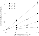
Normalized unwinding rates show a linear dependence on S1 concentration. The normalized unwinding rate is the ratio of the unwinding rate of each experimental condition relative to a reference condition: 21.3 pN force with 300 nM S1. All collected data are included. Black lines: linear fit (y = A·x + B) to each data set at one of the four force values. The R-squared values are all between 0.965 and 1.000, confirming the linearity of the dependence for all four data sets. The fitted value of the slope (A) increases with the force. The fitted values of the intercept (B) are 0.06 ± 0.02, 0.00 ± 0.05, 0.02 ± 0.03, 0.06 ± 0.02 for force values from 19.4 pN to 22.3 pN, respectively, indicating that essentially all four data sets show the dependence of y = A·x.
Fig. 4.
Fig. 4.
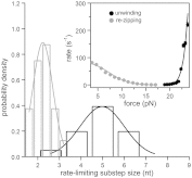
Histograms of the rate-limiting substep size for unwinding (black) and re-zipping (gray). The rate-limiting substep size was determined for each individual peak by fitting the force dependence of its unwinding or re-zipping rate to Eqns. 2a or 2b, respectively. Inset: The force dependence of the unwinding (black dots) and re-zipping (gray dots) rate of a single peak, which is centered at 155 nm RNA end-to-end extension change in Fig. 2A, and their best fit (black and gray lines).
Fig. 5.
Fig. 5.
Proposed scheme of hairpin unwinding and re-zipping by S1. The S1 RNA-binding domains (D3-6) bind to and dissociate from the single-strand RNA in a multi-step fashion, resulting in hairpin unwinding and re-zipping, respectively. The binding of domains D4-D5 is rate limiting for unwinding; re-zipping is rate limited by the releasing of a single domain (D3 or D6). The ribosome-binding domains (D1-2) do not bind to RNA.

References

    1. Draper DE, von Hippel PH. Nucleic acid binding properties of Escherichia coli ribosomal protein S1. I. Structure and interactions of binding site I. J Mol Biol. 1978;122:321–338. - PubMed
    1. Subramanian AR, van Duin J. Exchange of individual ribosomal proteins between ribosomes as studied by heavy isotope-transfer experiments. Mol Gen Genet. 1977;158:1–9. - PubMed
    1. Sengupta J, Agrawal RK, Frank J. Visualization of protein S1 within the 30S ribosomal subunit and its interaction with messenger RNA. Proc Natl Acad Sci USA. 2001;98:11991–11996. - PMC - PubMed
    1. Marzi S, et al. Structured mRNAs regulate translation initiation by binding to the platform of the ribosome. Cell. 2007;130:1019–1031. - PubMed
    1. Studer SM, Joseph S. Unfolding of mRNA secondary structure by the bacterial translation initiation complex. Mol Cell. 2006;22:105–115. - PubMed

Publication types

MeSH terms