. 2012 Sep 11;109(37):E2433-40.
doi: 10.1073/pnas.1207811109. Epub 2012 Aug 20.

Myosin IC generates power over a range of loads via a new tension-sensing mechanism

Affiliations

Myosin IC generates power over a range of loads via a new tension-sensing mechanism

Michael J Greenberg et al. Proc Natl Acad Sci U S A. .

Abstract

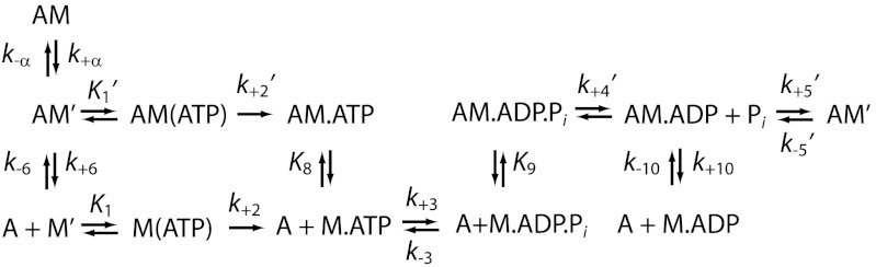
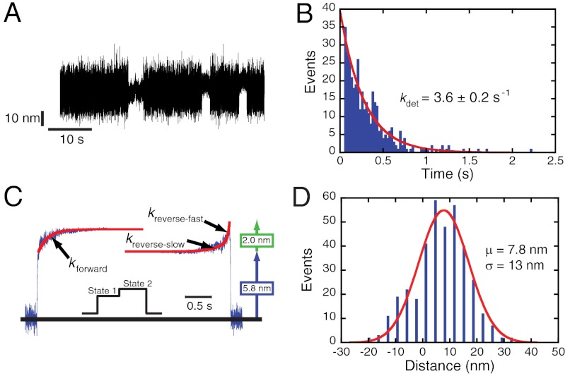
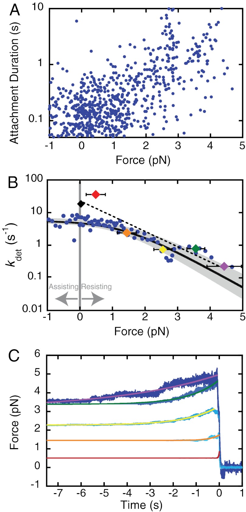
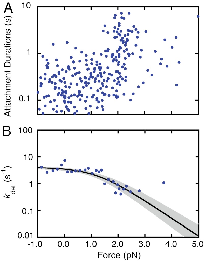
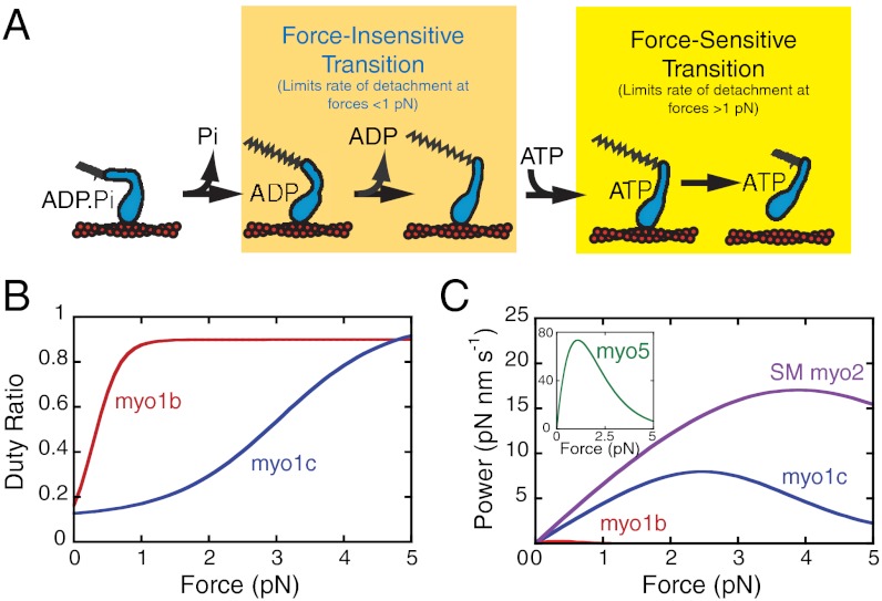
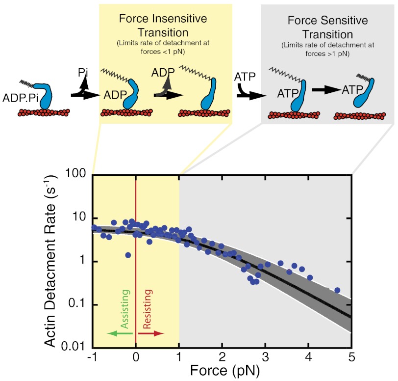
Myosin IC (myo1c), a widely expressed motor protein that links the actin cytoskeleton to cell membranes, has been associated with numerous cellular processes, including insulin-stimulated transport of GLUT4, mechanosensation in sensory hair cells, endocytosis, transcription of DNA in the nucleus, exocytosis, and membrane trafficking. The molecular role of myo1c in these processes has not been defined, so to better understand myo1c function, we utilized ensemble kinetic and single-molecule techniques to probe myo1c's biochemical and mechanical properties. Utilizing a myo1c construct containing the motor and regulatory domains, we found the force dependence of the actin-attachment lifetime to have two distinct regimes: a force-independent regime at forces < 1 pN, and a highly force-dependent regime at higher loads. In this force-dependent regime, forces that resist the working stroke increase the actin-attachment lifetime. Unexpectedly, the primary force-sensitive transition is the isomerization that follows ATP binding, not ADP release as in other slow myosins. This force-sensing behavior is unique amongst characterized myosins and clearly demonstrates mechanochemical diversity within the myosin family. Based on these results, we propose that myo1c functions as a slow transporter rather than a tension-sensitive anchor.


Conflict of interest statement

The authors declare no conflict of interest.

Figures

Scheme 1.
Scheme 1.
Pathway for the actomyosin ATPase cycle.
Fig. 1.
Fig. 1.
Optical-trapping experiments performed in the absence of an applied load (369 binding events). (A) Sample data trace of single myosin interactions with actin acquired in the presence of 5 mM ATP. (B) The distribution of attachment durations in the presence of 5 mM ATP. A single exponential function was fit to the data, giving a detachment rate (kdet) of 3.6 ± 0.2 s-1. (C) Ensemble averages of single-molecule interactions were constructed as described in Materials and Methods. Fitting a single exponential function to the time-forward average yields a rate (kforward) of 4.3 s-1. Two exponential functions were summed to fit the time-reverse average, with the major amplitude component (67 ± 4% of the total amplitude) having a rate (kreverse-fast) of 27 ± 3 s-1 and the minor component having a rate (kreverse-slow) of 3.8 ± 0.2 s-1. The size of the working stroke is equal to 7.8 nm, comprised of a first substep of 5.8 nm followed by a 2.0-nm substep. (Inset) A cartoon of the displacement generated by the myo1c3IQ working stroke where the myosin sequentially undergoes a substep from state 1 to 2 before detaching. (D) Distribution of the sizes of the total working stroke for myo1c3IQ. A single Gaussian function was fit to the data, and the reported error (σ) is the standard deviation of the Gaussian function. The total step has a size of 7.8 ± 13 nm. Note that the large variance is caused by Brownian motion–driven fluctuations of the actin filament position.
Fig. 2.
Fig. 2.
Force sensitivity of myo1c3IQ in the presence of 5 mM ATP. (A) Using the isometric optical clamp to apply a load to the myosin, the force on the myosin and the actomyosin-attachment duration were measured for each single-molecule interaction (n = 670). The 90% confidence intervals were calculated by maximum-likelihood estimation (MLE) of 1,000 bootstrap simulations of the data (see Materials and Methods). The overall detachment rate as a function of force was modeled as the sum of two rates, one force-dependent and one force-independent (Eq. 3). The rate of the force-independent transition (ki) is equal to 5.6(+1.6/-0.8) s-1. The rate of the force-dependent transition in the absence of force (kf0) is equal to 29(+9/-6) s-1 with a distance parameter (ddet) of 5.2(+0.5/-0.6) nm. (B) For the sake of visualization, events were ordered by the average force on the myosin, and sets of 10 points were binned and converted to detachment rates. The thick black line shows the calculated detachment rate as a function of force based on the MLE fitting of the unaveraged data (Fig. 2A). The shaded regions show the 90% confidence interval. Colored diamonds show the rate of the time-reverse ensemble averages binned by force (C). The force bins are 0–1 pN (red), 1–2 pN (orange), 2–3 pN (yellow), 3–4 pN (green), and > 4 pN (purple). The black diamond is the rate (kreverse-fast) at zero force. The dotted line is a fit of the Bell equation to the rates of the time-reverse ensemble averages as a function of force, giving a rate of kf0-ens avg = 26 ± 2 s-1 with a distance parameter (dens avg) of 4.6 ± 0.8 nm. (C) Time-reverse ensemble averages binned by average interaction force. The force bin ranges and colors are as in (B). Single exponential functions were fit to the data, and these rates are plotted (B).
Fig. 3.
Fig. 3.
Force sensitivity of myo1c3IQ in the presence of ADP. The isometric optical clamp was used to examine the force sensitivity of myo1c3IQ at 50 μM ADP and 1 mM ATP (n = 282). (A) Scatter plot of individual single-molecule attachment durations as a function of force . The overall detachment rate as a function of force was modeled as the sum of two rates, one force-dependent and one force-independent (Eq. 3). The best-fit parameters were calculated using MLE fitting and the 90% confidence intervals were calculated by bootstrapping. The force-independent rate (ki) is equal to 4.0(-0.5) s-1. The force-dependent rate (kf0) is equal to 25(+5/-2) s-1 with a distance parameter (ddet) of 6.3(+0.6/-0.5) nm. (B) For the sake of visualization, the detachment rates (the reciprocal of the attachment durations) were binned by force and averaged. The thick black line shows the best-fit curve and the grey-shaded regions show the 90% confidence intervals. All MLE fitting and bootstrapping was performed on the unaveraged data (Fig. 3A).
Fig. 4.
Fig. 4.
Comparison of myo1c3IQ and the closely related isoform, myo1b (splice isoform “a”). (A) Model of myo1c (blue) interacting with actin (red) in the presence of force. The transition corresponding to ADP release [the primary force-sensitive transition in myo1b (20), smooth muscle myosin-II (27), and myosin-V (25)] is force-insensitive for myo1c3IQ and rate-limiting for actomyosin detachment at forces less than 1 pN. At forces in excess of 1 pN, the primary force-sensitive transition (the isomerization that follows ATP binding) becomes rate-limiting for detachment. (B) The duty ratios of myo1c3IQ (blue) and myo1b (red), calculated from the transient-kinetics and optical-trapping experiments (20), show that the duty ratio of myo1c3IQ is substantially less force-sensitive than myo1b. (C) The calculated average power output (see Materials and Methods) of myo1c3IQ (blue), myo1b (red) (20), smooth muscle myosin-II (purple) (27), and myosin-V (Inset) (18, 25) as a function of force. Note that myo1c3IQ is substantially different from myo1b. Myo1b has a very low power output that approaches zero with very little force, consistent with myo1b acting as a tension-sensitive anchor. Myo1c3IQ, on the other hand, has a higher power output and is able to generate power over a range of forces, more consistent with its functioning as a transporter rather than as a tension-sensing anchor. The power output of myo1c3IQ is lower than smooth muscle myosin-II or myosin-V because of its slower overall kinetics; however, all three of these myosins are able to generate power over a range of forces.
Fig. P1.
Fig. P1.
(Top) Cartoon showing the interaction of myo1c (blue) with actin (red) during its mechanochemical cycle. At forces < 1 pN, ADP release limits actomyosin detachment, similar to other myosins. At forces > 1 pN, the primary force-dependent transition (i.e., the isomerization following ATP binding) limits detachment. (Bottom) The rate of actomyosin detachment as a function of force measured using an isometric optical clamp. The data clearly show two regimes, one force-independent (yellow) and one force-dependent (grey), corresponding to the highlighted mechanochemical transitions above. This force-sensing behavior is unique amongst characterized myosins.

References

    1. Sokac AM, Schietroma C, Gundersen CB, Bement WM. Myosin-1c couples assembling actin to membranes to drive compensatory endocytosis. Dev Cell. 2006;11:629–640. - PMC - PubMed
    1. Brandstaetter H, Kendrick-Jones J, Buss F. Myo1c regulates lipid raft recycling to control cell spreading, migration and Salmonella invasion. J Cell Sci. 2012;125:1991–2003. - PMC - PubMed
    1. Bose A, et al. Unconventional myosin Myo1c promotes membrane fusion in a regulated exocytic pathway. Mol Cell Biol. 2004;24:5447–5458. - PMC - PubMed
    1. Bose A, et al. Glucose transporter recycling in response to insulin is facilitated by myosin Myo1c. Nature. 2002;420:821–824. - PubMed
    1. Chen XW, Leto D, Chiang SH, Wang Q, Saltiel AR. Activation of RalA is required for insulin-stimulated Glut4 trafficking to the plasma membrane via the exocyst and the motor protein Myo1c. Dev Cell. 2007;13:391–404. - PubMed

Publication types

LinkOut - more resources