. 2012 Sep 4;109(36):14393-8.
doi: 10.1073/pnas.1206734109. Epub 2012 Aug 20.

Molecular basis for negative regulation of the glucagon receptor

Affiliations

Molecular basis for negative regulation of the glucagon receptor

Christopher M Koth et al. Proc Natl Acad Sci U S A. .

Abstract

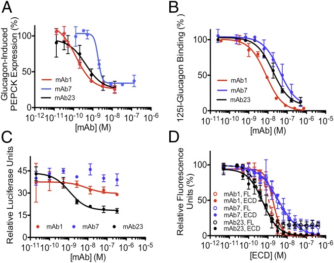
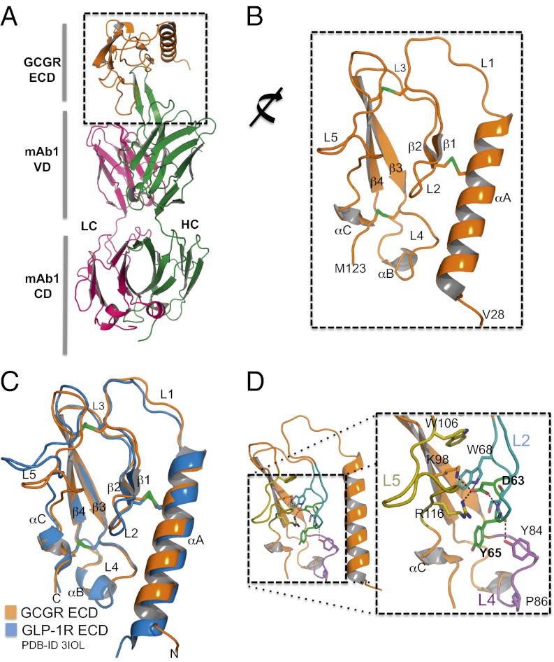
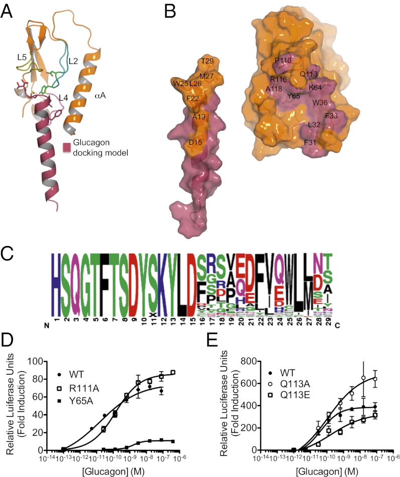
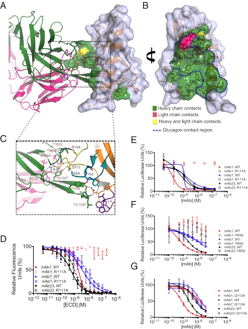
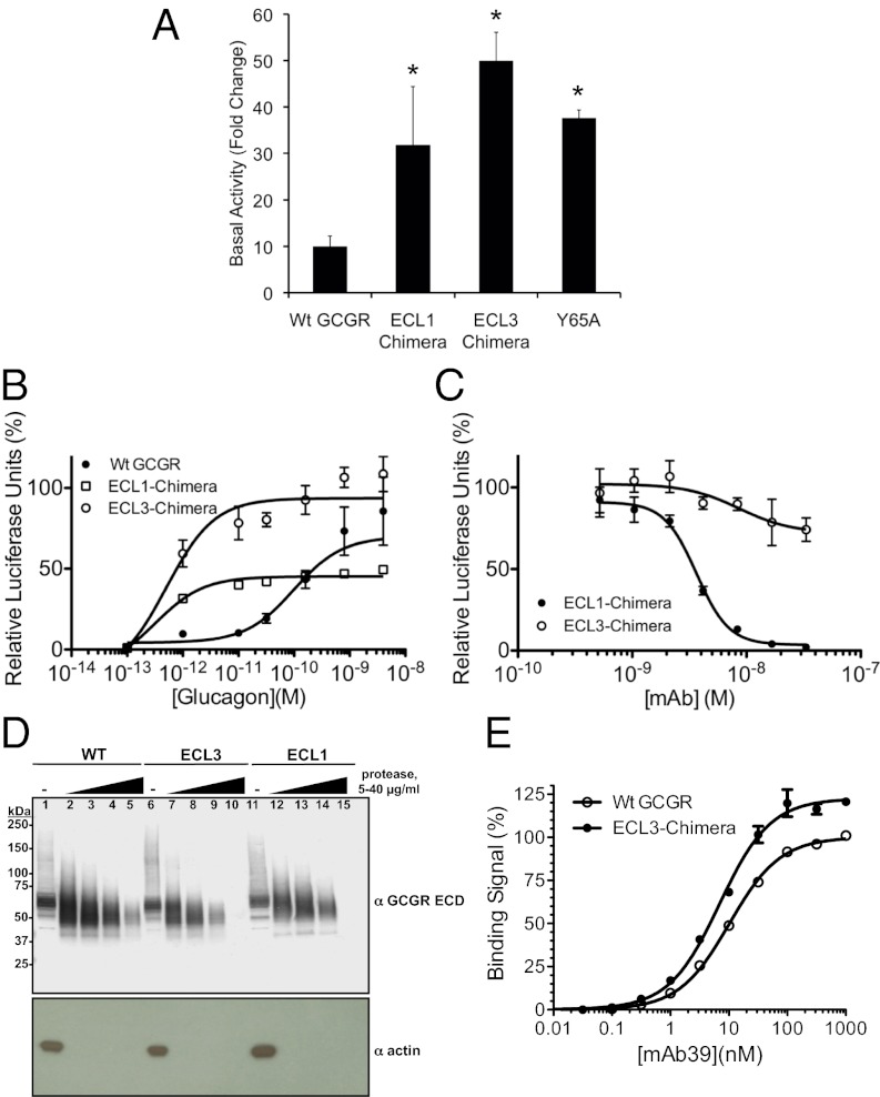
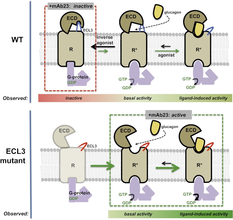
Members of the class B family of G protein-coupled receptors (GPCRs) bind peptide hormones and have causal roles in many diseases, ranging from diabetes and osteoporosis to anxiety. Although peptide, small-molecule, and antibody inhibitors of these GPCRs have been identified, structure-based descriptions of receptor antagonism are scarce. Here we report the mechanisms of glucagon receptor inhibition by blocking antibodies targeting the receptor's extracellular domain (ECD). These studies uncovered a role for the ECD as an intrinsic negative regulator of receptor activity. The crystal structure of the ECD in complex with the Fab fragment of one antibody, mAb1, reveals that this antibody inhibits glucagon receptor by occluding a surface extending across the entire hormone-binding cleft. A second antibody, mAb23, blocks glucagon binding and inhibits basal receptor activity, indicating that it is an inverse agonist and that the ECD can negatively regulate receptor activity independent of ligand binding. Biochemical analyses of receptor mutants in the context of a high-resolution ECD structure show that this previously unrecognized inhibitory activity of the ECD involves an interaction with the third extracellular loop of the receptor and suggest that glucagon-mediated structural changes in the ECD accompany receptor activation. These studies have implications for the design of drugs to treat class B GPCR-related diseases, including the potential for developing novel allosteric regulators that target the ECDs of these receptors.


Conflict of interest statement

Conflict of interest statement: All authors are employees of Genentech Inc.

Figures

Fig. 1.
Fig. 1.
Anti-GCGR antibodies that inhibit GCGR activity target the ECD. (A) Antibodies block glucagon-induced PEPCK gene expression in human hepatocytes. Average IC50s (nM) from two experiments are 0.15, 1.5, and 0.4 for mAb1, mAb7, and mAb23, respectively. (B) Antibodies block 125I-glucagon binding to cells expressing GCGR. Kis (nM) are 5, 47, and 10 for mAb1, mAb7, and mAb23, respectively. The EC50 of glucagon binding is 70 nM. (C) Reduction of basal GCGR activity in cells expressing human GCGR by mAb23. (D) Alphascreen assay measuring the ability of ECD to compete with mAbs bound to acceptor beads for binding to full-length GCGR (dashed lines) or ECD (solid lines) bound to donor beads. IC50s (nM) of mAbs on full-length GCGR are 1.2 ± 0.2, 2.9 ± 1.0, and 0.2 ± 0.1, and on ECD are 1.9 ± 0.3, 3.6 ± 1.4, and 0.6 ± 0.2, for mAb1, mAb7, and mAb23, respectively. Data shown are from a single representative of three (A and C) or two (B and D) independent experiments. Error bars represent SD of duplicate or triplicate determinations.
Fig. 2.
Fig. 2.
Crystal structure of GCGR ECD in complex with mAb1. (A) Cartoon representation of the complex of GCGR ECD/mAb1. The HCs and LCs of mAb1 are colored blue and pink, respectively. The ECD is colored wheat. (B) The GCGR ECD adopts an α-β-β-α fold common to class B GPCR ECDs. Conserved disulfide bonds are shown as green sticks. (C) Comparison of GCGR ECD and GLP-1R structures illustrates high structural homology. (D) Asp63 and Tyr65 are key residues located in L2. Asp63 is involved in multiple H-bond interactions with residues throughout the ECD.
Fig. 3.
Fig. 3.
Identification of ECD and glucagon residues involved in the glucagon–GCGR interaction. (A) Docking model of glucagon binding to the GCGR ECD crystal structure. (B) Open book view of the docking model of glucagon binding to the GCGR ECD. ECD residues contacted by glucagon are colored raspberry and labeled; residues of glucagon that contact the ECD are colored wheat and labeled. (C) Consensus logo of glucagon showing allowed variation for binding to GCGR ECD (represented by height) in residues 16 through 29. (D) Substitution of Ala for Y65, but not R111, and (E) substitution of Q113 with Glu increases the EC50 of glucagon-induced activation. Data shown are from a single representative of three independent experiments. Error bars represent SD of triplicate determinations.
Fig. 4.
Fig. 4.
Understanding the structural basis of antagonism by mAb1. (A) The CDR-H3 of mAb1 occludes the glucagon-binding site. (B) Residues of the ECD contacted by mAb1 are mapped onto the surface of the ECD. The surface contacted by glucagon is outlined. (C) Extensive interactions between H2, H3, and L3 loops of mAb1 and the GCGR ECD. ECD residues are indicated in black; mAb1 residues are indicated in red. The loop location of each residue is indicated in superscript. (D) Alphascreen assay measuring the ability of WT ECD to compete with mAbs bound to acceptor beads for binding to WT or R111A ECD bound to donor beads. Data are from a single representative of two independent experiments. Error bars represent SD of duplicate determinations. The IC50s (nM) for WT ECD are 1.9 ± 0.3, 3.6 ± 1.4, and 0.6 ± 0.2 for mAb1, mAb7, and mAb23, respectively. The IC50s (nM) for R111A ECD are not determinable, 3.6 ± 1.4, and 1.0 ± 0.2 for mAb1, mAb7, and mAb23, respectively. (E) R111A or (G) Q113A mutations prevent mAb1 inhibition of glucagon-induced GCGR activation, whereas (F) Y65A mutation prevents mAb1 and mAb23 from inhibiting glucagon-induced GCGR activation. Data shown are from a single representative of three independent experiments. Error bars represent SD of triplicate determinations.
Fig. 5.
Fig. 5.
GCGR activity and structural effects of Y65A ECD mutant and ECL chimeras. (A) ECL1 and ECL3 chimeras and Y65A GCGR have increased basal activity. Data are mean ± SE, n = 4. *P < 0.05. (B) ECL3 chimeric receptor has increased glucagon-induced activity. (C) mAb23 fails to inhibit activation of ECL3 chimeric receptor. (D) Cells expressing WT GCGR or ECL3 or ECL1 chimeric receptors were incubated with 0 (lanes 1, 6, and 7), 5 (lanes 2, 7, and 12), 10 (lanes 3, 8, and 13), 20 (lanes 4, 9, and 14), or 40 (lanes 5, 10, and 15) μg/mL LysC. The proteolytic products were resolved by nonreducing denaturing gel electrophoresis and probed by Western blotting with mAb1. (E) Increased binding of mAb39 to the ECL3 chimera compared with WT GCGR. Data shown are mean ± SD from two independent experiments, normalized to binding to WT GCGR. For B and C, data shown are from a single representative of three independent experiments. Error bars represent SD of triplicate determinations.
Fig. 6.
Fig. 6.
In the absence of agonist, GCGR is predominantly in an inactivated state. Basal activity indicates that the receptor is capable of adopting an active conformation, enabling signaling through heterotrimeric G protein nucleotide exchange. Agonist binding stabilizes an active conformation to enable G protein coupling. The ECL3 chimeric receptor is uncoupled from the ECD and more readily adopts an active conformation, even in the absence of agonist. Higher basal and ligand-induced activities are observed in the ECL3 chimera. An inactive conformation of the WT receptor is stabilized by mAb23, an effect lost on the ECL3 chimera.

References

    1. Jelinek LJ, et al. Expression cloning and signaling properties of the rat glucagon receptor. Science. 1993;259:1614–1616. - PubMed
    1. Unger RH, Cherrington AD. Glucagonocentric restructuring of diabetes: A pathophysiologic and therapeutic makeover. J Clin Invest. 2012;122:4–12. - PMC - PubMed
    1. Dallas-Yang Q, et al. Hepatic glucagon receptor binding and glucose-lowering in vivo by peptidyl and non-peptidyl glucagon receptor antagonists. Eur J Pharmacol. 2004;501:225–234. - PubMed
    1. Unson CG, Andreu D, Gurzenda EM, Merrifield RB. Synthetic peptide antagonists of glucagon. Proc Natl Acad Sci USA. 1987;84:4083–4087. - PMC - PubMed
    1. Petersen KF, Sullivan JT. Effects of a novel glucagon receptor antagonist (Bay 27-9955) on glucagon-stimulated glucose production in humans. Diabetologia. 2001;44:2018–2024. - PubMed

Publication types

Substances

Associated data