. 2012 Sep 4;109(36):14574-9.
doi: 10.1073/pnas.1212806109. Epub 2012 Aug 20.

Blocking antibody to the β-subunit of FSH prevents bone loss by inhibiting bone resorption and stimulating bone synthesis

Affiliations

Blocking antibody to the β-subunit of FSH prevents bone loss by inhibiting bone resorption and stimulating bone synthesis

Ling-Ling Zhu et al. Proc Natl Acad Sci U S A. .

Abstract

Low estrogen levels undoubtedly underlie menopausal bone thinning. However, rapid and profuse bone loss begins 3 y before the last menstrual period, when serum estrogen is relatively normal. We have shown that the pituitary hormone FSH, the levels of which are high during late perimenopause, directly stimulates bone resorption by osteoclasts. Here, we generated and characterized a polyclonal antibody to a 13-amino-acid-long peptide sequence within the receptor-binding domain of the FSH β-subunit. We show that the FSH antibody binds FSH specifically and blocks its action on osteoclast formation in vitro. When injected into ovariectomized mice, the FSH antibody attenuates bone loss significantly not only by inhibiting bone resorption, but also by stimulating bone formation, a yet uncharacterized action of FSH that we report herein. Mesenchymal cells isolated from mice treated with the FSH antibody show greater osteoblast precursor colony counts, similarly to mesenchymal cells isolated from FSH receptor (FSHR)(-/-) mice. This suggests that FSH negatively regulates osteoblast number. We confirm that this action is mediated by signaling-efficient FSHRs present on mesenchymal stem cells. Overall, the data prompt the future development of an FSH-blocking agent as a means of uncoupling bone formation and bone resorption to a therapeutic advantage in humans.


Conflict of interest statement

Conflict of interest statement: M.Z. is a named inventor of a pending patent application related to osteoclastic bone resorption filed by the Mount Sinai School of Medicine (MSSM). In the event the pending or issued patent is licensed, he would be entitled to a share of any proceeds MSSM receives from the licensee.

Figures
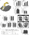
Fig. 1.
Fig. 1.
Antibody directed to the receptor-binding domain of the β-subunit of FSH (FSH Ab) prevents ovariectomy-induced bone loss. (A) FSH antibody blocks FSH binding to FSHR by steric hindrance. The crystal structure of the β-subunit of FSH (36) (ribbon cartoon highlighted with space-filled atoms in yellow and white) and the truncated FSHR ectodomain (space-filled atoms in gray) complex is shown (Protein Data Bank ID code: 1XWD). FSHβ residues buried at the receptor–ligand surface interface, underlined in this peptide sequence LVYKDPARPKIQK from which the FSH antibody is generated, are shown as yellow space-filled atoms. FSHβ residues, which are not buried, are shown as white space-filled atoms. Binding of the FSH antibody to the FSHβ subunit disrupts its interaction with the FSHR. For illustration purposes, the FSHα subunit is not shown in the complex. (B) Enzyme-linked immunoabsorbent assay (ELISA) demonstrating binding of the anti-FSH antibody (FSH Ab) or control IgG to either the FSH peptide fragment (a) or full-length FSH (b) (concentrations of plated peptide are shown). (C) Tartrate-resistant acid phosphatase-positive (TRAP+) osteoclasts formed in murine bone marrow cell cultures treated with FSH (30 ng/mL) with or without FSH Ab (1 or 10 μg/mL). Statistics: Student t test; comparisons against zero-dose control; *P ≤ 0.05; n = 8 wells per group. (D) cAMP responses in TSH receptor (TSHR) overexpressing CHO cells induced by TSH either without incubation (light bars) or following incubation (dark bars) with FSH Ab (10 μg/mL) or control goat IgG (1 μg/mL). Statistics: mean ± SEM; Student t test; comparisons against TSH alone. Overall, the data show that FSH Ab specifically detects FSH and not TSH and inhibits the osteoclastogenesis induced by FSH. (E) Effect of FSH Ab injections (100 μg/d), beginning the day of ovariectomy (OVX), on bone loss, measured by micro-CT, and shown as bone volume/total volume (BV/TV), trabecular number (TbN), trabecular thickness (TbTh, trabecular spacing (TbS), and connectivity density (ConnD). Statistics: Student t test with Bonferroni’s correction; comparisons as shown, *P ≤ 0.05; **P ≤ 0.01; n = 8 mice per group. (F) Mean plasma estradiol (E2) and FSH levels in sham-operated and OVX mice treated with IgG or FSH Ab (100 μg/d). Statistics: Student t test; P values as shown; n = 8 mice per group. (G) Effect of preincubating FSH Ab or IgG (concentrations as noted) with FSH (1 μg per well) on the ability of the ELISA to detect FSH (absorbance at 450 nm). Notably, binding of FSH Ab to FSH does not reduce the levels of FSH detectable by the ELISA antibody. Statistics: Student t test; comparisons against zero-dose control; **P ≤ 0.01; in duplicate.
Fig. 2.
Fig. 2.
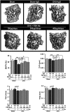
FSH antibody (FSH Ab) attenuates hypogonadal bone loss even at extended dosing intervals. The effect of FSH Ab injections (doses as noted over 6 wk) or IgG on trabecular architecture seen on micro-CT, or on estimates of bone volume/total volume (BV/TV), trabecular thickness (TbTh), trabecular number (TbN), and trabecular spacing (TbSp) (Fig. 1E shows a lack of responsiveness of TbTh in the 4-wk treatment protocol) are shown. Statistics: ANOVA with Bonferroni’s correction; comparisons as shown, *P ≤ 0.05; **P ≤ 0.01; mean ± SEM is shown, n = 5 mice per group.
Fig. 3.
Fig. 3.
Anti-FSH antibody (FSH Ab) inhibits bone resorption and stimulates bone synthesis. Tartrate-resistant acid phosphatase-labeled surfaces (pink) (A) and xylelol orange/calcein double-labeled surfaces (B) in bone sections (Materials and Methods) from mice treated with FSH Ab (100 μg/d) or IgG for 4 wk following sham operation (Sham) or ovariectomy (OVX). Representative double-labeled surfaces are magnified to show differences in interlabel distances. Bone resorption and formation parameters, namely resorption surfaces (resorbed S/BPm) (C), mineralizing surface (MS), mineral apposition rates (MAR), and bone formation rates (BFR) (D) are shown. Statistics by Student t test with Bonferroni’s correction; comparisons of FSH Ab-treated mice against IgG, *P ≤ 0.05, **P ≤ 0.01; other comparisons noted; ^P ≤ 0.05, ^^P ≤ 0.01; mean ± SEM is shown, n = 8 mice per group.
Fig. 4.
Fig. 4.
Inhibition of FSH signaling by FSH antibody (FSH Ab) or FSHR deletion stimulates osteoblastogenesis. (A) Representative images and quantification of alkaline phosphatase-positive colony forming units-fibroblastic (CFU-f) in bone marrow mesenchymal cell cultures isolated from wild-type (FSHR+/+) and FSHR−/− mice. Statistics by Student t test; **P ≤ 0.01; three wells per group. (B) Representative photomicrographs of labeled surfaces and calculated mineralizing surface (MS) in FSHR+/+ and FSHR−/− mice. Statistics by Student t test; *P ≤ 0.05. (C) Representative images and counting alkaline phosphatase-positive colony forming units-fibroblastic (CFU-f) and von Kossa-positive colony forming units-osteoblastic (CFU-ob) in bone marrow mesenchymal cell cultures isolated from sham-operated (Sham) or ovariectomized (OVX) mice treated with FSH Ab (100 μg/d) or IgG for 4 wk. Statistics by Student t test; *P ≤ 0.05, **P ≤ 0.01; three wells per group; cells from eight mice pooled. (D) RT-PCR showing the expression of a FSHR transcript in human mesenchymal stem cells (MSC) and in ovarian cells (COV), but not in mature human osteoblasts (hOB). Western immunoblotting shows an ∼60-kDa band detected by an anti-FSHR antibody in human MSCs and COV cells, but not in hOBs. (E) Western blots showing the phosphorylation of ERK1/2 in MSCs by FSH or control vehicle. This response is similar to that seen with oleoyl serine (34). (F) Effect of FSH, human CG (hCG), and forskolin (positive control) on cAMP production in MSCs. Statistics by Student t test; **P ≤ 0.01, compared with vehicle (Ctr).

Similar articles

Cited by

References

    1. Randolph JF, Jr, et al. Change in estradiol and follicle-stimulating hormone across the early menopausal transition: Effects of ethnicity and age. J Clin Endocrinol Metab. 2004;89:1555–1561. - PubMed
    1. Sowers MR, et al. Hormone predictors of bone mineral density changes during the menopausal transition. J Clin Endocrinol Metab. 2006;91:1261–1267. - PubMed
    1. Sowers MR, et al. Amount of bone loss in relation to time around the final menstrual period and follicle-stimulating hormone staging of the transmenopause. J Clin Endocrinol Metab. 2010;95:2155–2162. - PMC - PubMed
    1. Zaidi M. Skeletal remodeling in health and disease. Nat Med. 2007;13:791–801. - PubMed
    1. Pazianas M, Epstein S, Zaidi M. Evaluating the antifracture efficacy of bisphosphonates. Rev Recent Clin Trials. 2009;4:122–130. - PubMed

Publication types