

Autonomous phagosomal degradation and antigen presentation in dendritic cells
- PMID: 22908282
- PMCID: PMC3437883
- DOI: 10.1073/pnas.1203912109
Autonomous phagosomal degradation and antigen presentation in dendritic cells
Abstract
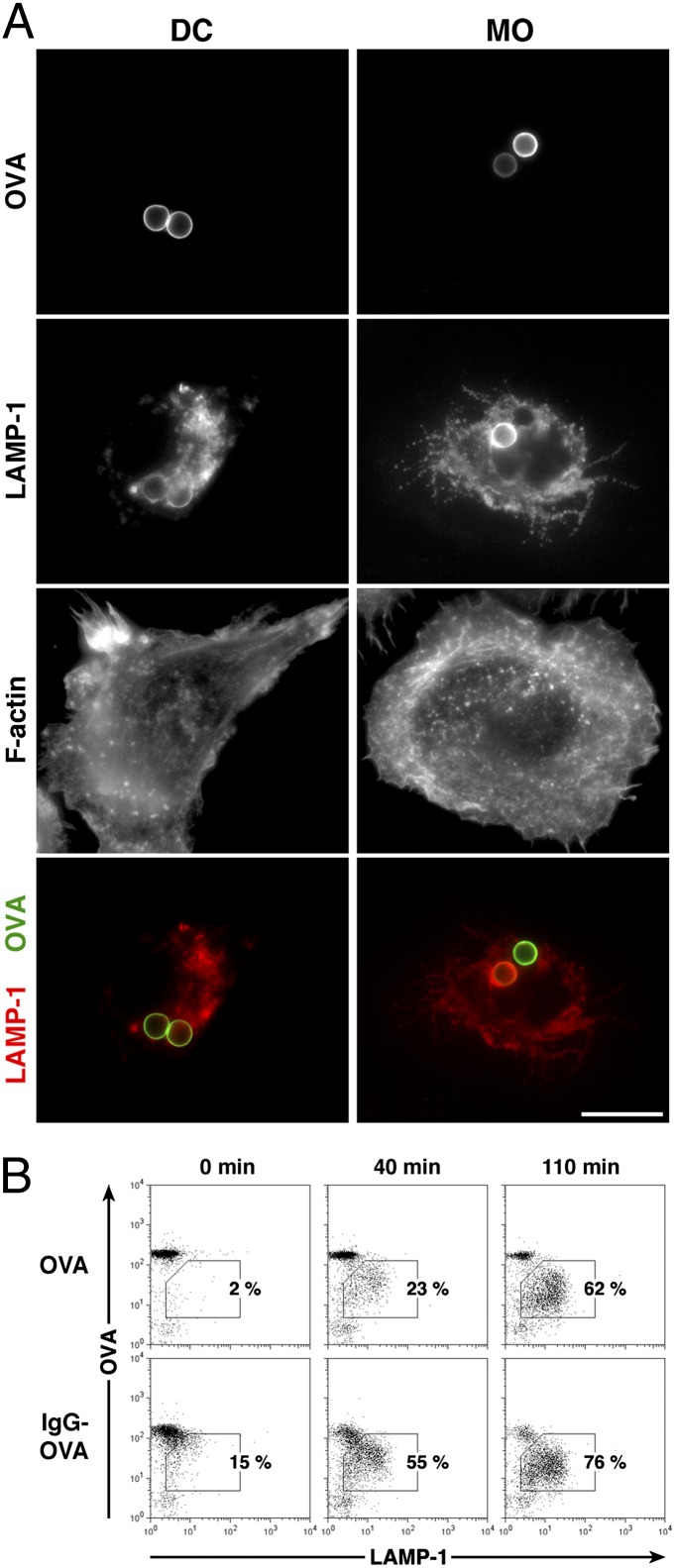
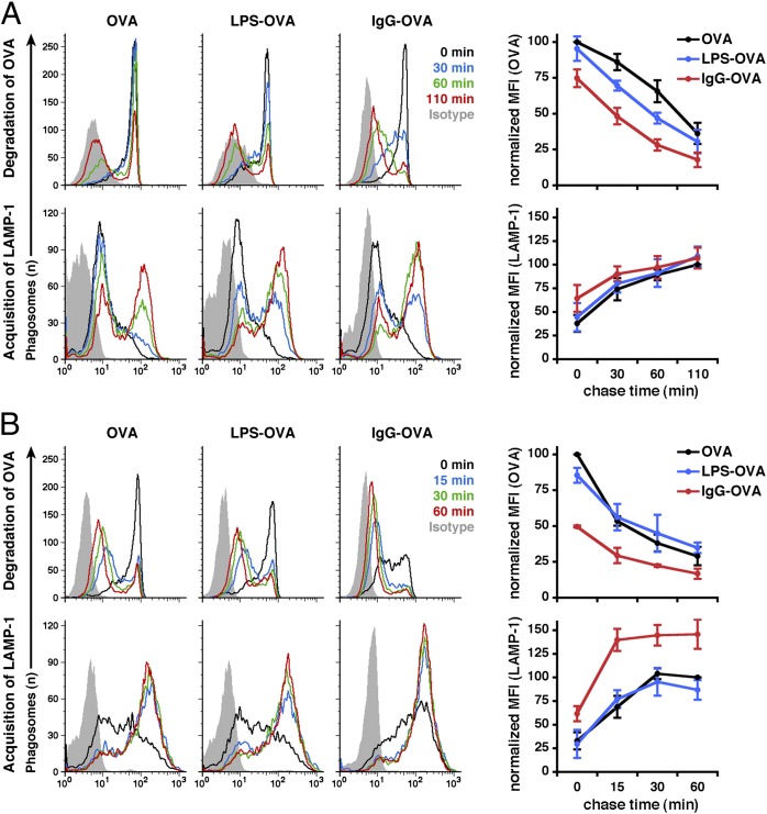
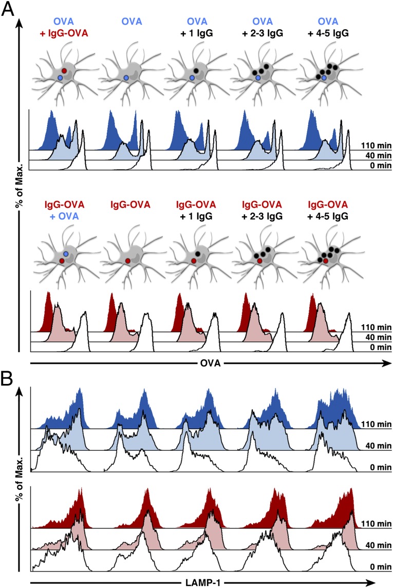
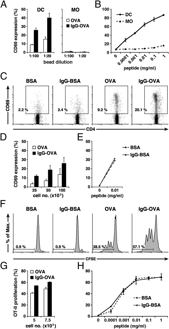
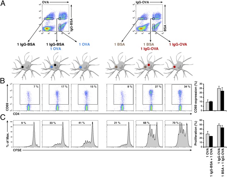
Phagocytosis plays a critical role in both innate and adaptive immunity. Phagosomal fusion with late endosomes and lysosomes enhances proteolysis, causing degradation of the phagocytic content. Increased degradation participates in both innate protection against pathogens and the production of antigenic peptides for presentation to T lymphocytes during adaptive immune responses. Specific ligands present in the phagosomal cargo influence the rate of phagosome fusion with lysosomes, thereby modulating both antigen degradation and presentation. Using a combination of cell sorting techniques and single phagosome flow cytometry-based analysis, we found that opsonization with IgG accelerates antigen degradation within individual IgG-containing phagosomes, but not in other phagosomes present in the same cell and devoid of IgG. Likewise, IgG opsonization enhances antigen presentation to CD4(+) T lymphocytes only when antigen and IgG are present within the same phagosome, whereas cells containing phagosomes with either antigen or IgG alone failed to present antigen efficiently. Therefore, individual phagosomes behave autonomously, in terms of both cargo degradation and antigen presentation to CD4(+) T cells. Phagosomal autonomy could serve as a basis for the intracellular discrimination between self and nonself antigens, resulting in the preferential presentation of peptides derived from opsonized, nonself antigens.
Conflict of interest statement
The authors declare no conflict of interest.
Figures
Similar articles
-
The neonatal FcR-mediated presentation of immune-complexed antigen is associated with endosomal and phagosomal pH and antigen stability in macrophages and dendritic cells.J Immunol. 2011 Apr 15;186(8):4674-86. doi: 10.4049/jimmunol.1003584. Epub 2011 Mar 14. J Immunol. 2011. PMID: 21402891
-
TLR-dependent phagosome tubulation in dendritic cells promotes phagosome cross-talk to optimize MHC-II antigen presentation.Proc Natl Acad Sci U S A. 2014 Oct 28;111(43):15508-13. doi: 10.1073/pnas.1412998111. Epub 2014 Oct 13. Proc Natl Acad Sci U S A. 2014. PMID: 25313083 Free PMC article.
-
Adaptor protein-3 in dendritic cells facilitates phagosomal toll-like receptor signaling and antigen presentation to CD4(+) T cells.Immunity. 2012 May 25;36(5):782-94. doi: 10.1016/j.immuni.2012.02.018. Epub 2012 May 3. Immunity. 2012. PMID: 22560444 Free PMC article.
-
A guide to measuring phagosomal dynamics.FEBS J. 2021 Mar;288(5):1412-1433. doi: 10.1111/febs.15506. Epub 2020 Aug 16. FEBS J. 2021. PMID: 32757358 Free PMC article. Review.
-
Phagocytosis and antigen presentation in dendritic cells.Immunol Rev. 2007 Oct;219:143-56. doi: 10.1111/j.1600-065X.2007.00552.x. Immunol Rev. 2007. PMID: 17850487 Review.
Cited by
-
To stay or not to stay intact as an allergen: the endolysosomal degradation assay used as tool to analyze protein immunogenicity and T cell epitopes.Front Allergy. 2024 Jul 12;5:1440360. doi: 10.3389/falgy.2024.1440360. eCollection 2024. Front Allergy. 2024. PMID: 39071040 Free PMC article. Review.
-
Anti-COVID-19 Nanomaterials: Directions to Improve Prevention, Diagnosis, and Treatment.Nanomaterials (Basel). 2022 Feb 25;12(5):783. doi: 10.3390/nano12050783. Nanomaterials (Basel). 2022. PMID: 35269270 Free PMC article. Review.
-
Fc-FcγR interactions during infections: From neutralizing antibodies to antibody-dependent enhancement.Immunol Rev. 2024 Nov;328(1):221-242. doi: 10.1111/imr.13393. Epub 2024 Sep 13. Immunol Rev. 2024. PMID: 39268652 Free PMC article. Review.
-
Diversification of IgG effector functions.Int Immunol. 2017 Jul 1;29(7):303-310. doi: 10.1093/intimm/dxx025. Int Immunol. 2017. PMID: 28472280 Free PMC article. Review.
-
Patterns, Receptors, and Signals: Regulation of Phagosome Maturation.Trends Immunol. 2017 Jun;38(6):407-422. doi: 10.1016/j.it.2017.03.006. Epub 2017 Apr 14. Trends Immunol. 2017. PMID: 28416446 Free PMC article. Review.
References
-
- Savina A, Amigorena S. Phagocytosis and antigen presentation in dendritic cells. Immunol Rev. 2007;219:143–156. - PubMed
-
- Delamarre L, Pack M, Chang H, Mellman I, Trombetta ES. Differential lysosomal proteolysis in antigen-presenting cells determines antigen fate. Science. 2005;307:1630–1634. - PubMed
Publication types
MeSH terms
Substances
LinkOut - more resources
Full Text Sources
Other Literature Sources
Molecular Biology Databases
Research Materials
