. 2012 Sep 4;109(36):14598-603.
doi: 10.1073/pnas.1206937109. Epub 2012 Aug 20.

Receptor interacting protein kinase mediates necrotic cone but not rod cell death in a mouse model of inherited degeneration

Affiliations

Receptor interacting protein kinase mediates necrotic cone but not rod cell death in a mouse model of inherited degeneration

Yusuke Murakami et al. Proc Natl Acad Sci U S A. .

Abstract

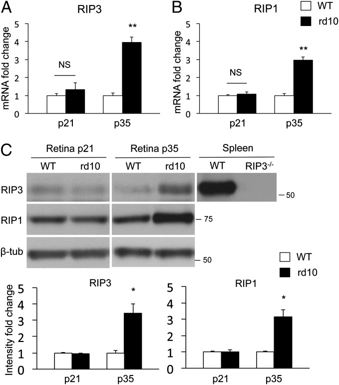
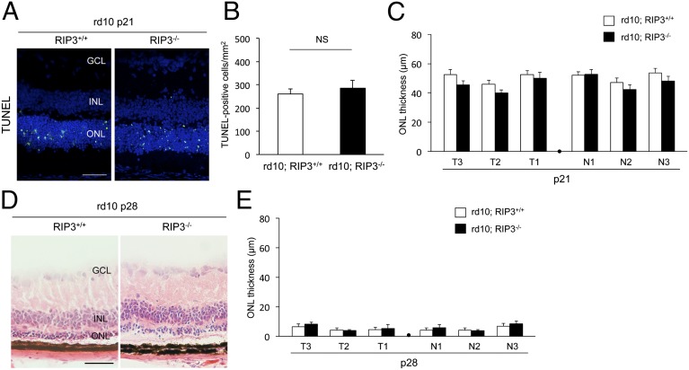
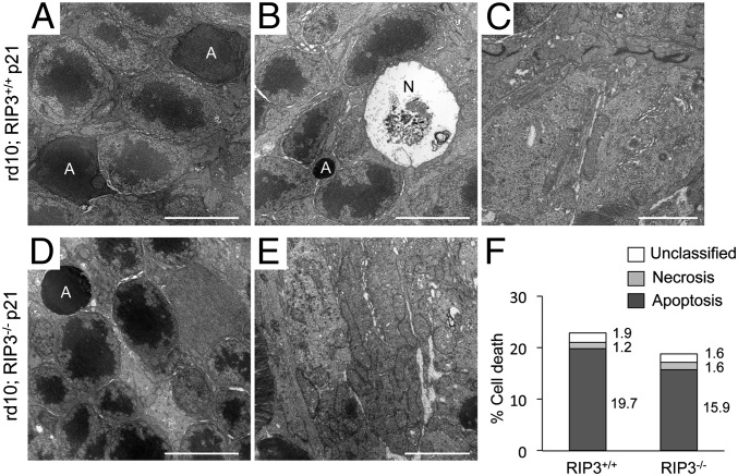
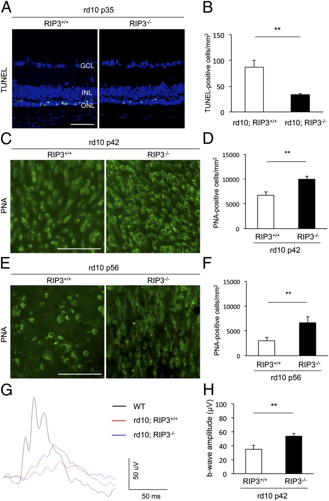
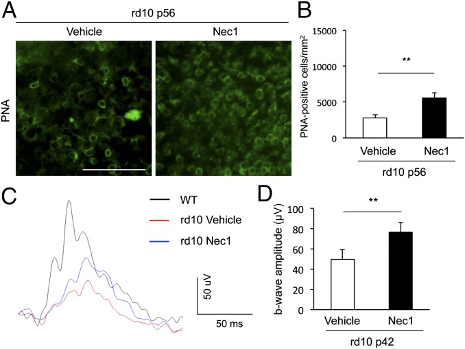
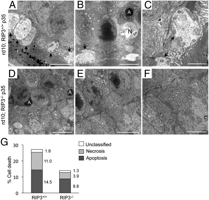
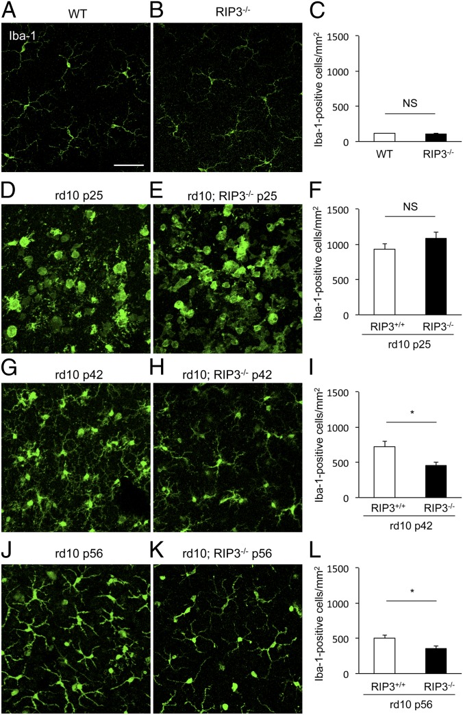
Retinitis pigmentosa comprises a group of inherited retinal photoreceptor degenerations that lead to progressive loss of vision. Although in most cases rods, but not cones, harbor the deleterious gene mutations, cones do die in this disease, usually after the main phase of rod cell loss. Rod photoreceptor death is characterized by apoptotic features. In contrast, the mechanisms and features of subsequent nonautonomous cone cell death remain largely unknown. In this study, we show that receptor-interacting protein (RIP) kinase mediates necrotic cone cell death in rd10 mice, a mouse model of retinitis pigmentosa caused by a mutation in a rod-specific gene. The expression of RIP3, a key regulator of programmed necrosis, was elevated in rd10 mouse retinas in the phase of cone but not rod degeneration. Although rd10 mice lacking Rip3 developed comparable rod degeneration to control rd10 mice, they displayed a significant preservation of cone cells. Ultrastructural analysis of rd10 mouse retinas revealed that a substantial fraction of dying cones exhibited necrotic morphology, which was rescued by Rip3 deficiency. Additionally, pharmacologic treatment with a RIP kinase inhibitor attenuated histological and functional deficits of cones in rd10 mice. Thus, necrotic mechanisms involving RIP kinase are crucial in cone cell death in inherited retinal degeneration, suggesting the RIP kinase pathway as a potential target to protect cone-mediated central and peripheral vision loss in patients with retinitis pigementosa.


Conflict of interest statement

Conflict of interest statement: The Massachusetts Eye and Ear Infirmary Institution has filed patents on the subject of neuroprotection in retinal degenerations. Y.M., J.W.M., and D.G.V. are named inventors.

Figures

Fig. 1.
Fig. 1.
Increased RIP3 and RIP1 expression in the late phase of retinal degeneration in rd10 mice. (A and B) Quantitative real-time PCR analysis for RIP3 (A) and RIP1 (B) in WT and rd10 retinas at P21 and P35 (n = 5–7 each). **P < 0.01. (C) Western blot analysis for RIP3 and RIP1 at P21 and P35 of WT and rd10 retinas (n = 4 each). Levels normalized to β-tubulin. For RIP3 analysis, spleen samples from WT and Rip3−/− animals were used as positive and negative controls, respectively. The bar graphs indicate the relative level of RIP3 and RIP1 to β-tubulin by densitometric analysis. *P < 0.05.
Fig. 2.
Fig. 2.
Rip3 deficiency does not prevent rod photoreceptor degeneration in rd10 mice. (A) TUNEL (green) and DAPI (blue) staining at P21 of rd10;Rip3+/+ and rd10;Rip3−/− mice. GCL, ganglion cell layer; INL, inner nuclear layer; ONL, outer nuclear layer. (Scale bar, 50 μm.) (B and C) Quantification of TUNEL-positive photoreceptors (B) and ONL thickness (C) at P21 of rd10;Rip3+/+ and rd10;Rip3−/− mice (n = 6 each). NS, not significant. T3–T1: retinal thickness at 1,200 μm, 800 μm, and 400 μm from the optic nerve in the temporal hemisphere. N1–N3: retinal thickness at 400 μm, 800 μm, and 1,200 μm from the optic nerve in the nasal hemisphere. There was no significant difference in either TUNEL-positive cells or ONL thickness between rd10;Rip3+/+ and rd10;Rip3−/− mice. (D and E) Retinal histology (D) and quantification of ONL thickness (E) at P28 of rd10; Rip3+/+ and rd10; Rip3−/− mice (n = 6 each). (Scale bar, 50 μm.) No significant difference was observed between rd10;Rip3+/+ and rd10;Rip3−/− mice.
Fig. 3.
Fig. 3.
Rod photoreceptor cell death is mainly associated with apoptotic morphology. (AE) TEM photomicrographs in the ONL (A, B, and D) and in the inner segment (C and E) at P21 of rd10;Rip3+/+ (AC) and rd10;Rip3−/− mice (D and E). A, apoptotic cell; N, necrotic cell. [Scale bars, 5 μm (A, B, and D), 2 μm (C and E).] Apoptotic photoreceptor nuclei were frequently observed in both rd10;Rip3+/+ (A) and rd10;Rip3−/− mice (D). Necrotic photoreceptor cell death was rarely seen (B). The mitochondrial structure was well preserved in both genotypes (C and E). (F) Quantification of apoptotic and necrotic photoreceptor cell death (n = 4 each). There was no significant difference in rod cell death morphology between rd10;Rip3+/+ and rd10;Rip3−/− mice. The percentage of each cell death was shown on the right of bar.
Fig. 4.
Fig. 4.
Rip3 deficiency decreases cone cell death and preserves cone function in rd10 mice. (A and B) TUNEL staining (A) and quantification of TUNEL-positive photoreceptors (B) at P35 of rd10;Rip3+/+ and rd10;Rip3−/− mice (n = 6 each). **P = 0.0062. GCL, ganglion cell layer; INL, inner nuclear layer; ONL, outer nuclear layer. (Scale bar, 50 μm.) (CF) PNA staining (C and E) and quantification of PNA-positive cone photoreceptors (D and F) at P42 (C and D) and P56 (E and F) of rd10;Rip3+/+ and rd10;Rip3−/− mice (n = 6 each). **P = 0.0039 each. (Scale bars, 50 μm.) Rip3 deficiency decreased cone photoreceptor cell loss in rd10 mice. (G and H) Photopic ERG (G) and quantification of b-wave amplitude (H) at P42 of rd10;Rip3+/+ and rd10;Rip3−/− mice (n = 12 each). **P = 0.0022. The mean b-wave amplitude in rd10;Rip3 −/− mice was significantly higher than those of rd10;Rip3+/+ mice.
Fig. 5.
Fig. 5.
RIP1 kinase inhibitor Nec-1 suppresses cone degeneration in rd10 mice. (A and B) PNA staining (A) and quantification of PNA-positive cone photoreceptors (B) at P56 of rd10 mice. The Alzet osmotic pumps containing Nec-1 (15 mg⋅kg⋅d, delivery for 28 d) or vehicle were implanted subcutaneously at P28 (n = 8 and 6). **P < 0.01. (Scale bar, 50 μm.) (C and D) Photopic ERG (C) and quantification of b-wave amplitude (D) at P42 of rd10 mice treated with Nec-1 or vehicle (n = 10 each). **P < 0.01.
Fig. 6.
Fig. 6.
Necrotic morphology of cone photoreceptor cell death in rd10 mice. (A–F) TEM photomicrographs in the ONL and inner segment at P35 of rd10;Rip3+/+ (AC) and rd10;Rip3−/− (DF) mice. A, apoptotic cell; N, necrotic cell. Arrow: cytoplasmic swelling of the inner segment. [Scale bar, 5 μm (A, B, D and E), 2 μm (C and F).] Cytoplasmic swelling of the inner segment and plasma membrane rupture were observed in rd10;Rip3+/+ mice. The swelling of the cytoplasm and mitochondria was decreased in rd10;Rip3−/− mice. (G) Quantification of apoptotic and necrotic photoreceptor cell death (rd10;Rip3+/+ mice: n = 5 and rd10;Rip3-/ mice: n = 6). The appearance of necrotic photoreceptors was significantly decreased in rd10;Rip3−/− mice (P < 0.05). The percentage of each cell death was shown on the right of bar.
Fig. 7.
Fig. 7.
Effect of RIP kinase on microglial activation during rod and cone cell death. (AI) Immunofluorescence for Iba-1 (A, B, D, E, G, H, J, and K) and quantification of Iba-1+ microglia (C, F, I, and L) in WT (A), Rip3−/− (B), P25 rd10;Rip3+/+ (D), P25 rd10;Rip3−/− (E), P42 rd10;Rip3+/+ (G), P42 rd10;Rip3−/− (H), P56 rd10;Rip3+/+ (J), and P56 rd10;Rip3−/− (K) mice (n = 6 each). *P < 0.05; NS, not significant. Whereas no difference in microglial activation was observed between P25 rd10;Rip3+/+ and rd10;Rip3−/− mouse retinas (F), microglial infiltration was significantly reduced in rd10;Rip3−/− retinas compared with rd10;Rip3+/+ retinas at both P42 and P56 (I and L). (Scale bar, 50 μm.)

References

    1. Hartong DT, Berson EL, Dryja TP. Retinitis pigmentosa. Lancet. 2006;368:1795–1809. - PubMed
    1. Berson EL, Rosner B, Sandberg MA, Weigel-Difranco C, Willett WC. ω-3 Intake and visual acuity in patients with retinitis pigmentosa receiving vitamin A. Arch Ophthalmol. 2012;130:707–711. - PMC - PubMed
    1. Kerr JF, Wyllie AH, Currie AR. Apoptosis: A basic biological phenomenon with wide-ranging implications in tissue kinetics. Br J Cancer. 1972;26:239–257. - PMC - PubMed
    1. Kroemer G, et al. Nomenclature Committee on Cell Death 2009 Classification of cell death: Recommendations of the Nomenclature Committee on Cell Death 2009. Cell Death Differ. 2009;16:3–11. - PMC - PubMed
    1. Holler N, et al. Fas triggers an alternative, caspase-8-independent cell death pathway using the kinase RIP as effector molecule. Nat Immunol. 2000;1:489–495. - PubMed

Publication types

MeSH terms

Substances