An RNA virus hijacks an incognito function of a DNA repair enzyme
- PMID: 22908287
- PMCID: PMC3437895
- DOI: 10.1073/pnas.1208096109
An RNA virus hijacks an incognito function of a DNA repair enzyme
Abstract
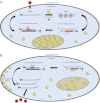
A previously described mammalian cell activity, called VPg unlinkase, specifically cleaves a unique protein-RNA covalent linkage generated during the viral genomic RNA replication steps of a picornavirus infection. For over three decades, the identity of this cellular activity and its normal role in the uninfected cell had remained elusive. Here we report the purification and identification of VPg unlinkase as the DNA repair enzyme, 5'-tyrosyl-DNA phosphodiesterase-2 (TDP2). Our data show that VPg unlinkase activity in different mammalian cell lines correlates with their differential expression of TDP2. Furthermore, we show that recombinant TDP2 can cleave the protein-RNA linkage generated by different picornaviruses without impairing the integrity of viral RNA. Our results reveal a unique RNA repair-like function for TDP2 and suggest an unusual role in host-pathogen interactions for this cellular enzyme. On the basis of the identification of TDP2 as a potential antiviral target, our findings may lead to the development of universal therapeutics to treat the millions of individuals afflicted annually with diseases caused by picornaviruses, including myocarditis, aseptic meningitis, encephalitis, hepatitis, and the common cold.
Conflict of interest statement
The authors declare no conflict of interest.
Figures


Similar articles
-
Effects of TDP2/VPg Unlinkase Activity on Picornavirus Infections Downstream of Virus Translation.Viruses. 2020 Jan 31;12(2):166. doi: 10.3390/v12020166. Viruses. 2020. PMID: 32023921 Free PMC article.
-
VPg unlinkase/TDP2 in cardiovirus infected cells: Re-localization and proteolytic cleavage.Virology. 2018 Mar;516:139-146. doi: 10.1016/j.virol.2018.01.010. Epub 2018 Jan 30. Virology. 2018. PMID: 29353210 Free PMC article.
-
Divergent Requirement for a DNA Repair Enzyme during Enterovirus Infections.mBio. 2015 Dec 29;7(1):e01931-15. doi: 10.1128/mBio.01931-15. mBio. 2015. PMID: 26715620 Free PMC article.
-
Tyrosyl-DNA-phosphodiesterases (TDP1 and TDP2).DNA Repair (Amst). 2014 Jul;19:114-29. doi: 10.1016/j.dnarep.2014.03.020. Epub 2014 May 22. DNA Repair (Amst). 2014. PMID: 24856239 Free PMC article. Review.
-
Mammalian Tyrosyl-DNA Phosphodiesterases in the Context of Mitochondrial DNA Repair.Int J Mol Sci. 2019 Jun 20;20(12):3015. doi: 10.3390/ijms20123015. Int J Mol Sci. 2019. PMID: 31226795 Free PMC article. Review.
Cited by
-
Recent Insights into the Control of Human Papillomavirus (HPV) Genome Stability, Loss, and Degradation.J Clin Med. 2015;4(2):204-30. doi: 10.3390/jcm4020204. J Clin Med. 2015. PMID: 25798290 Free PMC article.
-
Metabolism and function of hepatitis B virus cccDNA: Implications for the development of cccDNA-targeting antiviral therapeutics.Antiviral Res. 2015 Oct;122:91-100. doi: 10.1016/j.antiviral.2015.08.005. Epub 2015 Aug 10. Antiviral Res. 2015. PMID: 26272257 Free PMC article. Review.
-
Tyrosyl-DNA phosphodiesterases: rescuing the genome from the risks of relaxation.Nucleic Acids Res. 2018 Jan 25;46(2):520-537. doi: 10.1093/nar/gkx1219. Nucleic Acids Res. 2018. PMID: 29216365 Free PMC article. Review.
-
Is it time to switch to a formulation other than the live attenuated poliovirus vaccine to prevent poliomyelitis?Front Public Health. 2024 Jan 8;11:1284337. doi: 10.3389/fpubh.2023.1284337. eCollection 2023. Front Public Health. 2024. PMID: 38259741 Free PMC article. Review.
-
A Conserved Interaction between a C-Terminal Motif in Norovirus VPg and the HEAT-1 Domain of eIF4G Is Essential for Translation Initiation.PLoS Pathog. 2016 Jan 6;12(1):e1005379. doi: 10.1371/journal.ppat.1005379. eCollection 2016 Jan. PLoS Pathog. 2016. PMID: 26734730 Free PMC article.
References
-
- Rozovics JM, Semler BL. Genome replication I: the players. In: Ehrenfeld E, Domingo E, Roos RP, editors. The Picornaviruses. Washington, DC: ASM; 2010. pp. 107–125.
-
- Ambros V, Pettersson RF, Baltimore D. An enzymatic activity in uninfected cells that cleaves the linkage between poliovirion RNA and the 5′ terminal protein. Cell. 1978;15:1439–1446. - PubMed
-
- Ambros V, Baltimore D. Purification and properties of a HeLa cell enzyme able to remove the 5′-terminal protein from poliovirus RNA. J Biol Chem. 1980;255:6739–6744. - PubMed
-
- Yusupova RA, Gulevich AY, Drygin YF. Isolation from ascites carcinoma Krebs II cells of an unlinking enzyme hydrolyzing a covalent bond between picornavirus RNA and VPg. Biochemistry (Mosc) 2000;65:1219–1226. - PubMed
Publication types
MeSH terms
Substances
Grants and funding
LinkOut - more resources
Full Text Sources
Molecular Biology Databases
Research Materials

