. 2012 Sep 4;109(36):14604-9.
doi: 10.1073/pnas.1209367109. Epub 2012 Aug 20.

Nonviral delivery of self-amplifying RNA vaccines

Affiliations

Nonviral delivery of self-amplifying RNA vaccines

Andrew J Geall et al. Proc Natl Acad Sci U S A. .

Abstract

Despite more than two decades of research and development on nucleic acid vaccines, there is still no commercial product for human use. Taking advantage of the recent innovations in systemic delivery of short interfering RNA (siRNA) using lipid nanoparticles (LNPs), we developed a self-amplifying RNA vaccine. Here we show that nonviral delivery of a 9-kb self-amplifying RNA encapsulated within an LNP substantially increased immunogenicity compared with delivery of unformulated RNA. This unique vaccine technology was found to elicit broad, potent, and protective immune responses, that were comparable to a viral delivery technology, but without the inherent limitations of viral vectors. Given the many positive attributes of nucleic acid vaccines, our results suggest that a comprehensive evaluation of nonviral technologies to deliver self-amplifying RNA vaccines is warranted.


Conflict of interest statement

Conflict of interest statement: All authors are Novartis shareholders and employees of Novartis Vaccines and Diagnostics and Novartis Institutes for BioMedical Research.

Figures
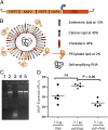
Fig. 1.
Fig. 1.
Characterization of self-amplifying RNA vaccines. (A) Schematic illustration of a self-amplifying RNA derived from an alphavirus contains a 5′cap, nonstructural genes (NSP1–4), 26S subgenomic promoter (grey arrow), the gene of interest (GOI), and a 3′ polyadenylated tail. (B) Schematic illustration of a lipid nanoparticle (LNP) encapsulating self-amplifying RNA, with the percent molar ratios of lipid components as indicated. (C) RNA agarose gel electrophoresis: RNA ladder (lane 1), self-amplifying RNA (lane 2), self-amplifying RNA after exposure to RNase A (lane 3), phenol-chloroform extraction of self-amplifying RNA from an LNP (lane 4), phenol-chloroform extraction of self-amplifying RNA from an LNP after exposure to RNase A (lane 5). (D) In vivo expression of secreted alkaline phosphatase (SEAP) 6 d after intramuscular (i.m.) injection of 1 μg self-amplifying RNA in PBS (RNA), 0.1 μg self-amplifying RNA encapsulated in an LNP (LNP/RNA), and 0.1 μg self-amplifying RNA mixed (not encapsulated) with LNP (LNP + RNA). Data are from individual mice (five per group, depicted as dots), and the geometric mean is represented by a solid line. NS, not significant.
Fig. 2.
Fig. 2.
Whole-mouse in vivo bioluminescence imaging after bilateral i.m. injection with RNA or DNA encoding firefly luciferase. (A) Bioluminescence recorded from individual animals on day 7 after administration of viral replicon particles (VRPs, 1 × 10⁶ infectious units) (IU), self-amplifying RNA (1 μg), self-amplifying RNA encapsulated in lipid nanoparticles (LNP/RNA, 1 μg), plasmid DNA (pDNA, 10 μg), or a pDNA-launched self-amplifying RNA (10 μg). Each dot represents the whole-mouse photon count expressed as photons per second per square centimeter per steradian (p/s/cm2/sr); the solid line represents the geometric mean; and the dotted line represents the limit of detection (1 × 10³ p/s/cm2/sr). (B) Average radiance over 63 d after administration of VRPs (1 × 10⁶ IU), with representative bioluminescence images at day 7. (C) Average radiance over 63 d after administration of self-amplifying RNA (1 μg) encapsulated in LNP, with representative bioluminescence images at day 7. (D) Average radiance over 63 d after administration of electroporated pDNA (10 μg), with representative bioluminescence images at day 7. Data are from individual mice (five per group, gray circles); the geometric mean is represented by a solid line; the limit of detection is indicated by the dashed line (1 × 10³ p/s/cm2/sr). Additional images from the 63-d time course can be found in Fig. S2.
Fig. 3.
Fig. 3.
Comparative mouse immunogenicity studies of a lipid nanoparticle formulated self-amplifying RNA (LNP/RNA) vaccine candidate encoding RSV-F. Groups of eight mice (except LNP/DNA, with four mice per group) were vaccinated intramuscularly (i.m.) on days 0 and 21 with viral replicon particles (VRPs, 1 × 10⁶ IU), plasmid DNA delivered using electroporation (DNA + EP, 20 μg), pDNA formulated with LNP (LNP/DNA, 0.1 μg), naked self-amplifying RNA (RNA, 1.0 μg) or self-amplifying RNA formulated in LNP (LNP/RNA, 0.1 μg). Sera were collected on day 35, and F-specific IgG titers were determined by ELISA. Data are from individual mice (depicted as dots), and the geometric mean titers (GMTs) are solid lines. Dotted line indicates the limit of titer quantification (25 titer limit). To calculate GMTs, titers <25 were assigned a value of 5. F-specific CD4+ and CD8+ T cell frequencies can be found in Fig. S4. NS, not significant.
Fig. 4.
Fig. 4.
Mouse immunogenicity studies of a lipid nanoparticle formulated self-amplifying RNA (LNP/RNA) candidate vaccine encoding RSV-F. Groups of eight mice were vaccinated intramuscularly (i.m.) on days 0 and 21 with naked self-amplifying RNA (0.01–1 μg), self-amplifying RNA formulated in lipid nanoparticles (LNP/RNA, 0.01–10 μg), or viral replicon particles (VRPs, 1 × 10⁶ IU). Sera were collected on day 35, and F-specific IgG (A), IgG1 (B), and IgG2a (C) titers were determined by ELISA. Dots depict measurements from individual mice and solid lines, the geometric mean titers of eight mice per group. Dotted lines indicate the limit of 25 for quantification. For determination of GMTs, a titer <25 was assigned a value of 5. (D) Frequencies of RSV-F antigen-specific, cytokine-producing CD8+4 T cells in spleens of BALB/c mice vaccinated on days 0 and 21 with RNA, RNA/LNP, or VRP. Spleens were collected 4 wk after the second vaccination and pooled (four spleens per pool) before antigen stimulation in vitro and flow cytometry analysis. Error bars indicate the 95% confidence upper limits. No IL2+ TNFα+, IL2+IFNγ+, IL2+, or IL5+ CD8 T cells were detected. Serum IgG titers 2 wk after the first vaccination can be found in Fig. S3. NS, not significant.
Fig. 5.
Fig. 5.
Cotton rat immunogenicity and protection studies of a lipid nanoparticle-formulated self-amplifying RNA (LNP/RNA) candidate vaccine encoding RSV-F. Groups of eight rats were vaccinated i.m. on days 0 and 21 with naked self-amplifying RNA (1 μg), self-amplifying RNA formulated in LNPs (LNP/RNA, 1 μg), viral replicon particles (VRPs, 5 × 10⁶ IU), or alum-formulated RSV-F subunit (10 μg); or they were not vaccinated. All animals were challenged intranasally with 1 × 10⁵ pfu of RSV on day 49. (A) Serum RSV neutralization titers and (B) serum F-specific IgG titers 2 wk after the second vaccination (day 35). (C) Lung viral load 5 d after the RSV challenge (day 54). Data are from individual rats (depicted as dots), and the geometric mean titers (GMTs) are solid lines. Dotted lines indicate assay limits of detection (A = 20, B = 25, C = 195). For determination of GMTs, a titer below the limit of detection was assigned a value of 10 (A), 5 (B), or 100 (C). NS, not significant.

Comment in

References

    1. Rappuoli R, Mandl CW, Black S, De Gregorio E. Vaccines for the twenty-first century society. Nat Rev Immunol. 2011;11:865–872. - PMC - PubMed
    1. Kutzler MA, Weiner DB. DNA vaccines: Ready for prime time? Nat Rev Genet. 2008;9:776–788. - PMC - PubMed
    1. Kofler RM, et al. Mimicking live flavivirus immunization with a noninfectious RNA vaccine. Proc Natl Acad Sci USA. 2004;101:1951–1956. - PMC - PubMed
    1. Smerdou C, Liljeström P. Non-viral amplification systems for gene transfer: Vectors based on alphaviruses. Curr Opin Mol Ther. 1999;1:244–251. - PubMed
    1. Wolff JA, et al. Direct gene transfer into mouse muscle in vivo. Science. 1990;247:1465–1468. - PubMed

Publication types

LinkOut - more resources