. 2012 Sep 4;109(36):14640-5.
doi: 10.1073/pnas.1206098109. Epub 2012 Aug 20.

Structural insights into Helicobacter pylori oncoprotein CagA interaction with β1 integrin

Affiliations

Structural insights into Helicobacter pylori oncoprotein CagA interaction with β1 integrin

Burcu Kaplan-Türköz et al. Proc Natl Acad Sci U S A. .

Abstract

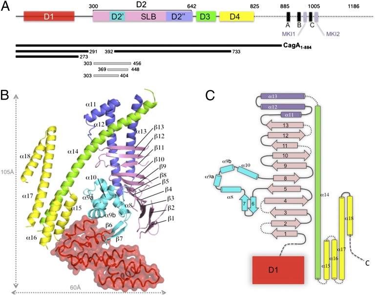
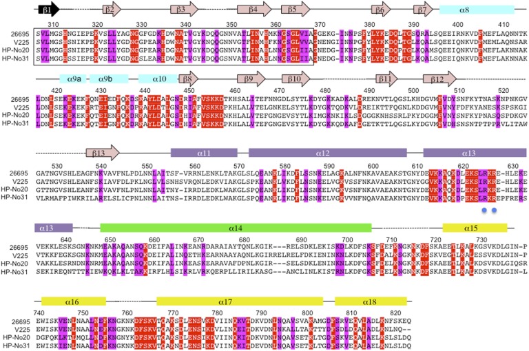
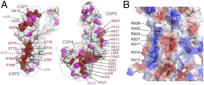
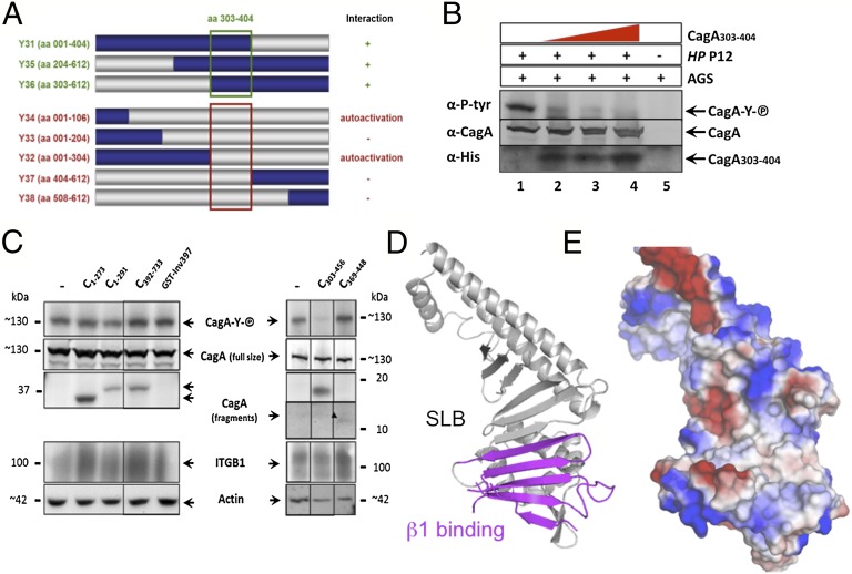
Infection with the gastric pathogen Helicobacter pylori is a risk factor for the development of gastric cancer. Pathogenic strains of H. pylori carry a type IV secretion system (T4SS) responsible for the injection of the oncoprotein CagA into host cells. H. pylori and its cag-T4SS exploit α5β1 integrin as a receptor for CagA translocation. Injected CagA localizes to the inner leaflet of the host cell membrane, where it hijacks host cell signaling and induces cytoskeleton reorganization. Here we describe the crystal structure of the N-terminal ~100-kDa subdomain of CagA at 3.6 Å that unveils a unique combination of folds. The core domain of the protein consists of an extended single-layer β-sheet stabilized by two independent helical subdomains. The core is followed by a long helix that forms a four-helix helical bundle with the C-terminal domain. Mapping of conserved regions in a set of CagA sequences identified four conserved surface-exposed patches (CSP1-4), which represent putative hot-spots for protein-protein interactions. The proximal part of the single-layer β-sheet, covering CSP4, is involved in specific binding of CagA to the β1 integrin, as determined by yeast two-hybrid and in vivo competition assays in H. pylori cell-culture infection studies. These data provide a structural basis for the first step of CagA internalization into host cells and suggest that CagA uses a previously undescribed mechanism to bind β1 integrin to mediate its own translocation.


Conflict of interest statement

The authors declare no conflict of interest.

Figures

Fig. 1.
Fig. 1.
Overall structure of CagA N-terminal domain. (A) Schematic representation of CagA (Western strain 26695). Boxes indicate the domain definition derived from the CagA1–884 X-ray structure, with D1 colored in red, the SLB in pink, subdomain D2′ in cyan, D2′′ in blue, D3 in green, and D4 in yellow. The positions of the EPIYA motifs are indicated by A, B, and C and those of PAR1-MARK kinase binding motifs (14) by MKI1 and MKI2. Bars indicate the positions of the protein fragments used in the study. (B) Ribbon representation of the structure of CagA1–884 colored according to A. (C) Topology diagram of CagA1–884 structure.
Fig. 2.
Fig. 2.
Structure-based sequence alignment of CagA1–884. Sequences of CagA from strain 26695 (46), V225 (47), HP-No20, and HP-No31 (48), were selected from the multiple alignment of 80 sequences generated by CONSURF (49) to illustrate CagA diversity. Sequence numbering corresponds to CagA from strain 26695. Conserved and invariant residues (among 80 CagA sequences) exposed on the surface of CagA1–884 crystal structure are shaded in magenta and red, respectively. The exact sequence of strand β1 could not be determined; the mostly likely candidate sequence is indicated in black. Secondary structure elements are indicated above the sequences and colored according to the domain: D2′, cyan; SLB, pink; D2′′, purple; D3, green; and D4, yellow. Blue dots are positioned below residues R624 and R626, involved in PS binding (25).
Fig. 3.
Fig. 3.
Potential functional sites in CagA structure. (A) Semitransparent surface representation of the CagA domain D3-D4 (Left) and D2 (Right) colored in gray with strictly conserved and conserved residues colored in red and pink, respectively. The location of the conserved surface exposed patch “CSP1–4” are indicated. (B) Surface representation of α12 and α13 colored by electrostatic potential. Residues forming the basic patch potentially involved in PS binding are indicated.
Fig. 4.
Fig. 4.
Mapping the CagA-β1 integrin interaction domains. (A) Schematic representation of CagA fragments interacting with β1 integrin in a YTH screen. Construction of systematically shortened CagA1–612 clones (Y31–Y38) in the bait vector (gray represents deleted sequences). The green square denotes the CagA amino acid sequence essential for interaction of CagA with β1 integrin. Clones Y32 and Y34 grew on selective medium without interaction partner (autoactivation). (B) Recombinant CagA303–404 interferes with CagA translocation. AGS cells were incubated with 4, 8, or 12 nMol purified CagA303–404 (lanes 2–4) and infected with H. pylori P12 or not (lane 5). Lysates were run on SDS/PAGE, blotted, and reacted with antibodies as indicated. (C) AGS cells were incubated with purified CagA fragments (His-tagged; see Fig. 1B for their exact location) or recombinant Yersinia invasin (GST-Inv397), and the structure-derived fragments CagA303–456 or CagA369–448 and infected with H. pylori P12 strain. Lysates were run on SDS/PAGE or single gel system, blotted, and reacted with antibodies, as indicated. (D) Ribbon diagram of CagA1–884 domain D2 (gray) showing the β1 integrin interacting portion (magenta). (E) Surface representation of the structure in the same orientation colored according to the electrostatic potential.

Similar articles

Cited by

References

    1. Peek RM, Jr, Blaser MJ. Helicobacter pylori and gastrointestinal tract adenocarcinomas. Nat Rev Cancer. 2002;2:28–37. - PubMed
    1. Ohnishi N, et al. Transgenic expression of Helicobacter pylori CagA induces gastrointestinal and hematopoietic neoplasms in mouse. Proc Natl Acad Sci USA. 2008;105:1003–1008. - PMC - PubMed
    1. Odenbreit S, et al. Translocation of Helicobacter pylori CagA into gastric epithelial cells by type IV secretion. Science. 2000;287:1497–1500. - PubMed
    1. Rohde M, Püls J, Buhrdorf R, Fischer W, Haas R. A novel sheathed surface organelle of the Helicobacter pylori Cag type IV secretion system. Mol Microbiol. 2003;49:219–234. - PubMed
    1. Terradot L, Waksman G. Architecture of the Helicobacter pylori Cag-type IV secretion system. FEBS J. 2011;278:1213–1222. - PubMed

Publication types

MeSH terms

Associated data

LinkOut - more resources