. 2012 Aug 20;198(4):491-9.
doi: 10.1083/jcb.201205090.

Polo-like kinase-1 regulates kinetochore-microtubule dynamics and spindle checkpoint silencing

Affiliations

Polo-like kinase-1 regulates kinetochore-microtubule dynamics and spindle checkpoint silencing

Dan Liu et al. J Cell Biol. .

Abstract

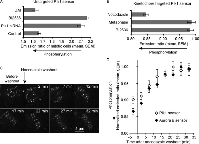
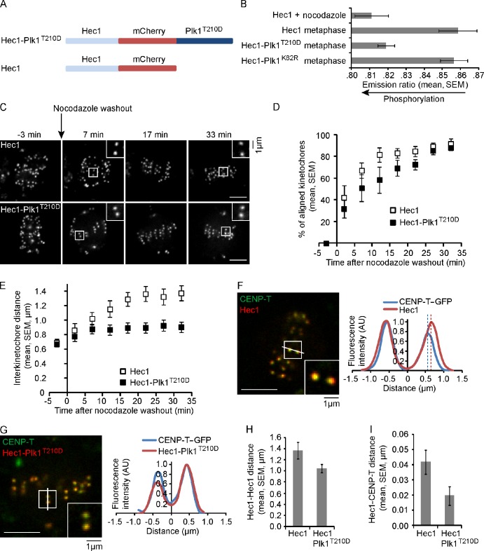
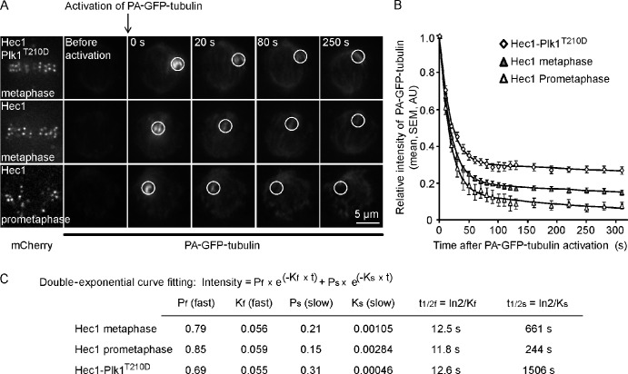
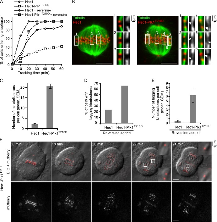
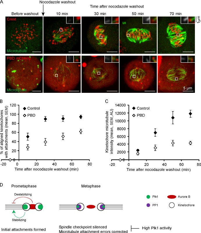
Polo-like kinase-1 (Plk1) is a highly conserved kinase with multiple mitotic functions. Plk1 localizes to prometaphase kinetochores and is reduced at metaphase kinetochores, similar to many checkpoint signaling proteins, but Plk1 is not required for spindle checkpoint function. Plk1 is also implicated in stabilizing kinetochore-microtubule attachments, but these attachments are most stable when kinetochore Plk1 levels are low at metaphase. Therefore, it is unclear how Plk1 function at kinetochores can be understood in the context of its dynamic localization. In this paper, we show that Plk1 activity suppresses kinetochore-microtubule dynamics to stabilize initial attachments in prometaphase, and Plk1 removal from kinetochores is necessary to maintain dynamic microtubules in metaphase. Constitutively targeting Plk1 to kinetochores maintained high activity at metaphase, leading to reduced interkinetochore tension and intrakinetochore stretch, a checkpoint-dependent mitotic arrest, and accumulation of microtubule attachment errors. Together, our data show that Plk1 dynamics at kinetochores control two critical mitotic processes: initially establishing correct kinetochore-microtubule attachments and subsequently silencing the spindle checkpoint.


Figures

Figure 1.
Figure 1.
A FRET-based biosensor for Plk1 activity at kinetochores is dephosphorylated as chromosomes align at metaphase. (A) The YFP/CFP emission ratio was averaged over multiple mitotic cells (n ≥ 9) expressing an untargeted Plk1 phosphorylation sensor and treated with an inhibitor for Aurora B (ZM) or Plk1 (BI2536) or with Plk1 siRNA, as indicated. (B) The YFP/TFP emission ratio was analyzed in cells expressing a kinetochore-targeted Plk1 sensor at metaphase or treated with nocodazole or BI2536, as indicated (n ≥ 10 cells, n ≥ 15 kinetochores per cell). (C and D) Cells expressing a kinetochore-targeted Plk1 sensor (C and D) or a kinetochore-targeted Aurora B sensor (D) were imaged live after nocodazole washout. Images (C) of the Plk1 sensor (YFP emission) show kinetochore alignment after washout. To compare FRET changes for the two sensors (D), the ratios for each were normalized by dividing by the maximum value for that sensor (n ≥ 10 cells, n ≥ 15 kinetochores per cell).
Figure 2.
Figure 2.
Persistent Plk1 activity at kinetochores disrupts both interkinetochore tension and intrakinetochore stretch. (A) Schematic of Hec1 and Hec1-Plk1T210D constructs. (B) The YFP/TFP emission ratio was analyzed in cells expressing a kinetochore-targeted Plk1 phosphorylation sensor, together with either Hec1, Hec1-Plk1T210D, or Hec1-Plk1K82R (kinase-inactive mutant), under the conditions indicated (n ≥ 10 cells, n ≥ 15 kinetochores per cell). (C–E) Cells expressing either Hec1 or Hec1-Plk1T210D were imaged live after nocodazole washout. Images (C) are maximal intensity projections of confocal z series. Insets are optical sections showing individual kinetochore pairs. Note that Hec1-Plk1T210D localizes to both kinetochores and spindle poles. Metaphase alignment (D) and interkinetochore distance (E) were calculated at each time point (n ≥ 40 kinetochores per time point from multiple cells). (F–I) Cells expressing CENP-T–GFP, together with either Hec1 or Hec1-Plk1T210D, were imaged live at metaphase. Images (F and G) are single confocal planes, and insets show individual kinetochore pairs used for the line scans. Dashed lines indicate estimated Hec1 and CENP-T positions. Distances were calculated between sister kinetochores (H) or between Hec1 and CENP-T within a kinetochore (n ≥ 80 kinetochore pairs from multiple cells; I). AU, arbitrary unit. (C, F, and G) Bars, 5 µm.
Figure 3.
Figure 3.
Persistent Plk1 activity at kinetochores suppresses microtubule dynamics. (A) Cells expressing PA-GFP-tubulin together with either Hec1 or Hec1-Plk1T210D were imaged live before and after photoactivation of a spot near the metaphase plate at t = 0. Images show PA-GFP-tubulin or Hec1 or Hec1-Plk1T210D visualized with mCherry. (B) GFP intensity in the activated spot (white circles in A) was calculated at each time point as the intensity relative to the initial value after activation. The relative GFP intensities were corrected for photobleaching based on a taxol control, averaged over multiple cells (n ≥ 8), and fit with double-exponential decay curves. AU, arbitrary unit. (C) Curve-fitting parameters Kf and Ks represent the fast and slow time constants, respectively.
Figure 4.
Figure 4.
Kinetochores with persistent Plk1 activity accumulate microtubule attachment errors and fail to silence the spindle checkpoint. (A) Cells expressing either Hec1 or Hec1-Plk1T210D were selected at metaphase and followed for 60 min to determine the time of anaphase onset (n > 20). The Mps1 inhibitor reversine was added, as indicated, at t = 0 to override the spindle checkpoint. (B and C) Cells expressing Hec1 or Hec1-Plk1T210D were briefly permeabilized and treated with calcium to remove nonkinetochore microtubules, fixed, and stained for microtubules. Images (B) are maximal intensity projections of confocal z series. Insets show sister kinetochore pairs in optical sections. Images are scaled differently in the insets to show merotelic attachments (arrows) more clearly. The number of kinetochores with microtubules attached from both directions (merotelic errors) was determined (n = 20 cells; C). (D–F) For cells treated with reversine at metaphase, as in A, the fraction of cells with lagging kinetochores in anaphase (D) and the number of laggers per cell (E) were determined (n > 20). Images (F) show a representative cell expressing Hec1-Plk1T210D with lagging kinetochores in anaphase. Insets show lagging kinetochores at higher magnification. Note that Hec1-Plk1T210D localizes to kinetochores and spindle poles and to the anaphase spindle midzone. DIC, differential interference contrast. (B and F) Bars, 5 µm.
Figure 5.
Figure 5.
Plk1 activity at kinetochores is required for efficient formation of stable kinetochore–microtubule attachments. (A–C) Cells expressing PBD-mCherry or untransfected controls were fixed at the indicated time points after nocodazole washout and analyzed for cold-stable microtubules. Images (A) are maximal intensity projections of confocal z series. Insets are optical sections showing individual kinetochores. The PBD-mCherry images are scaled differently in the insets to show kinetochores more clearly. The fraction of aligned kinetochores with cold-stable attachments (B) and the microtubule staining intensities adjacent to kinetochores (C) were determined at each time point (n ≥ 10 cells, n ≥ 30 kinetochores per cell). AU, arbitrary unit. (D) A model showing that Aurora B and Plk1 activities are both high in prometaphase and have opposite effects on kinetochore microtubules, with Aurora B destabilizing and Plk1 stabilizing. In metaphase, both Aurora B and Plk1 activities are reduced at kinetochores, whereas PP1 is recruited. The reduction of Plk1 activity is important for maintaining dynamic microtubules, establishing intrakinetochore stretch and interkinetochore tension, silencing the spindle checkpoint, and correcting attachment errors (which can also occur in prometaphase).

References

    1. Archambault V., Glover D.M. 2009. Polo-like kinases: Conservation and divergence in their functions and regulation. Nat. Rev. Mol. Cell Biol. 10:265–275 10.1038/nrm2653 - DOI - PubMed
    1. Bakhoum S.F., Genovese G., Compton D.A. 2009a. Deviant kinetochore microtubule dynamics underlie chromosomal instability. Curr. Biol. 19:1937–1942 10.1016/j.cub.2009.09.055 - DOI - PMC - PubMed
    1. Bakhoum S.F., Thompson S.L., Manning A.L., Compton D.A. 2009b. Genome stability is ensured by temporal control of kinetochore-microtubule dynamics. Nat. Cell Biol. 11:27–35 10.1038/ncb1809 - DOI - PMC - PubMed
    1. Carmena M., Pinson X., Platani M., Salloum Z., Xu Z., Clark A., Macisaac F., Ogawa H., Eggert U., Glover D.M., et al. 2012. The chromosomal passenger complex activates Polo kinase at centromeres. PLoS Biol. 10:e1001250 10.1371/journal.pbio.1001250 - DOI - PMC - PubMed
    1. Cimini D., Howell B., Maddox P., Khodjakov A., Degrassi F., Salmon E.D. 2001. Merotelic kinetochore orientation is a major mechanism of aneuploidy in mitotic mammalian tissue cells. J. Cell Biol. 153:517–527 10.1083/jcb.153.3.517 - DOI - PMC - PubMed

Publication types

MeSH terms

Substances