. 2012 Aug 20;198(4):623-36.
doi: 10.1083/jcb.201111105.

Hsp110 is required for spindle length control

Affiliations

Hsp110 is required for spindle length control

Taras Makhnevych et al. J Cell Biol. .

Abstract

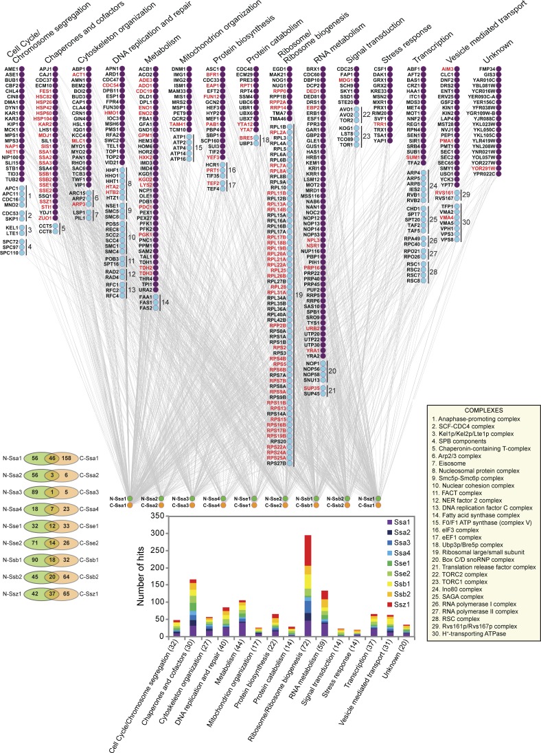
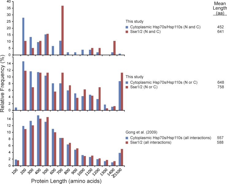
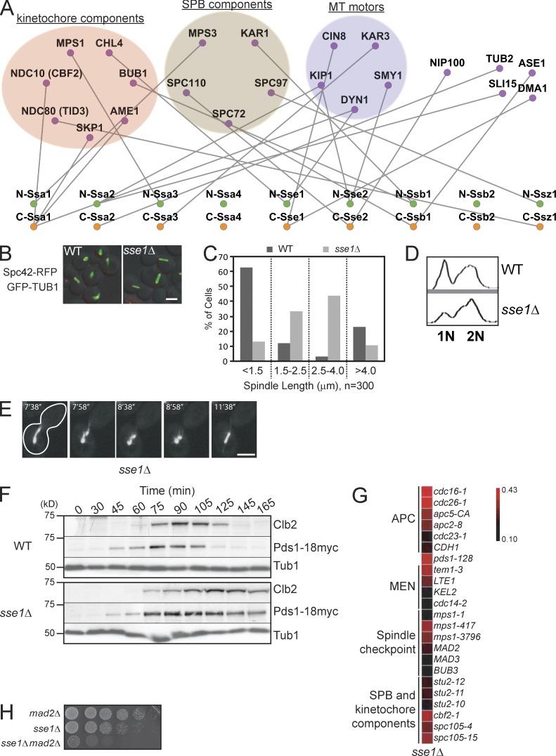
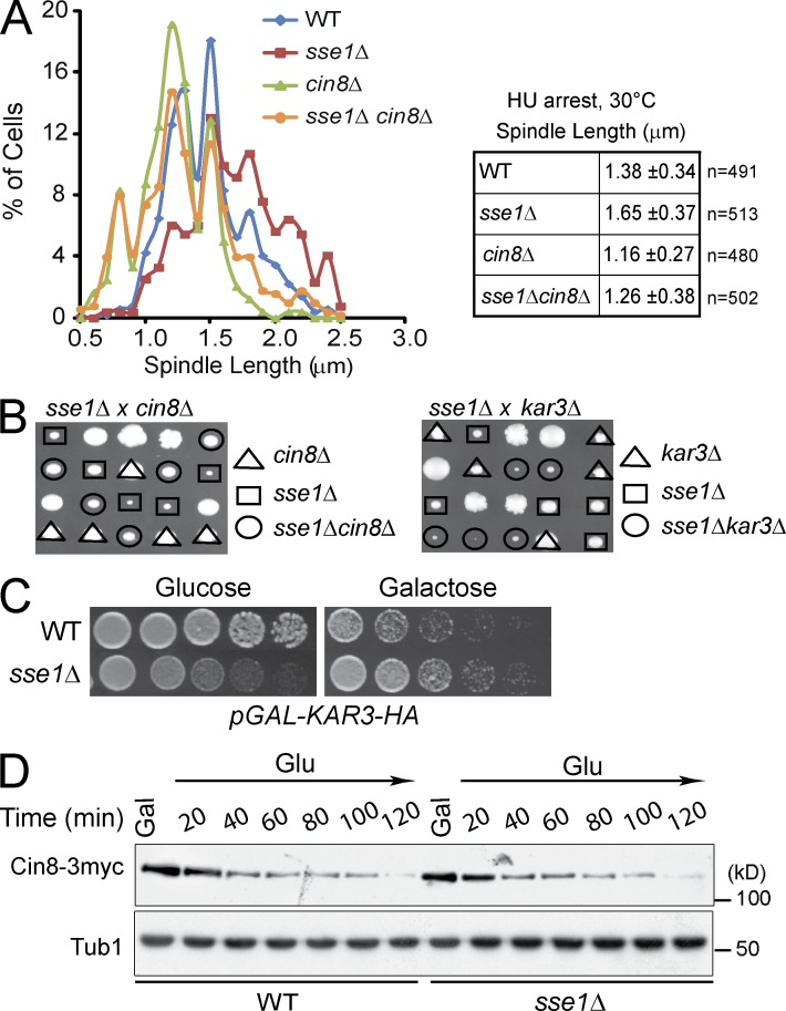
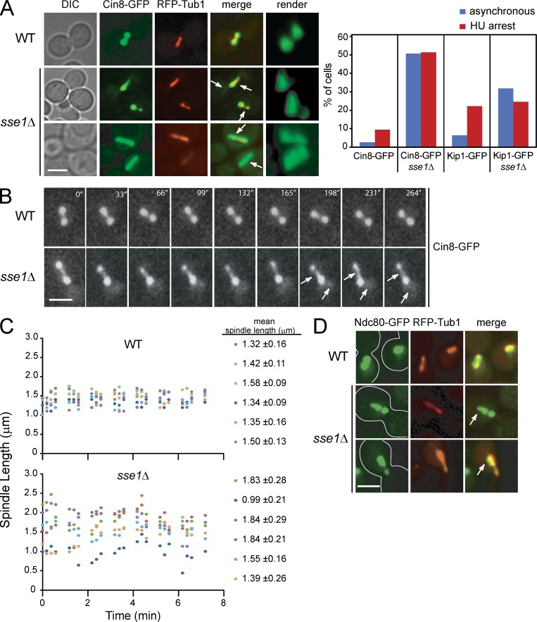
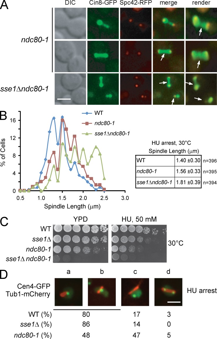
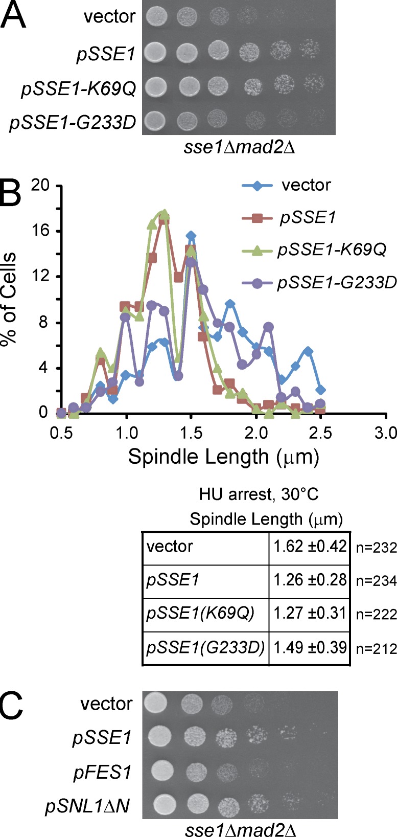
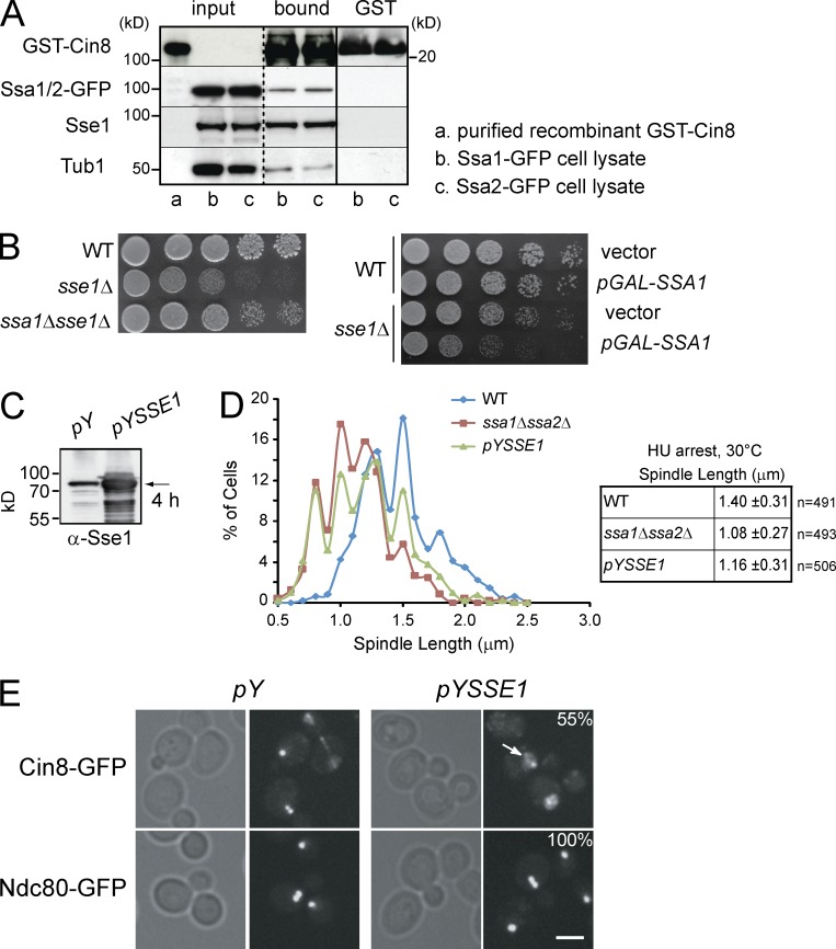
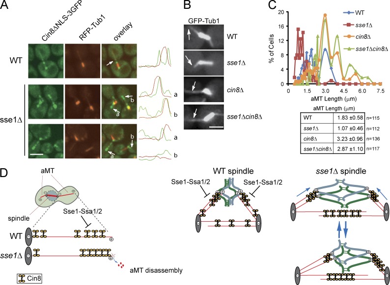
Systematic affinity purification combined with mass spectrometry analysis of N- and C-tagged cytoplasmic Hsp70/Hsp110 chaperones was used to identify new roles of Hsp70/Hsp110 in the cell. This allowed the mapping of a chaperone-protein network consisting of 1,227 unique interactions between the 9 chaperones and 473 proteins and highlighted roles for Hsp70/Hsp110 in 14 broad biological processes. Using this information, we uncovered an essential role for Hsp110 in spindle assembly and, more specifically, in modulating the activity of the widely conserved kinesin-5 motor Cin8. The role of Hsp110 Sse1 as a nucleotide exchange factor for the Hsp70 chaperones Ssa1/Ssa2 was found to be required for maintaining the proper distribution of kinesin-5 motors within the spindle, which was subsequently required for bipolar spindle assembly in S phase. These data suggest a model whereby the Hsp70-Hsp110 chaperone complex antagonizes Cin8 plus-end motility and prevents premature spindle elongation in S phase.


Figures

Figure 1.
Figure 1.
The TAP tag–based Hsp70/Hsp110 interaction network. The main figure shows the hits obtained from the TAP tag pull-down of the nine Hsp70s/Hsp110s. The hits are grouped according to Gene Ontology categories (also shown in the bottom graph; The Gene Ontology Consortium, 2000) and are organized into complexes as defined by Pu et al. (2009; bottom right inset). The overlap in the hits between N- and C-tagged chaperones is shown in the bottom left. Data are from experiments completed once. Complexes identified are shown in the top right inset. FACT, facilitates chromatin transcription; NER, nucleotide excision repair; snoRNP, small nucleolar ribonucleoprotein; SCF, Skp, Cullin, F-box–containing complex.
Figure 2.
Figure 2.
Size distribution of chaperone hits. The size distribution of the hits obtained in this work for the cytoplasmic Hsp70s/Hsp110s compared with those obtained for the Hsp110s (Sse1/2) alone is shown as a bar graph. (N and C) refers to the hits that overlapped between N- and C-TAP tag pull-down experiments, whereas (N or C) refers to the hits obtained in either N- or C-TAP tag pull-down experiments. The size distribution of the hits obtained in our previous study (Gong et al., 2009) is also shown. Proteins interacting with Sse1/2 were found to be longer than proteins interacting with the cytoplasmic Hsp70/Hsp110 chaperones (Mann–Whitney test, P < 0.04). Data are from experiments completed once.
Figure 3.
Figure 3.
The role of Hsp70/Hsp110 in spindle organization. (A) A subnetwork of the Hsp70/Hsp110 protein interactions (also refer to Fig. 1 and Table S2) highlighting TAP tag–based physical interactions between N- and C-tagged Hsp70/Hsp110 and components of SPB, kinetochore, and MT motors. (B) The fluorescence images show spindle morphology examined by confocal microscopy in logarithmically growing WT and sse1Δ strains at 30°C containing an SPB marker, endogenous Spc42-RFP (red), and harboring the pGFP-TUB1 (green) plasmid. Bar, 5 µm. (C) Bar graph showing the distribution of spindle lengths observed in WT and sse1Δ cells. The data shown are from a single representative experiment out of three repeats. (D) FACS profiles of WT and sse1Δ cells. 3 ml of early log–phase culture was used. (E) The spindle of an sse1Δ cell was visualized using plasmid-borne GFP-Tub1 and examined by time-lapse confocal microscopy. The cell outline is traced by the white line. Bar, 5 µm. (F) Logarithmically growing cultures of WT and sse1Δ expressing Pds1-18myc were synchronized in G1 with α-factor (time = 0 min). α-factor was then removed, and cells were grown at 26°C in YPD. Samples were taken at the indicated time points, and total cell lysates were analyzed by Western blot analysis using antibodies directed against cMyc, Clb2, and tubulin. Molecular mass markers are shown on the left of the gels. (G) A synthetic genetic array subnetwork highlighting genetic interactions between sse1Δ and genes involved in chromosome segregation and cell cycle progression. APC and MEN refer to anaphase-promoting complex and mitotic exit network, respectively. The intensity of the red color correlates with the strength of the genetic interaction (refer to Costanzo et al. [2010]). (H) 10× serial dilutions of log-phase cells of the indicated genotypes spotted onto YPD and incubated at 26°C for 2 d are shown.
Figure 4.
Figure 4.
Deletion of Sse1 promotes Cin8-dependent spindle elongation in S phase. (A) Logarithmically growing cultures of WT, sse1Δ, cin8Δ, and sse1Δcin8Δ and cells expressing a plasmid-borne copy of GFP-Tub1 and/or containing Spc42-RFP were synchronized in S phase with 100 mM HU, and spindle length was measured by confocal microscopy. The data shown are from a single representative experiment out of three repeats. (B) The dissection of sse1Δcin8Δ and sse1Δkar3Δ diploid strains demonstrating the presence of alleviating genetic interaction between sse1Δ and cin8Δ and aggravating genetic interaction between sse1Δ and kar3Δ is shown. (C) 10× serial dilutions of log-phase WT and sse1Δ cells harboring pGAL-KAR3-HA were spotted onto glucose or galactose medium and incubated at 26°C for 2 d. (D) WT and sse1Δ cells expressing Cin8-3myc from a CEN plasmid were grown in yeast extract peptone + raffinose at 26°C and then arrested using α-factor. Cells were then released from arrest into yeast extract peptone + raffinose + galactose (Gal) medium containing 100 mM HU to induce the expression of Cin8-3myc expression and arrest cells in S phase. After 120 min, cells were transferred to glucose (Glu) medium containing 100 mM HU and 1 µg/ml cycloheximide to turn off the GAL promoter and prevent further translation, and the level of Cin8 was monitored by Western blot analysis using antibodies directed against cMyc. Molecular mass markers are shown on the right of the gels.
Figure 5.
Figure 5.
Localization of GFP-tagged kinesin-5 motors in WT and sse1Δ cells. (A) WT and sse1Δ cells expressing RFP-Tub1 and Cin8-GFP were synchronized in S phase with HU for 2.5 h at 26°C. Representative images (extended focus and 3D render) of Cin8-GFP (green) and RFP-Tub1 (red) obtained using fluorescence confocal microscopy are shown. Arrows point to the presence of Cin8 in the midzone and nucleoplasm. Bar, 5 µm. The bar graph shows the percentage of cells of the indicated genotype that have asymmetric distribution of Cin8 in either asynchronous or HU-arrested cultures. About 200 cells were observed. The data shown are from a single representative experiment out of three repeats. DIC, differential interference contrast. (B) WT and sse1Δ cells expressing Cin8-GFP were incubated with HU for 2.5 h, and time-lapse microscopy was performed. The images show that deletion of SSE1 results in redistribution of Cin8 localization and, hence, in varied spindle length. The arrows show the presence of Cin8 in the midzone and nucleoplasm. Bar, 5 µm. (C) Spindle length is plotted for six HU-arrested WT or sse1Δ cells as a function of time. The mean spindle length measured for each cell over the indicated time period is given on the right. (D) WT and sse1Δ cells expressing Ndc80-GFP (green) and RFP-Tub1 (red) were synchronized in S phase with HU for 2.5 h at 26°C. Images were obtained using fluorescence confocal microscopy. Arrows point to the tighter clustering of Ndc80-GFP near the SPB. Bar, 5 µm.
Figure 6.
Figure 6.
Cin8-GFP localization pattern in kinetochore mutant ndc80-1 mirrors its localization in the sse1Δ strain. (A) Logarithmically growing cultures of ndc80-1 and sse1Δndc80-1 expressing Cin8-GFP and the SPB marker Spc42-RFP were synchronized in S phase with HU for 2.5 h at 30°C. Images were then obtained using fluorescence confocal microscopy. Arrows point to Cin8 accumulation in the midzone area and the nucleoplasm. DIC, differential interference contrast. Bar, 5 µm. (B) Spindle length in HU-arrested ndc80-1 and sse1Δndc80-1 cells grown at 30°C was measured using Spc42-RFP fluorescence by confocal microscopy. The data shown are from a single representative experiment out of three repeats. (C) 10× serial dilutions of log-phase cells of the indicated genotypes were spotted onto YPD and incubated at 30°C for 2 d. (D) The localization of Cen4-GFP and Tub1-mCherry in HU-arrested WT sse1Δ and ndc80-1 cells is shown. A single GFP dot near one end of the spindle might represent cases in which chromosome IV is attached to one SPB, as not all centromeres are replicated in the presence of HU (a). Cen4-GFP was also observed in the middle of the spindle (b), displaced from the spindle, which might represent detached chromosomes (c), and duplicated on both ends of the spindle (d). The percentages were obtained based on observing 100 cells. Bar, 5 µm.
Figure 7.
Figure 7.
Nucleotide exchange activity of Sse1 is required for proper spindle assembly. (A) 10× serial dilutions of log-phase sse1Δmad2Δ cells harboring either empty vector, pSSE1, pSSE1-K69Q, or pSSE1-G233D plasmid were spotted onto YPD and incubated at 30°C for 2 d. (B) Logarithmically growing sse1Δ cells expressing Spc42-RFP as an SPB marker and harboring either empty vector, pSSE1, pSSE1-K69Q, or pSSE1-G233D plasmid were arrested in S phase with 100 mM HU for 2.5 h at 30°C. Spindle length was measured using Spc42-RFP fluorescence. The data shown are from a single representative experiment out of three repeats. (C) 10× serial dilutions of log-phase sse1Δmad2Δ cells harboring either empty vector, pSSE1, pFES1, or pSNL1ΔN plasmid were spotted onto YPD and incubated at 30°C for 2 d.
Figure 8.
Figure 8.
The effect of Sse1 on spindle assembly is mediated through Ssa1/Ssa2. (A) Recombinant GST-Cin8 or GST alone was incubated with yeast cytosol obtained from strains expressing endogenous Ssa1-GFP or Ssa2-GFP (see Materials and methods). After extensive washing, proteins bound to GST-Cin8 and GST-alone were eluted with SDS sample buffer, separated by SDS-PAGE, and analyzed by Western blot using antibodies directed against GFP, Sse1, Tub1, and GST. Molecular mass markers are shown on both sides of the gels. The broken vertical line indicates that intervening lanes of the gel have been spliced out. The solid vertical line indicates that the samples on the left of the line are from a different gel than those on the right. (B) 10× serial dilutions of log-phase cells of the indicated genotypes were spotted onto YPD and incubated at 30°C for 2 d. The right image shows WT and sse1Δ cells harboring either empty vector or pGAL-SSA1 plasmid spotted onto selective plates containing galactose to induce expression from GAL promoter. Tub1 is used as a control. (C) Western blot analysis using anti-Sse1 antibodies of WT cells harboring empty vector or pYSSE1 expressing Sse1 under the control of inducible CUP1 promoter. Sse1 (indicated by the arrow) was induced with 0.5 mM copper sulfate for 4 h. (D) Spindle length in HU-arrested cells at 30°C measured using GFP-Tub1 fluorescence. The data shown are from a single representative experiment out of three repeats. (E) The localization of Cin8 and Ndc80 in WT cells containing empty vector (pY) or overexpressing Sse1 (pYSSE1). Cells were grown to early log phase, and Sse1 overexpression was induced for 4 h by the addition of copper sulfate to a final concentration of 0.5 mM. Arrow points to the presence of Cin8-GFP in the nucleoplasm. 55% of the cells overexpressing Sse1 (n = 60) showed significant mislocalization of Cin8-GFP, as shown. Bar, 5 µm.
Figure 9.
Figure 9.
The effect of Sse1 on Cin8 oligomerization. (A) Representative images of Cin8ΔNLS-3GFP (green) and RFP-Tub1 (red) expressed in WT and sse1Δ cells. Arrows indicate the localization of Cin8ΔNLS-3GFP to aMT plus ends. The curves on the right show the intensity of the GFP and RFP signals along the spindle. Bar, 5 µm. (B and C) Logarithmically growing cultures of WT, sse1Δ, cin8Δ, and sse1Δcin8Δ cells expressing a plasmid-borne copy of GFP-Tub1 were synchronized in S phase with 100 mM HU for 2.5 h at 26°C. aMT length was measured using GFP-Tub1 and/or Spc42-RFP fluorescence by confocal microscopy. Arrows in B point to aMT in the cytoplasm. Bar, 5 µm. The data shown in C are from a single representative experiment out of three repeats. (D) A model for the regulation of Cin8 motility on MT by Sse1-Ssa1/2. Refer to the text for further details.

Similar articles

Cited by

References

    1. Albanèse V., Yam A.Y., Baughman J., Parnot C., Frydman J. 2006. Systems analyses reveal two chaperone networks with distinct functions in eukaryotic cells. Cell. 124:75–88 10.1016/j.cell.2005.11.039 - DOI - PubMed
    1. Alberti S., Gitler A.D., Lindquist S. 2007. A suite of Gateway cloning vectors for high-throughput genetic analysis in Saccharomyces cerevisiae. Yeast. 24:913–919 10.1002/yea.1502 - DOI - PMC - PubMed
    1. Allen J.B., Zhou Z., Siede W., Friedberg E.C., Elledge S.J. 1994. The SAD1/RAD53 protein kinase controls multiple checkpoints and DNA damage-induced transcription in yeast. Genes Dev. 8:2401–2415 10.1101/gad.8.20.2401 - DOI - PubMed
    1. Andréasson C., Fiaux J., Rampelt H., Mayer M.P., Bukau B. 2008. Hsp110 is a nucleotide-activated exchange factor for Hsp70. J. Biol. Chem. 283:8877–8884 10.1074/jbc.M710063200 - DOI - PubMed
    1. Brachmann C.B., Davies A., Cost G.J., Caputo E., Li J., Hieter P., Boeke J.D. 1998. Designer deletion strains derived from Saccharomyces cerevisiae S288C: A useful set of strains and plasmids for PCR-mediated gene disruption and other applications. Yeast. 14:115–132 10.1002/(SICI)1097-0061(19980130)14:2<115::AID-YEA204>3.0.CO;2-2 - DOI - PubMed

Publication types

MeSH terms