. 2012 Aug 20;198(4):731-48.
doi: 10.1083/jcb.201206008.

Hierarchical assembly of the eggshell and permeability barrier in C. elegans

Affiliations

Hierarchical assembly of the eggshell and permeability barrier in C. elegans

Sara K Olson et al. J Cell Biol. .

Abstract

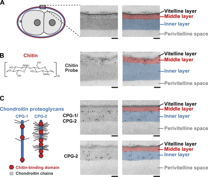
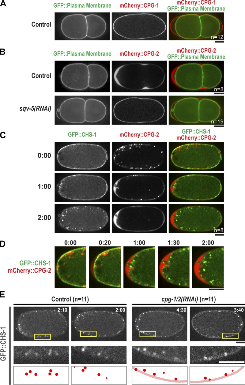
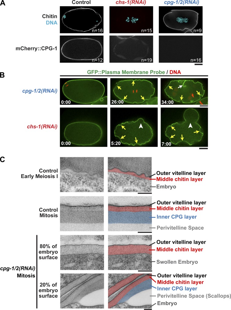
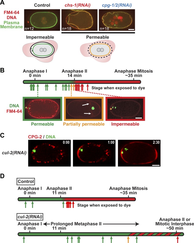
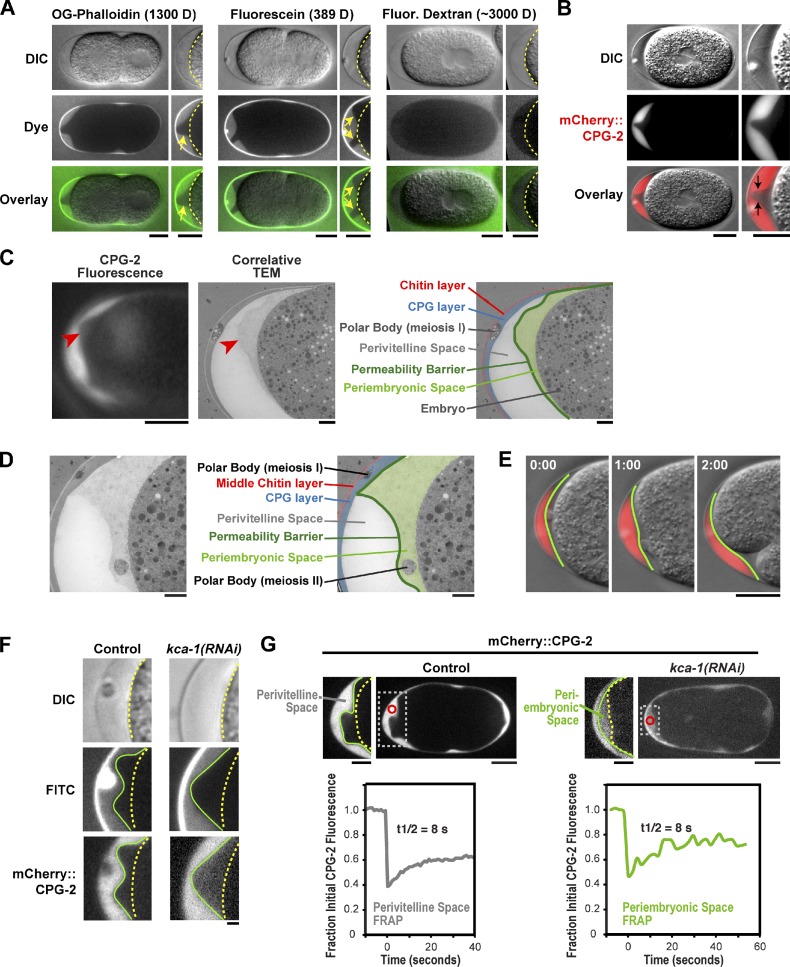
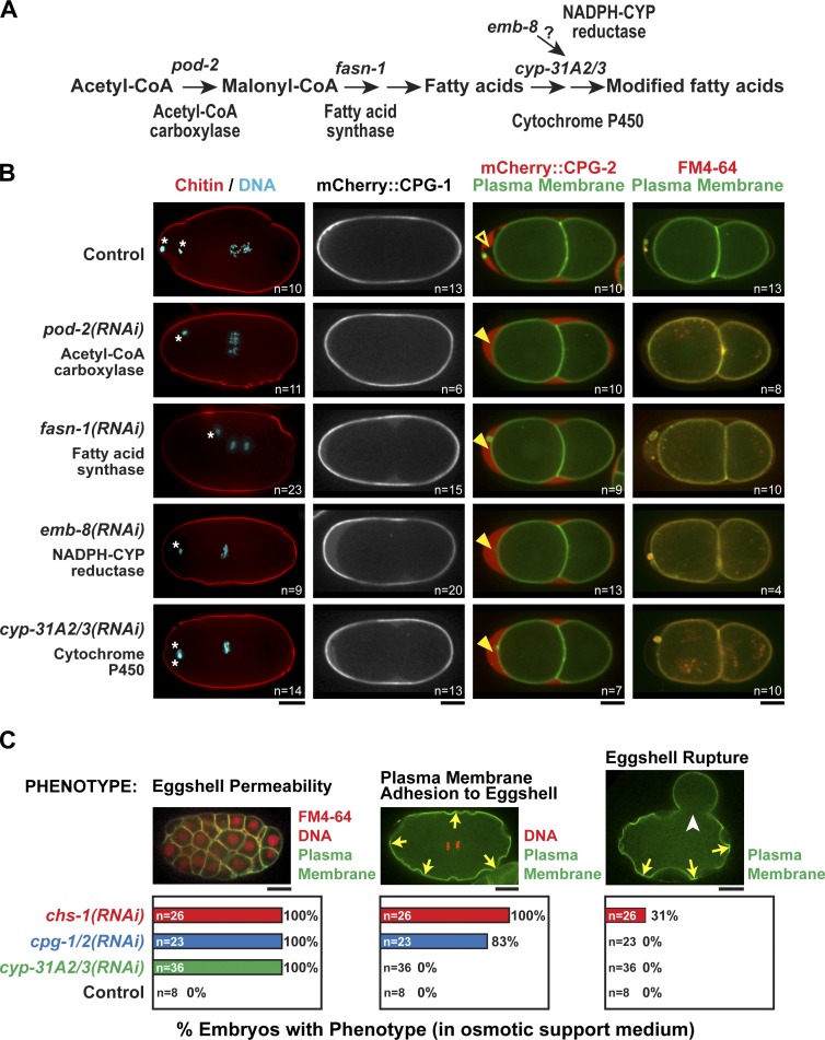
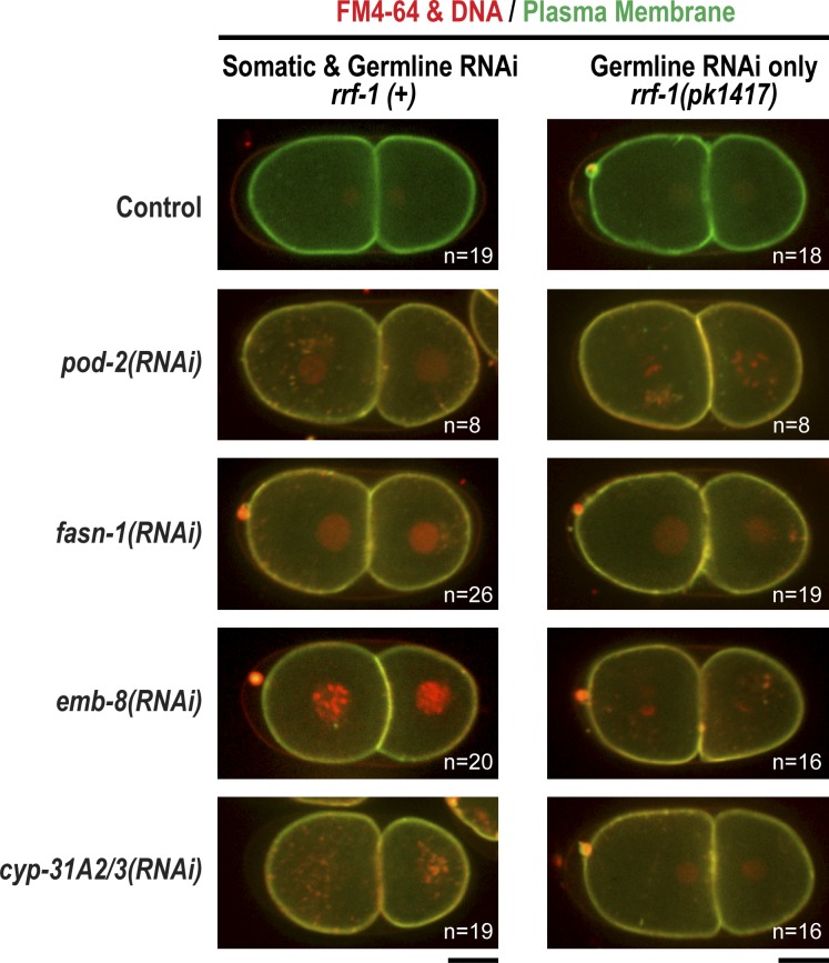
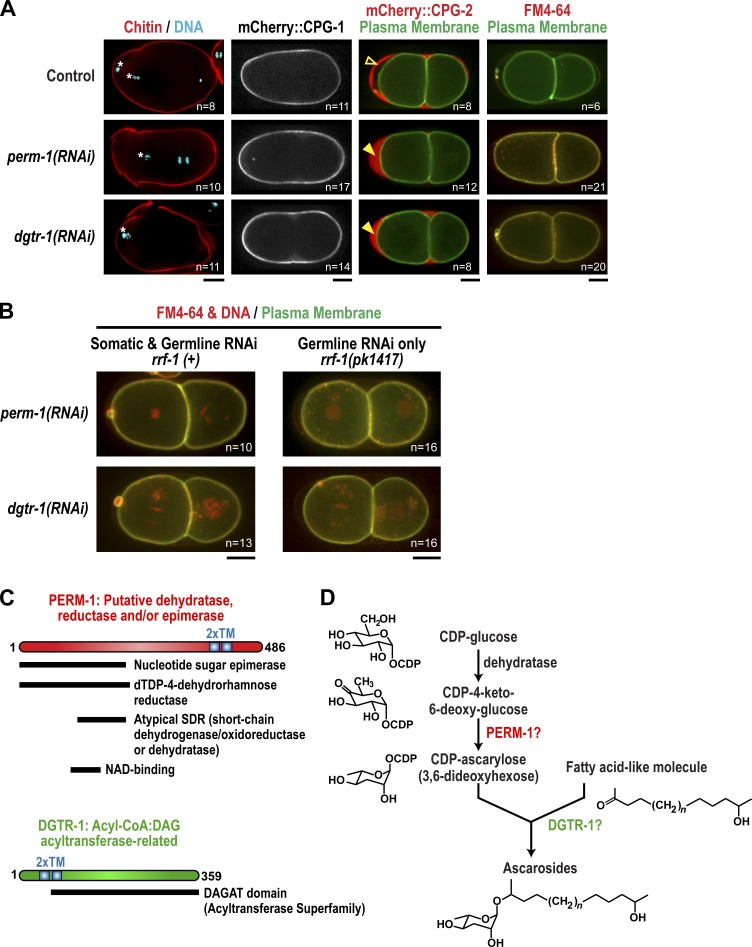
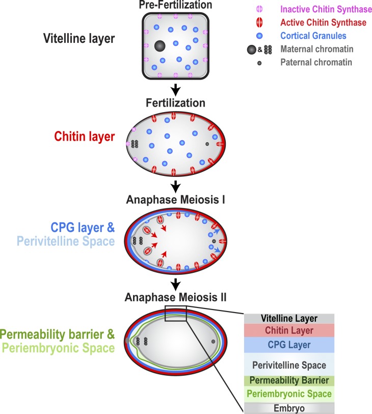
In metazoans, fertilization triggers the assembly of an extracellular coat that constitutes the interface between the embryo and its environment. In nematodes, this coat is the eggshell, which provides mechanical rigidity, prevents polyspermy, and is impermeable to small molecules. Using immunoelectron microscopy, we found that the Caenorhabditis elegans eggshell was composed of an outer vitelline layer, a middle chitin layer, and an inner layer containing chondroitin proteoglycans. The switch between the chitin and proteoglycan layers was achieved by internalization of chitin synthase coincident with exocytosis of proteoglycan-containing cortical granules. Inner layer assembly did not make the zygote impermeable as previously proposed. Instead, correlative light and electron microscopy demonstrated that the permeability barrier was a distinct envelope that formed in a separate step that required fatty acid synthesis, the sugar-modifying enzyme PERM-1, and the acyl chain transfer enzyme DGTR-1. These findings delineate the hierarchy of eggshell assembly and define key molecular mechanisms at each step.


Figures

Figure 1.
Figure 1.
Chitin and the CPG-1/2 localize to the middle and inner layers of the trilaminar eggshell, respectively. (middle) Transmission electron micrographs of the eggshell in high-pressure frozen embryos. (right) Pseudocolored micrographs illustrate the location of the outer (black), middle, and inner eggshell layers. Beneath the inner layer is the perivitelline space between the eggshell and embryo plasma membrane. (A, left) A schematic is provided for orientation, with the black box highlighting the imaged region. (B) Chitin is composed of repeating units of β1,4-linked N-acetylglucosamine. Image is an electron micrograph of an eggshell after immunogold labeling (10-nm gold beads) with a chitin-binding probe. (C) CPG-1 and CPG-2 are secreted CPGs composed of a protein core (blue) with covalently linked chondroitin side chains (thin black lines are putative chondroitin attachment sites based on sequence consensus; Olson et al., 2006) and chitin-binding domains. Images are electron micrographs of eggshells after immunogold labeling (10-nm beads) using antibodies recognizing both CPG-1 and CPG-2 (top) or specifically CPG-2 (bottom). Bars, 100 nm.
Figure 2.
Figure 2.
Chitin synthase is internalized concurrent with cortical granule exocytosis, facilitating the deposition of chitin and CPG-1/2 in sequential layers. (A) Confocal images of a two-cell stage embryo expressing a GFP-labeled plasma membrane probe (left) and mCherry::CPG-1 (middle). (B) Confocal images of two-cell stage control (top) and sqv-5(RNAi) (bottom) embryos expressing a GFP-labeled plasma membrane probe (left) and mCherry::CPG-2 (middle). (C) Time-lapse confocal images of a one-cell stage embryo expressing GFP::CHS-1 (left) and mCherry::CPG-2 (middle). Times are minutes and seconds after anaphase I. Embryo anterior is on the left. (D) Higher magnification view of the anterior of embryo in C. (E) Confocal images of two control (left) and two cpg-1/2(RNAi) (right) embryos expressing GFP::CHS-1. Times are minutes and seconds past anaphase I, when CHS-1 normally starts to internalize from the plasma membrane into subcortical puncta. Boxed regions are magnified below. Schematics highlight GFP::CHS-1 localization. By 2 min after anaphase I, the majority of GFP::CHS-1 in control embryos has been internalized into puncta (red spheres). In cpg-1/2(RNAi) embryos at a later time point, ∼4 min after anaphase I, some GFP::CHS-1 have internalized into puncta, but some also remain associated with the plasma membrane (red line). Bars, 10 µm. n = number of imaged embryos.
Figure 3.
Figure 3.
Formation of the inner CPG layer requires prior deposition of the chitin layer. (A, top) Immunofluorescence images of embryos stained with a chitin-binding probe (white) and for DNA. (bottom) Confocal images of embryos expressing mCherry::CPG-1. (B) Confocal images of cpg-1/2(RNAi) (top; n = 23) and chs-1(RNAi) (bottom; n = 26) embryos expressing a GFP-labeled plasma membrane probe and mCherry::histone H2B. Locations where the plasma membrane is able to separate from the eggshell (scallops; yellow arrows), a failed attempt at cytokinesis (white arrow) and a point where the eggshell lost integrity (rupture; white arrowheads) are marked. Times are minutes and seconds after the first frame. (C) Transmission electron micrographs of high-pressure frozen embryos. (right) Pseudocolored micrographs illustrate the location of the vitelline, chitin, and CPG layers. The mitotic control image is reproduced from Fig. 1 A for comparison. Bars: (A and B) 10 µm; (C) 200 nm. n = number of imaged embryos.
Figure 4.
Figure 4.
Formation of a functional permeability barrier requires passage through anaphase of meiosis II. (A) Confocal images of control (n = 12), chs-1(RNAi) (n = 18), and cpg-1/2(RNAi) (n = 18) embryos expressing a GFP-labeled plasma membrane probe and mCherry::histone H2B (red) that were dissected into FM4-64 dye (illustrated in schematics). (B) Embryos expressing GFP::histone H2B (green) were dissected in FM4-64 dye at different stages between anaphase of meiosis I and anaphase of the first mitotic division. Arrow positions indicate the times when that embryo was exposed to dye, and the color indicates whether it was permeable. White arrows highlight weak FM4-64 staining of the plasma membrane in a partially permeable embryo. (C) Time-lapse confocal images of a cul-2(RNAi) embryo expressing mCherry::CPG-2 and GFP::histone H2B (n = 19). Times are minutes and seconds past anaphase I. (D) Control (top; n = 8) and cul-2(RNAi) (bottom; n = 44) embryos expressing GFP::histone H2B and GFP::γ-tubulin were exposed to FM4-64 dye at different stages to assess eggshell permeability. Arrow positions indicate the time when that embryo was exposed to dye, and the color indicates permeability status. Embryos observed during this experiment whose temporal staging was imprecise are represented by the green and red striped region of the timeline rather than arrows (21 permeable, 2 partially permeable, and 11 impermeable embryos). Bars, 10 µm.
Figure 5.
Figure 5.
The permeability barrier is a distinct envelope that forms between the embryo surface and trilaminar eggshell. (A) Embryos placed in Oregon green (OG) phalloidin (left; n = 14), fluorescein (middle; n = 7), or 3,000-D fluorescein dextran (right; n = 10) were imaged by differential interference contrast (DIC; top row) and fluorescence (middle row; green in merge) microscopy. Magnified views of the embryo anterior are shown on the right (yellow dashed lines mark the embryo plasma membrane; yellow arrows point to the edge of the permeability barrier). (B) DIC (top) and fluorescence (middle; red in merge) images of a one cell–stage mitotic embryo expressing mCherry::CPG-2 (n = 9). The embryo anterior is magnified on the right. The contrast of the top DIC image has been adjusted to visualize the edge of the permeability barrier (indicated by the black arrows in the merge). (C, left) A fluorescence image of CPG-2 was acquired immediately before cryoimmobilization and processing for transmission electron microscopy (n = 3). (middle) Alignment of the resulting correlative transmission electron micrograph (TEM) image allows visualization of the edge of the permeability barrier (red arrowheads). (right) A pseudocolored micrograph illustrates the location of the chitin and CPG eggshell layers along with the edge of the permeability barrier. The periembryonic and perivitelline spaces and the first polar body are also labeled. (D) Transmission electron micrographs of a control embryo showing the location of the polar bodies extruded during anaphase of meiosis I (embedded in the CPG layer; see also C) and during anaphase of meiosis II (in the periembryonic space). (E) Merged DIC and fluorescence images of the anterior of an embryo at the two-cell stage expressing mCherry::CPG-2. Green lines mark the location of the permeability barrier. Time indicates minutes and seconds past the first frame. (F) DIC and confocal fluorescence images of control (n = 12) and partial kca-1(RNAi) (n = 12) embryos expressing mCherry::CPG-2. (middle row) Embryos were also exposed to FITC dye to monitor the integrity of the permeability barrier (green lines). The embryo surface is also marked (dashed yellow lines). (G) Confocal fluorescence images of control (left) and partial kca-1(RNAi) (right) embryos expressing mCherry::CPG-2. The mCherry::CPG-2 in the perivitelline (left) or periembryonic (right) spaces was photobleached (red circle inside dashed white box, magnified on the left) at 0 s, and the embryo was continuously imaged every 1 or 2 s (left and right, respectively) to monitor recovery (quantified in graphs below). Bars: (A–C [left], E, and G) 10 µm; (C [middle and right], D, and F) 1 µm.
Figure 6.
Figure 6.
Genes involved in fatty acid synthesis are required to form the permeability barrier. (A) Schematic outline of some of the genes in the fatty acid biosynthetic and modification pathway. CYP, Cytochrome P450. (B) Confocal images of control embryos and embryos in which the indicated proteins were inhibited by RNAi. (first column) The middle chitin layer was visualized in fixed embryos by staining for chitin and DNA. White asterisks mark extruded polar bodies. (second column) The inner CPG layer was visualized in embryos expressing mCherry::CPG-1. (third column) The presence of the permeability barrier was assessed in embryos expressing mCherry::CPG-2 and a GFP-tagged plasma membrane marker. In control embryos, the permeability barrier prevents diffusion of mCherry::CPG-2 to the embryo surface (open arrowhead). When fatty acid synthesis is inhibited, the permeability barrier is disrupted, and mCherry::CPG-2 fills the entire space between the eggshell and embryo surface (closed arrowheads). (last column) Embryos expressing a GFP-tagged plasma membrane probe were placed in FM4-64 dye to test their permeability. (C) The phenotypic consequences of disrupting the eggshell permeability barrier (cyp-31A2/3(RNAi)), the inner CPG layer (cpg-1/2(RNAi)), and the middle chitin layer (chs-1(RNAi)) were compared by analyzing the percentage of embryos exhibiting each of the indicated phenotypes. Data were pooled from >10 independent imaging sessions for each condition. Plasma membrane adhesion (yellow arrows) was also usually accompanied by cytokinesis failure (59% of cpg-1/2(RNAi) embryos failed cytokinesis). In addition to the quantified phenotypes, chitin layer disruption also led to polyspermy (24% of chs-1(RNAi) embryos were polyspermic). The images of embryos illustrating membrane adhesion and eggshell rupture (white arrowhead) are reproduced from Fig. 3 B. The left image is of an embryo expressing a GFP-tagged plasma membrane marker and mCherry::histone H2B (red) that was placed in FM4-64 dye. Bars, 10 µm. n = number of imaged embryos.
Figure 7.
Figure 7.
Permeability barrier formation requires germline expression of fatty acid synthesis genes. Confocal images of control embryos and embryos in which the indicated components of the fatty acid biosynthesis pathway were inhibited by RNAi in either an rrf-1(+) (left) or rrf-1(pk1417) mutant background (right). Embryos expressing a GFP-tagged plasma membrane probe and mCherry::histone H2B (red) were placed in FM4-64 dye. All RNAi-treated embryos were permeable in the rrf-1(pk1417) background, indicating that germline expression of these genes is required. n = number of imaged embryos. Bars, 10 µm.
Figure 8.
Figure 8.
A requirement for PERM-1 and DGTR-1 suggests that an ascaroside may be important for permeability barrier formation. (A) Confocal images of control embryos and embryos in which PERM-1 and DGTR-1 were depleted by RNAi. (first column) The middle chitin layer was visualized in fixed embryos by staining for chitin and DNA. White asterisks mark extruded polar bodies. (second column) The inner CPG layer was visualized in embryos expressing mCherry::CPG-1. (third column) The presence of the permeability barrier was assessed in embryos expressing mCherry::CPG-2 and a GFP-tagged plasma membrane marker. In control embryos, the permeability barrier prevents diffusion of mCherry::CPG-2 to the embryo surface (open arrowhead). When PERM-1 and DGTR-1 are inhibited, mCherry::CPG-2 fills the entire space between the eggshell and embryo surface (closed arrowheads). (last column) Embryos expressing a GFP-tagged plasma membrane probe were placed in FM4-64 dye to test their permeability. (B) Confocal images of embryos in which PERM-1 and DGTR-1 were inhibited by RNAi in either an rrf-1(+) (left column) or rrf-1(pk1417) mutant background (right column). Embryos expressing a GFP-tagged plasma membrane probe and mCherry::histone H2B (red) were placed in FM4-64 dye to test their permeability. For A and B, n = number of imaged embryos. Bars, 10 µm. (C) Schematics of PERM-1 and DGTR-1 showing the location of domains predicted by BLAST (Basic Local Alignment Search Tool). The TOPCONS program (Stockholm Bioinformatics Center) predicted both proteins to contain two transmembrane domains (2×TM). dTDP, deoxythymidine diphosphate. (D) A putative pathway for ascaroside synthesis in which activated CDP-ascarylose (left side) combines with a long-chain fatty acid–like molecule (right side) to generate ascaroside glycolipid. The reactions proposed to convert CDP-glucose into CDP-ascarylose are based on chemical reactions identified in prokaryotes (Thibodeaux et al., 2007). Points where PERM-1 and DGTR-1 might function in this pathway are indicated.
Figure 9.
Figure 9.
Model for assembly of the C. elegans eggshell and permeability barrier. Schematic outline of sequential steps in assembly of the eggshell and permeability barrier. The plasma membrane of prefertilization oocytes is coated with an electron-dense vitelline layer. Fertilization activates chitin synthase, which deposits a chitin layer between the vitelline layer and plasma membrane. At anaphase of meiosis I, cortical granules containing CPG-1 and CPG-2 are exocytosed in a wave that proceeds from the anterior (left) to the posterior (right) of the embryo. As cortical granules are exocytosed, the CPG layer is deposited, and a perivitelline space opens up between the plasma membrane and eggshell. Chitin synthase is internalized from the embryo surface after the onset of cortical granule exocytosis. At anaphase of meiosis II, fatty acid derivatives, possibly ascarosides, assemble between the eggshell and the embryo surface to form the permeability barrier. Between the permeability barrier and the embryo surface is the periembryonic space, which receives the contents of vesicles exocytosed after anaphase of meiosis II.

Similar articles

Cited by

References

    1. Anya A.O. 1976. Physiological aspects of reproduction in nematodes. Adv. Parasitol. 14:267–351 10.1016/S0065-308X(08)60516-3 - DOI - PubMed
    1. Audhya A., Hyndman F., McLeod I.X., Maddox A.S., Yates J.R., III, Desai A., Oegema K. 2005. A complex containing the Sm protein CAR-1 and the RNA helicase CGH-1 is required for embryonic cytokinesis in Caenorhabditis elegans. J. Cell Biol. 171:267–279 10.1083/jcb.200506124 - DOI - PMC - PubMed
    1. Bartley J.P., Bennett E.A., Darben P.A. 1996. Structure of the ascarosides from Ascaris suum. J. Nat. Prod. 59:921–926 10.1021/np960236+ - DOI - PubMed
    1. Bembenek J.N., Richie C.T., Squirrell J.M., Campbell J.M., Eliceiri K.W., Poteryaev D., Spang A., Golden A., White J.G. 2007. Cortical granule exocytosis in C. elegans is regulated by cell cycle components including separase. Development. 134:3837–3848 10.1242/dev.011361 - DOI - PMC - PubMed
    1. Benenati G., Penkov S., Müller-Reichert T., Entchev E.V., Kurzchalia T.V. 2009. Two cytochrome P450s in Caenorhabditis elegans are essential for the organization of eggshell, correct execution of meiosis and the polarization of embryo. Mech. Dev. 126:382–393 10.1016/j.mod.2009.02.001 - DOI - PubMed

Publication types

MeSH terms

LinkOut - more resources