

Osteosarcoma following single fraction radiation prophylaxis for heterotopic ossification
- PMID: 22908888
- PMCID: PMC3488033
- DOI: 10.1186/1748-717X-7-140
Osteosarcoma following single fraction radiation prophylaxis for heterotopic ossification
Abstract
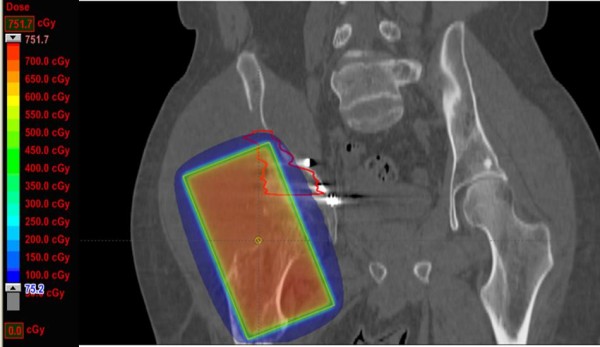
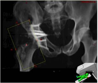
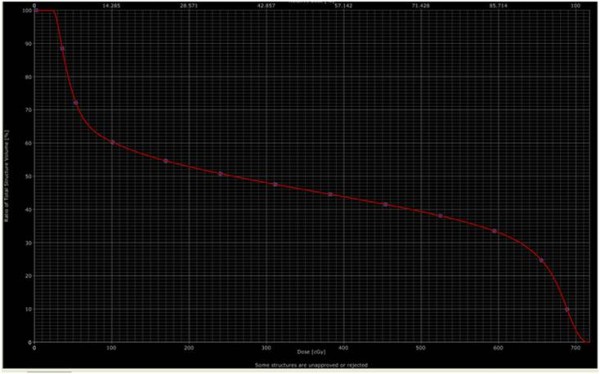
Radiotherapy for prophylaxis of heterotopic ossification (HO) is commonly used in high risk patients following orthopedic surgery. While treatment is effective and can prevent morbidity associated with HO, with any dose of radiation there is a concern of a radiation induced malignancy. Here we a report a case of radiation induced osteosarcoma which developed 11 years after a single fraction of 700 cGy. We performed dosimetric analysis by superimposing the patient's original treatment field on a CT scan performed after the diagnosis. The radiotherapy dose for this patient is lower than classically reported for radiation induced sarcomas. We identified greatest bony destruction that was thought to be the epicenter of the tumor, and this was specially contoured on the diagnostic CT scan. This volume appears to be located at the edge of the radiotherapy field. Fifty percent of the treated volume received 240 cGy, the mean dose was 333 cGy. There was a variation across the treatment volume, between 21.8 cGy and 717 cGy. While a rare complication, we stress the importance of informing regarding the risk of a radiation induced malignancy following HO prophylaxis.
Figures
Similar articles
-
Reliable Radiation Technique to Minimize Ovarian Dose During Radiation Prophylaxis of Heterotopic Ossification.Anticancer Res. 2017 Dec;37(12):6929-6935. doi: 10.21873/anticanres.12157. Anticancer Res. 2017. PMID: 29187475
-
Testicular Dose During Prophylaxis of Heterotopic Ossification with Radiation Therapy.In Vivo. 2017 May-Jun;31(3):461-466. doi: 10.21873/invivo.11084. In Vivo. 2017. PMID: 28438880 Free PMC article.
-
Heterotopic Ossification Prophylaxis After Total Hip Arthroplasty: Randomized Trial of 400 vs 700 cGy.J Arthroplasty. 2017 Apr;32(4):1328-1334. doi: 10.1016/j.arth.2016.10.030. Epub 2016 Nov 1. J Arthroplasty. 2017. PMID: 27884418 Clinical Trial.
-
Advanced therapeutic strategy for radiation-induced osteosarcoma in the skull base: a case report and review.Radiat Oncol. 2012 Aug 10;7:136. doi: 10.1186/1748-717X-7-136. Radiat Oncol. 2012. PMID: 22883312 Free PMC article. Review.
-
Chemotherapy for osteosarcoma and Ewing's sarcoma.Acta Orthop Scand Suppl. 1997 Feb;273:120-5. doi: 10.1080/17453674.1997.11744716. Acta Orthop Scand Suppl. 1997. PMID: 9057601 Review. No abstract available.
Cited by
-
Neurological heterotopic ossification: novel mechanisms, prognostic biomarkers and prophylactic therapies.Bone Res. 2020 Dec 9;8(1):42. doi: 10.1038/s41413-020-00119-9. Bone Res. 2020. PMID: 33298867 Free PMC article. Review.
-
NSAIDs for Prophylaxis for Heterotopic Ossification After Total Hip Arthroplasty: A Bayesian Network Meta-analysis.Calcif Tissue Int. 2021 Feb;108(2):196-206. doi: 10.1007/s00223-020-00763-7. Epub 2020 Oct 12. Calcif Tissue Int. 2021. PMID: 33044630 Free PMC article.
-
The role of miRNA and lncRNA in heterotopic ossification pathogenesis.Stem Cell Res Ther. 2022 Dec 15;13(1):523. doi: 10.1186/s13287-022-03213-3. Stem Cell Res Ther. 2022. PMID: 36522666 Free PMC article. Review.
-
Radiation therapy for the prevention of heterotopic ossification: Efficacy and toxicity of single fraction radiotherapy.Orthop Rev (Pavia). 2020 Aug 18;12(2):8577. doi: 10.4081/or.2020.8577. eCollection 2020 Aug 6. Orthop Rev (Pavia). 2020. PMID: 32922703 Free PMC article.
-
Radiation induced sarcoma: Everything comes with a price.Urol Ann. 2014 Jul;6(3):250-1. Urol Ann. 2014. PMID: 25125902 Free PMC article. No abstract available.
References
-
- Riedel B. Demonstration line durch ach Hagiges Umhergehen total destruirten kniegelenkes von einem patienten mit stichverletzing des ruckans. Verh Dtsch Gesellschaft Chirurg. 1883;12:93.
-
- Naraghi FF, DeCoster TA, Moneim MS, Miller RA, Rivero D. Heterotopic ossification. Orthopedics. 1996;19:145–151. - PubMed
-
- Ahrengart L, Lindgren U, Reinholt FP. Comparative study of the effects of radiation, indomethacin, prednisolone, and ethane-1-hydroxy-1,1-diphosphonate (EHDP) in the prevention of ectopic bone formation. Clin Orthop Relat Res. 1988;229:265–273. - PubMed
Publication types
MeSH terms
Substances
LinkOut - more resources
Full Text Sources
Medical
