. 2012 Aug 21;43(1):61.
doi: 10.1186/1297-9716-43-61.

The role of glycoprotein H of equine herpesviruses 1 and 4 (EHV-1 and EHV-4) in cellular host range and integrin binding

Affiliations

The role of glycoprotein H of equine herpesviruses 1 and 4 (EHV-1 and EHV-4) in cellular host range and integrin binding

Walid Azab et al. Vet Res. .

Abstract

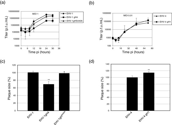
Equine herpesvirus type 1 and 4 (EHV-1 and EHV-4) glycoprotein H (gH) has been hypothesized to play a role in direct fusion of the virus envelope with cellular membranes. To investigate gH's role in infection, an EHV-1 mutant lacking gH was created and the gH genes were exchanged between EHV-1 and EHV-4 to determine if gH affects cellular entry and/or host range. In addition, a serine-aspartic acid-isoleucine (SDI) integrin-binding motif present in EHV-1 gH was mutated as it was presumed important in cell entry mediated by binding to α4β1 or α4β7 integrins. We here document that gH is essential for EHV-1 replication, plays a role in cell-to-cell spread and significantly affects plaque size and growth kinetics. Moreover, we could show that α4β1 and α4β7 integrins are not essential for viral entry of EHV-1 and EHV-4, and that viral entry is not affected in equine cells when the integrins are inaccessible.


Figures

Figure 1
Figure 1
Identification of recombinant viruses by RFLP and Western blotting. (a) Purified DNA from EHV-1, EHV-1gH4, EHV-1gH4Kan and EHV-1gHS440A (left panel) as well as EHV-4, EHV-4gH1Kan and EHV-4gH1 (right panel) were digested with SalI and SacII. Fragments in the mutants that appeared as a consequence of the deletion or insertion of gH sequence are marked by arrows. (b) Amino acid sequence alignment of EHV-1 and EHV-4 gHs’ N-terminal part. The alignment begins with aa49 to aa 327 (H1 domain homologue, that binds gL, in HSV-2) [20]. (c) Parental and mutant viruses as well as complementing RK13/gH1 cells express gH at similar levels. Cell lysates were prepared either from infected FHK cells or from RK13/gH1 and proteins were separated by SDS-10%-PAGE. The blots were incubated with EHV-1 polyclonal anti-gH antibody and detected with anti-rabbit IgG peroxidase conjugate. For RK13/gH1 cells (left panel), EHV-1 and related mutants (middle panel), and EHV-4 and related mutants (right panel), two bands of approximately 125 and 115kD were detected that are not present in mock-infected cells. β-actin was used as a loading control.
Figure 2
Figure 2
In vitro growth characterization of parental and mutant viruses. (a, b) For growth kinetics, NBL-6 cells were infected at MOI’s of 1 or 0.01. Infected cells and supernatants were collected and virus titers were determined at the indicated times pi. The data presented are means ± SD of triplicate measurements. The asterisk indicates a P < 0.05 for means when compared to the parental viruses. (c, d) Means ± SD of diameters of 50 plaques measured for each virus are shown. The plaque diameter of parental viruses was set to 100%. The asterisks indicate a P < 0.05 for means when compared to the parental viruses.
Figure 3
Figure 3
Infection of RK13 cells with EHV-1ΔgH. RK13 cells were transfected with either EHV-1ΔgH DNA (a) or EHV-1 parental DNA (b). EHV-1ΔgH was also transfected into RK13/gH1 cells (c). Recovered virus was collected from RK13/gH1 cells and used to infect naïve RK13 cells (d). Infected cells appear green as all viruses express EGFP. Cells were inspected with a fluorescent microscope (Zeiss Axiovert) and images were taken with a CCD camera (Zeiss Axiocam). The bar represents 100 μm.
Figure 4
Figure 4
The role of gH in EHV-1 and EHV-4 cellular tropism. RK13 (a), CHO-K1 (b), CHO-A (c), CHO-B (d), CHO-C (e), or Vero (f) cells were infected at an MOI of 0.5 with the engineered virus recombinants, all of which express EGFP. At 48 h pi, cells were inspected with a fluorescent microscope (Zeiss Axiovert) and images were taken with a CCD camera (Zeiss Axiocam). The bar represents 100 μm.
Figure 5
Figure 5
Infection of equine PBMC with recombinant viruses. PBMC were incubated with EHV-1 or EHV-4 at an MOI of 1 or 6 (a) for 1 h at 37°C. After 48 h, the percentage of infected cells was determined by flow cytometry. In another experiment, PBMC were infected with the engineered mutant viruses (b). The data represent the mean ± SD of at least three independent experiments.
Figure 6
Figure 6
Integrins have no role in EHV-1 or EHV-4 entry into equine cells. PBMC (a) NBL-6 (b) or FHK (c) cells were incubated with anti-α4β7 MAb DATK32 or α4β1 MAb P4C2 for 1 h at RT, followed by incubation with Alexa fluor 488-labeled goat anti-mouse IgG for 1 h at RT. As controls, cells were incubated with irrelevant MAbs of the same IgG isotype. Integrin expression was determined by flow cytometry. (d) PBMC were preincubated with either α4β7 MAb DATK32 or α4β1 MAb P4C2 for 1 h at 4°C, followed by infection with recombinant viruses at an MOI of 5 for 2 h at 37°C. At 48 h pi, cells were washed and the percentage of infected cells was determined by flow cytometry. The rate of infection of parental viruses was set to 100%. All data represent the mean ± SD of three independent experiments.

Similar articles

Cited by

References

    1. King AMQ, Adams MJ, Carstens EB, Lefkowitz EJ. Virus Taxonomy: Ninth Report of the International Committee on Taxonomy of Viruses. UK: Academic Press, Elsevier; 2012. pp. 114–115.
    1. Patel JR, Heldens J. Equine herpesviruses 1 (EHV-1) and 4 (EHV-4)–epidemiology, disease and immunoprophylaxis: a brief review. Vet J. 2005;170:14–23. doi: 10.1016/j.tvjl.2004.04.018. - DOI - PubMed
    1. Telford EA, Watson MS, Perry J, Cullinane AA, Davison AJ. The DNA sequence of equine herpesvirus-4. J Gen Virol. 1998;79:1197–1203. - PubMed
    1. Gryspeerdt AC, Vandekerckhove AP, Garre B, Barbe F, Van de Walle GR, Nauwynck HJ. Differences in replication kinetics and cell tropism between neurovirulent and non-neurovirulent EHV1 strains during the acute phase of infection in horses. Vet Microbiol. 2009;142:242–253. - PubMed
    1. Lunn DP, Davis-Poynter N, Flaminio MJ, Horohov DW, Osterrieder K, Pusterla N, Townsend HG. Equine herpesvirus-1 consensus statement. J Vet Intern Med. 2009;23:450–461. doi: 10.1111/j.1939-1676.2009.0304.x. - DOI - PubMed

Publication types

MeSH terms

LinkOut - more resources