

Comparison of histopathology to gene expression profiling for the diagnosis of metastatic cancer
- PMID: 22909314
- PMCID: PMC3541121
- DOI: 10.1186/1746-1596-7-110
Comparison of histopathology to gene expression profiling for the diagnosis of metastatic cancer
Abstract
Background: Determining the primary site of metastatic cancer with confidence can be challenging. Pathologists commonly use a battery of immunohistochemical (IHC) stains to determine the primary site. Gene expression profiling (GEP) has found increasing use, particularly in the most difficult cases. In this pilot study, a direct comparison between GEP and IHC-guided methods was performed.
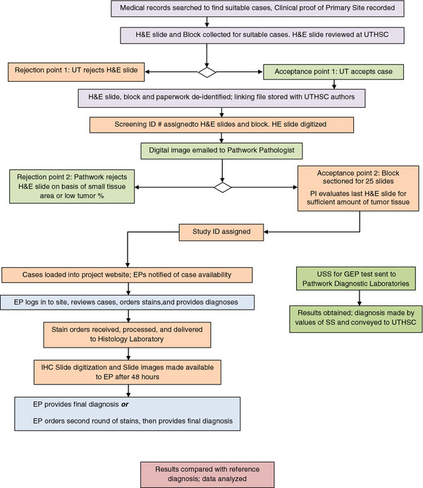
Methods: Ten archived formalin-fixed paraffin embedded metastatic tumor samples for which the primary site had been clinically determined were selected. Five pathologists who were blinded to the diagnosis were asked to determine the primary site using IHC and other stains selected from a panel of 84 stains. Each pathologist was provided patient sex, biopsy site and gross sample description only. Slides were digitized using ScanScope®XT at 0.25 μm/pixel. Each evaluating pathologist was allowed to provide a diagnosis in three stages: initial (after reviewing the H&E image), intermediate (after reviewing images from the first batch of stains) and final diagnosis (after the second batch of stains if requested). GEP was performed using the only FDA-cleared test for this intended use, the Pathwork Tissue of Origin Test. No sample information was provided for GEP testing except for patient sex. Results were reported as the tumor tissue type with the highest similarity score.
Results: In this feasibility study, GEP determined the correct primary site in 9 of the 10 cases (90%), compared to the IHC-guided method which determined the correct primary site for 32 of 50 case evaluations (average 64%, range 50% to 80%). The five pathologists directing the IHC-guided method ordered an average of 8.8 stains per case (range 1 to 18). GEP required an average of 3 slides per case (range 1 to 4).
Conclusions: Results of the pilot study suggest that GEP provides correct primary site identification in a higher percentage of metastatic cases than IHC-guided methods, and uses less tissue. A larger comparative effectiveness study using this study design is needed to confirm the results.
Virtual slides: The virtual slide(s) for this article can be found here: http://www.diagnosticpathology.diagnomx.eu/vs/1749854104745508.
Figures
References
-
- Abbruzzese JL, Lenzi R, Raber MN, Pathak S, Frost P. The biology of unknown primary tumors. Semin Oncol. 1993;20(3):238–243. - PubMed
Publication types
MeSH terms
Substances
LinkOut - more resources
Full Text Sources
Medical
