Review
. 2013 Feb 1;514(1):1-30.
doi: 10.1016/j.gene.2012.07.083. Epub 2012 Aug 15.

Function of alternative splicing

Affiliations
Review

Function of alternative splicing

Olga Kelemen et al. Gene. .

Abstract

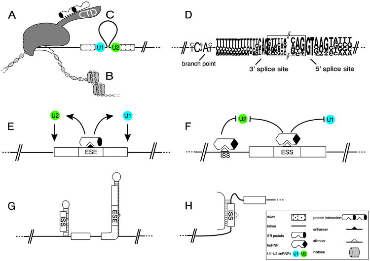
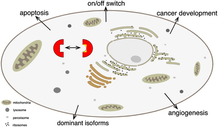
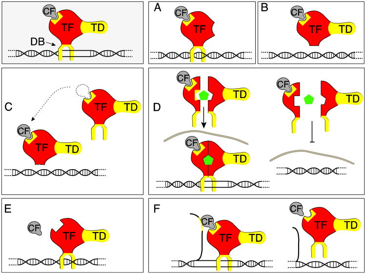
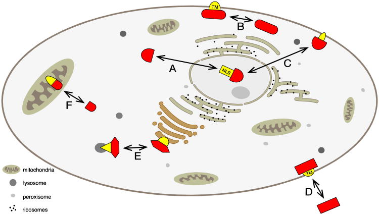
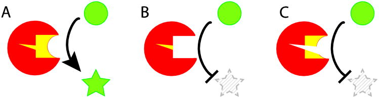
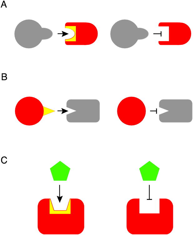
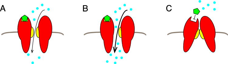
Almost all polymerase II transcripts undergo alternative pre-mRNA splicing. Here, we review the functions of alternative splicing events that have been experimentally determined. The overall function of alternative splicing is to increase the diversity of mRNAs expressed from the genome. Alternative splicing changes proteins encoded by mRNAs, which has profound functional effects. Experimental analysis of these protein isoforms showed that alternative splicing regulates binding between proteins, between proteins and nucleic acids as well as between proteins and membranes. Alternative splicing regulates the localization of proteins, their enzymatic properties and their interaction with ligands. In most cases, changes caused by individual splicing isoforms are small. However, cells typically coordinate numerous changes in 'splicing programs', which can have strong effects on cell proliferation, cell survival and properties of the nervous system. Due to its widespread usage and molecular versatility, alternative splicing emerges as a central element in gene regulation that interferes with almost every biological function analyzed.


Figures

Figure 1
Figure 1. Control elements of alternative exons
The usage of alternative exons is controlled by the combination of multiple factors. A: RNA polymerase II assembles splicing factors on its CTD, which can predetermine the splicing outcome of a nascent pre-mRNA. This example shows SR-proteins, but other factors assemble as well. B: Exon boundaries correlate with DNA nucleosome occupancy and alternative exon usage is influenced by chromatin marks. C: Large intron regions can loop out to bring exons together. D: An exon is defined by three crucial elements: the branch point, the 3′ splice site and the 5′ splice site, which follow consensus sequences indicated. E: Exons need additional factors to be recognized. These factors bind to exonic or intronic sequences. In this example, two SR-proteins bind to exonic enhancers (ESE: exonic splicing enhancer) and help stabilize binding of U2 and U1 snRNP. F: Exon usage can be repressed by exonic splicing silencers (ESS) and intronic splicing silencers (ISS) that prevent U1 or U2 snRNP binding. G: RNA secondary structures can mask exonic or intronic splicing enhancer and silencer, as proteins binding to these RNA elements typically recognize single stranded RNA. H: small RNAs can bind to splicing regulatory elements located on different mRNAs.
Figure 2
Figure 2. Overall changes in cellular properties due to alternative splicing
A eukaryotic cell is schematically depicted. Alternative splicing changes protein isoforms (red half squares) by introducing new protein sequences (yellow) that are encoded by alternative exons. Changes in global cellular processes are arranged similar to Table 1 that lists specific examples.
Figure 3
Figure 3. Changes in transcription factors
The gray box schematically depicts the general structure of a transcription factor complex. TD: transactivation domain; CF: Co-factor; TF: core transcription factor, DB: DNA binding domain. Components of transcription factors that undergo alternative splicing with well-studied effects are indicated in yellow. The numbering (A-F) of the Figure refers to Table 2 that lists specific examples. A. Change in transactivation domain B. Loss in DNA binding C. Generation of dominant negative isoforms, here a factor that lost its DNA binding domain, but can still compete for cofactors (indicated by arrow). D. Alternative splicing of nuclear recpetors. In this example, the ligand binding domain is subject to alternative splicing (yellow area) that interferes with ligand binding (green pentagon), which regulates translocation of the dimerized receptor complex into the nucleus (indicated by gray nuclear membrane). E. The binding of transcriptional cofactors is regulated by alternative splicing. F. ncRNAs can direct transcription factors to DNA, which is regulated by alternative splicing of the ncRNAs.
Figure 4
Figure 4. Changes in intracellular localization due to alternative splicing
A eukaryotic cell is schematically shown. Proteins that undergo alternative splicing are shown in red, parts encoded by alternative exons are shown in yellow. The Figure is arranged similar to Table 3, in clockwise arrangement. A. Change between cytosol and nucleus, typically by altering a nuclear localization signal (NLS). B. Change between plasma membrane and cytosol, typically by changing a transmembrane region (TM). C. Change between nucleus and membrane associated forms. D. Generation of soluble, secreted forms, typically by changing a transmembrane region (TM). E. Localization between different internal membranes. F. Localization in the mitochondria, typically by regulating a mitochondrial-targeting signal (MTS).
Figure 5
Figure 5. Change of enzymatic activity due to alternative splicing
The alternative exon is shown as a red circle, parts encoded by alternative exons are shown in yellow. A: The enzyme converts a substrate (circle) into a product (star). B: Alternative splicing influences the substrate binding, which prevents substrate formation (striped star). C: Alternative splicing changes the catalytic center (short triangle), which prevents substrate formation (examples in Table 4A).
Figure 6
Figure 6. Regulation of protein-protein and protein-ligand interaction by alternative splicing
Examples are given in Table 6, A-B. A. Alternative splicing of the domain-binding pocket regulates protein interactions. B. Alternative splicing of domains that bind to binding pockets regulates protein interactions. C. Alternative splicing changes binding of a ligand (green pentagon) to a protein (red).
Figure 7
Figure 7. Change of ion channels due to alternative splicing
The ion-channel is shown in red and the channel opening indicated by yellow half circles. The triangle indicates a ligand-binding site; the ligand is shown as a hexgon. A: Channel in its open conformation. Dots indicate the flow of ions. B: Alternative splicing changes the pore, which in this case leads to a larger flux. C: A change in the ligand-binding site prevents channel opening.

References

    1. Acevedo JM, Centanin L, Dekanty A, Wappner P. Oxygen sensing in Drosophila: multiple isoforms of the prolyl hydroxylase fatiga have different capacity to regulate HIFalpha/Sima. PLoS One. 2010;5:e12390. - PMC - PubMed
    1. Adams PJ, Garcia E, David LS, Mulatz KJ, Spacey SD, Snutch TP. Ca(V)2.1 P/Q-type calcium channel alternative splicing affects the functional impact of familial hemiplegic migraine mutations: implications for calcium channelopathies. Channels (Austin) 2009;3:110–21. - PubMed
    1. Adato A, Lefevre G, Delprat B, Michel V, Michalski N, Chardenoux S, Weil D, El-Amraoui A, Petit C. Usherin, the defective protein in Usher syndrome type IIA, is likely to be a component of interstereocilia ankle links in the inner ear sensory cells. Human molecular genetics. 2005;14:3921–32. - PubMed
    1. Ahn J, Febbraio M, Silverstein RL. A novel isoform of human Golgi complex-localized glycoprotein-1 (also known as E-selectin ligand-1, MG-160 and cysteine-rich fibroblast growth factor receptor) targets differential subcellular localization. J Cell Sci. 2005;118:1725–31. - PubMed
    1. Al-Balool HH, Weber D, Liu Y, Wade M, Guleria K, Nam PL, Clayton J, Rowe W, Coxhead J, Irving J, Elliott DJ, Hall AG, Santibanez-Koref M, Jackson MS. Post-transcriptional exon shuffling events in humans can be evolutionarily conserved and abundant. Genome research. 2011;21:1788–99. - PMC - PubMed

Publication types

LinkOut - more resources