. 2012 Oct 17;31(20):3961-75.
doi: 10.1038/emboj.2012.236. Epub 2012 Aug 21.

AATF/Che-1 acts as a phosphorylation-dependent molecular modulator to repress p53-driven apoptosis

Affiliations

AATF/Che-1 acts as a phosphorylation-dependent molecular modulator to repress p53-driven apoptosis

Katja Höpker et al. EMBO J. .

Abstract

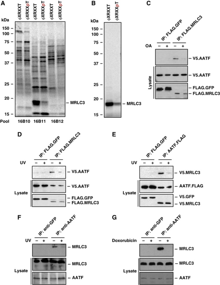
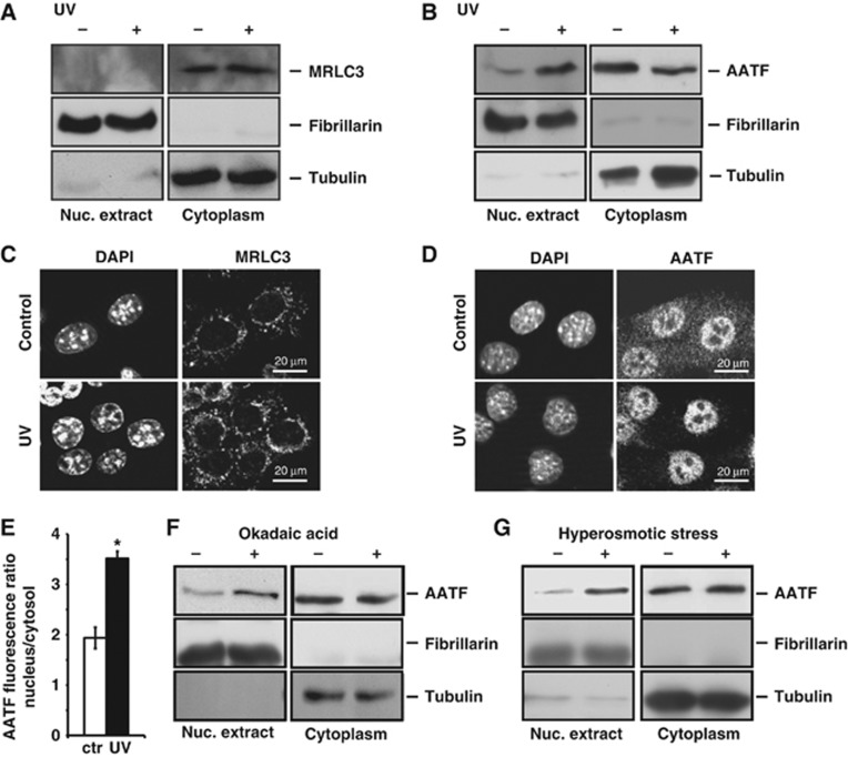
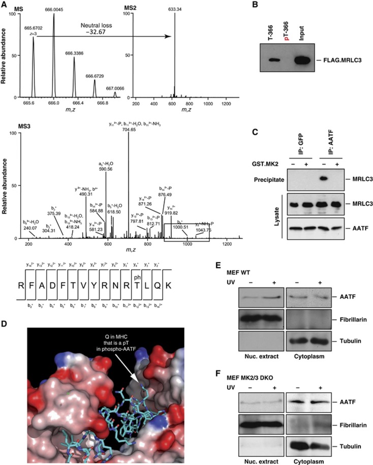
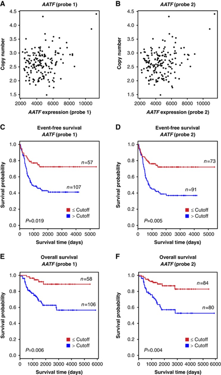
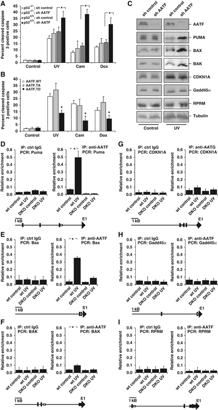
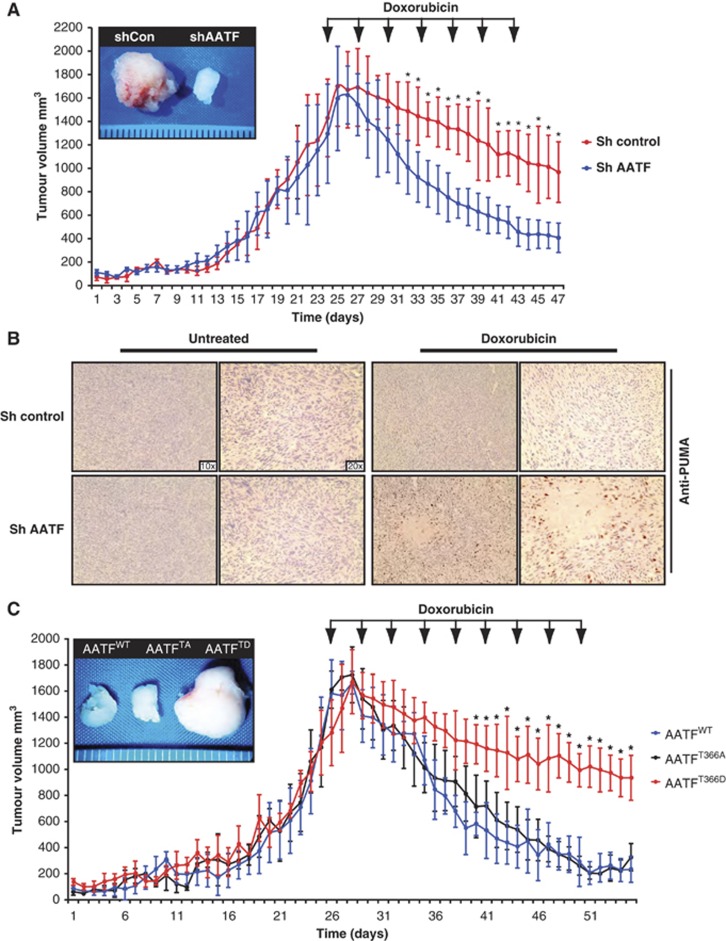
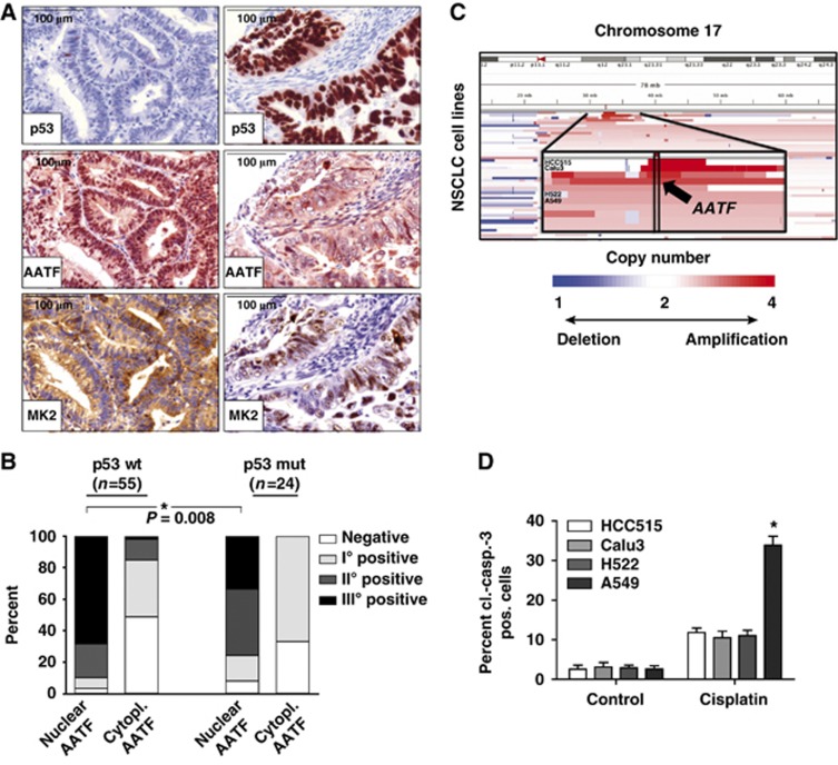
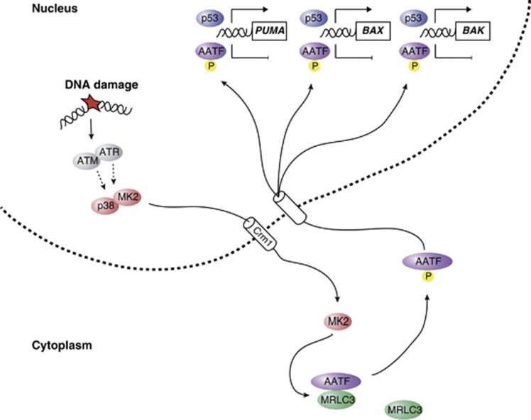
Following genotoxic stress, cells activate a complex signalling network to arrest the cell cycle and initiate DNA repair or apoptosis. The tumour suppressor p53 lies at the heart of this DNA damage response. However, it remains incompletely understood, which signalling molecules dictate the choice between these different cellular outcomes. Here, we identify the transcriptional regulator apoptosis-antagonizing transcription factor (AATF)/Che-1 as a critical regulator of the cellular outcome of the p53 response. Upon genotoxic stress, AATF is phosphorylated by the checkpoint kinase MK2. Phosphorylation results in the release of AATF from cytoplasmic MRLC3 and subsequent nuclear translocation where AATF binds to the PUMA, BAX and BAK promoter regions to repress p53-driven expression of these pro-apoptotic genes. In xenograft experiments, mice exhibit a dramatically enhanced response of AATF-depleted tumours following genotoxic chemotherapy with adriamycin. The exogenous expression of a phospho-mimicking AATF point mutant results in marked adriamycin resistance in vivo. Nuclear AATF enrichment appears to be selected for in p53-proficient endometrial cancers. Furthermore, focal copy number gains at the AATF locus in neuroblastoma, which is known to be almost exclusively p53-proficient, correlate with an adverse prognosis and reduced overall survival. These data identify the p38/MK2/AATF signalling module as a critical repressor of p53-driven apoptosis and commend this pathway as a target for DNA damage-sensitizing therapeutic regimens.


Conflict of interest statement

The authors declare that they have no conflict of interest.

Figures

Figure 1
Figure 1
Identification of a phosphorylation-sensitive protein complex consisting of AATF and MRLC3. (A) An oriented (pSer/pThr) phosphopeptide library, biased towards the basophilic phosphorylation motif of Chk1/2 and MK2, was immobilized on streptavidin beads. The phospho ϕXRXXpT and non-phosphorylated ϕXRXXT peptide libraries were screened for interaction against in vitro translated, 35S-Met-labelled proteins. (B) Identification of MRLC3 as a non-phospho binder occurred in pool 16B11 and through progressive subdivision to a single clone. (C) Yeast two-hybrid screening revealed AATF as an interactor of MRLC3. We further characterized this interaction through co-immunoprecipitation (co-IP), performed in the presence or absence of 1 μM okadaic acid (OA). FLAG.MRLC3 was immunoprecipitated from HEK293T cells co-expressing V5.AATF. FLAG.GFP served as a control. Lane 3 shows an interaction of FLAG.MRLC3 with V5.AATF, which was abolished by OA-mediated Ser/Thr phosphatase inhibition 1 h prior to lysis (lane 4). (D) The MRLC3:AATF complex is sensitive to UV-C-induced DNA damage. FLAG.MRLC3 and V5.AATF-expressing HEK293T cells were UV-C irradiated (20 J/m2) 30 min prior to lysis and IP with anti-FLAG beads. FLAG.GFP served as a negative control. While V5.AATF co-precipitated with FLAG.MRLC3 in the absence of UV-C, the interaction was abrogated in the presence of DNA damage. (E) Reversal of the co-IP experiment is shown in (D). Anti-FLAG IP reveals AATF.FLAG:V5.MRLC3 complexes that display strong sensitivity to UV-C-induced DNA damage. FLAG.GFP served as a negative control. (F) Endogenous AATF:MRLC complexes display UV-C sensitivity. AATF was immunoprecipitated from HCT116 cells that were mock-treated or exposed to UV-C (20 J/m2) 30 min prior to lysis and IP. GFP IP served as a negative control (lanes 1 and 2). While substantial amounts of MRLC co-immunoprecipitated with AATF (lane 3), this interaction was abolished by UV-C-induced DNA damage (lane 4). (G) Endogenous AATF:MRLC3 complexes are sensitive to the topoisomerase-II inhibitor doxorubicin. AATF was immunoprecipitated from HCT116 cells that were mock-treated or exposed to doxorubicin (1 μM) 1 h prior to IP. GFP antibody served as a negative control (lanes 1 and 2). Doxorubicin (lane 4) disrupted the interaction between AATF and MRLC (lane 3). Figure source data can be found with the Supplementary data.
Figure 2
Figure 2
The AATF:MRLC3 cytoplasmic complex is disrupted upon DNA damage. (A) Hypotonic lysis was used to generate nuclear and cytoplasmic fractions. The purity of the fractions was documented with immunoblotting for fibrillarin (nuclear) and tubulin (cytoplasmic). MRLC was exclusively detected in the cytoplasmic compartment. The subcellular distribution of MRLC was not affected by 20 J/m2 UV-C-induced genotoxic stress applied 30 min prior to lysis. (B) AATF resides in the cytoplasm of resting cells and translocates to the nucleus upon genotoxic stress. Nuclear and cytoplasmic fractions of mock- or UV-C-treated (20 J/m2) MEFs were generated as in (A). In the absence of UV, AATF is primarily cytoplasmic. Upon UV-C-induced DNA damage, substantial amounts of AATF are detectable in the nucleus. (C) Indirect immunofluorescence (IF) was performed to verify the subcellular localization of MRLC3 in MEFs 30 min following UV-C (20 J/m2). DAPI staining was used as a counterstain to provide a nuclear reference point. MRLC displayed a granular cytoplasmic staining pattern that was not affected by UV-C irradiation. No MRLC staining could be detected in the nuclei. (D) Indirect IF was performed to verify the subcellular distribution and spatial dynamics of AATF in MEFs 30 min following UV-C (20 J/m2). In resting cells, AATF staining revealed a dominant granular cytoplasmic pattern with some nuclear staining. 30 min following UV-C, only miniscule amounts of AATF remained detectable in the cytoplasm, while the bulk of AATF staining was now nuclear. (E) The DNA damage-induced nuclear re-localization of endogenous AATF was quantified using fluorescence microscopy. Error bars represent s.d. (*P<0.05). (F) Global Ser/Thr phosphatase inhibition promotes the nuclear accumulation of AATF. MEFs were mock-treated or pre-treated with 1 μM OA 60 min prior to lysis. Phosphatase inhibition resulted in nuclear accumulation of AATF. (G) Osmotic stress promotes the nuclear accumulation of AATF. MEFs were mock-treated or exposed to hypertonic Ringer solution 10 min prior to lysis. This non-genotoxic MK2-activating stimulus resulted in nuclear AATF accumulation similar to that seen after DNA damage. Figure source data can be found with the Supplementary data.
Figure 3
Figure 3
MK2-mediated AATF phosphorylation on Thr-366 disrupts cytoplasmic AATF:MRLC3 complexes to allow nuclear accumulation of AATF. (A) MK2 was subjected to in vitro kinase assays using AATF as a substrate. Reactions were quenched by the addition of SDS-buffer and proteins were resolved on SDS–PAGE before AATF-containing coomassie bands were isolated and proteins subjected to LC–MS/MS analysis after reduction, alkylation and Lys-C digestion. For MS analysis, up to four HCD and CID spectra (MS2) were acquired following each scan. When a neutral loss of phosphoric acid was detected, MS3 spectra were acquired in the linear ion trap to determine the peptide sequence. Mascot 2.2 was used for protein ID. A peptide spanning the phospho-Thr-366 residue was readily detected. (B) A biotinylated phospho-Thr-366-containing AATF peptide fails to interact with FLAG.MRLC3. In vitro pull down assays were performed from FLAG.MRLC3-expressing HEK293T cells using streptavidin-immobilized Thr-366 phosphorylated and non-phosphorylated peptides (AATF amino acid 357–374) as baits. While the non-phosphrylated peptide bound to FLAG.MRLC3, no interaction was observed between the phosphorylated peptide and FLAG.MRLC3. (C) Endogenous AATF:MRLC complexes are disrupted by MK2 in vitro. AATF:MRLC complexes were immunoprecipitated from HCT116 cells using AATF antibodies. GFP-immunoprecipitations served as a negative control. Precipitated material was subjected to a 30-min incubation with MK2 or left untreated before SDS–PAGE and immunoblotting. Incubation with MK2 (lane 4) disrupted the AATF:MRLC complexes immunoprecipitated in the control samples (lane 3). (D) A structural basis for phospho-dependent release of AATF from MRLC. The structure of MRLC bound to MHC is shown (PDB code 2OS8) with MRLC in a surface representation shaded by electrostatic potential (red: acidic, blue: basic). MHC is shown in stick representation with carbons coloured cyan, nitrogens blue, and oxygens red. The position of a Gln residue in MHC corresponding to the position of Thr-366 in AATF based on sequence and secondary structure alignments (Supplementary Figure 6) is indicated. (E) AATF re-localizes to the nucleus following UV-C-induced DNA damage in MK2wt/wt MEFs. Cells were either mock-treated or exposed to UV-C (20 J/m2) 30 min prior to biochemical fractionation. Fibrillarin and tubulin served to control the purity of the fractions. (F) DNA damage-induced nuclear localization of AATF is repressed in MK2/3−/− MEFs. Cells were treated as shown in (E). Figure source data can be found with the Supplementary data.
Figure 4
Figure 4
Association of AATF copy number, expression and clinical outcome in neuroblastoma. In all, 164 neuroblastoma samples were subjected to aCGH-based copy number and mRNA expression analyses to determine whether a correlation between AATF copy number and mRNA expression exists in human tumours. (A, B) Scatter plot showing the correlation between genomic copy number and the expression level of AATF for probe 1 (A, r=0.29, P<0.001) and probe 2 (B, r=0.30, P<0.001). (CF) Kaplan–Meier survival curves for event-free (C, probe 1; D, probe 2) and overall survival (E, probe 1; F, probe 2) of neuroblastoma patients separated according to AATF expression levels. Red, expression level lower than the cutoff value; blue, expression level higher than the cutoff value.
Figure 5
Figure 5
AATF prevents p53-driven apoptosis by repressing DNA damage-dependent induction of pro-apoptotic genes. (A) p53+/+ and p53−/− HCT116 cells expressing control or AATF-specific shRNA were left untreated or exposed to UV (40 J/m2), camptothecin (cam, 10 μM) or doxorubicin (dox, 1 μM) and harvested for quantification of apoptosis 24 h later. AATF depletion in p53+/+ cells resulted in a significant increase in the number of apoptotic cells following all treatment regimens with DNA-damaging agents. No increase in the number of apoptotic cells could be observed in p53−/− cells following DNA damage. Asterisk indicates statistical significance, error bars represent s.d., two-tailed Student’s t-test, P<0.05, n=16. (B) p53+/+ HCT116 cells expressing AATFWT, AATFTA or AATFTD were left untreated or exposed to UV (40 J/m2), camptothecin (10 μM) or doxorubicin (1 μM) and harvested for FACS-based quantification of apoptosis 24 h later. AATFTD expression significantly repressed apoptosis in response to all three genotoxic treatments. Asterisk indicates statistical significance (P<0.05), error bars represent s.d., two-tailed Student’s t-test, P<0.05, n=16. (C) RNAi-mediated AATF depletion in p53+/+ HCT116 cells promotes DNA damage-induced induction of pro-apoptotic p53 target genes, while expression of cell-cycle-regulating p53 target genes remains unaffected. Immunoblotting was used to detect the pro-apoptotic p53-target gene products PUMA, BAX and BAK, and the cell-cycle-arresting p53 target gene products CDKN1A, GADD45α and REPRIMO 12 h after 40 J/m2 UV-C. Tubulin served as a loading control. (DF) AATF binds to the promoters of the pro-apoptotic p53-target genes PUMA, BAX and BAK in a DNA damage and MK2-dependent manner. ChIP experiments were performed in MK2/3+/+ and MK2/3−/− MEFs that were left untreated or exposed to 40 J/m2 UV-C 60 min prior to cross-linking. DNA was precipitated using AATF antibodies. qPCR was used to quantify PUMA, BAX and BAK promoter-specific DNA in the precipitates. Unspecific IgG served as a control. Asterisk indicates statistical significance, error bars represent s.d., two-tailed Student’s t-test, P<0.05, n=9. (GI) AATF binding to the CDKN1Ap21, GADD45α and RPRM promoter is not regulated in a DNA damage or MK2-dependent manner. ChIP experiments were performed as in (DF) and promoter-specific primers were used for qPCR. Unspecific IgG served as a control. Asterisk indicates statistical significance, error bars represent s.d., two-tailed Student’s t-test, P<0.05, n=9. Primers to amplify the genomic DNA of the promoter regions were chosen to cover known p53-binding sites. Schematic drawings (DI) indicated localization of primer (box) and the known p53-binding sites (black bars). Figure source data can be found with the Supplementary data.
Figure 6
Figure 6
AATF promotes chemo-resistance in vivo. (A) AATF depletion results in a significant increase doxorubicin sensitivity of p53+/+ tumours. In all, 106 HCT116 cells expressing either control or AATF-specific shRNA were xenograftet into the flanks of NCRnu/nu mice (n=6 for each group). (B) Immunohistochemical analysis of control and AATF shRNA-expressing tumours excised at the termination point of the experiments shown in (A). Specimens were stained with PUMA-specific antibodies and counterstained with haematoxylin. (C) AATFTD expression promotes doxorubicin resistance in vivo. p53+/+ HCT116 cells were transduced to express either AATFWT, AATFTA or AATFTD. In all, 106 HCT116 cells of each group were injected into the flanks of NCRnu/nu-mice (n=6 for each group). Arrows indicate doxorubicin injections (5 mg/kg, i.p.). Asterisks indicate significance (Student’s t-test, two-tailed, P<0.05).
Figure 7
Figure 7
AATF is amplified in human tumours and displays a nuclear localization pattern in p53-proficient tumours. (A) Nuclear localization of AATF is selected for in p53-proficient tumours. Human endometrial tumours were stained for p53, AATF and MK2. p53-defective tumours show predominantly cytosolic AATF and nuclear MK2 staining (A—right) whereas tumours containing wild-type p53 display prominent nuclear enrichment of AATF accompanied by a prominent cytoplasmic MK2 signal (A—left). (B) Quantification of nuclear and cytoplasmic AATF staining was performed using a four-tier scoring system and subjected to statistical analysis (Fisher’s exact test). (C) AATF amplification is associated with increased resistance of lung cancer cell lines against cisplatin. Copy-number alterations as determined by SNP-arrays (blue=deletion; white=copy number neutral; red=amplification) at chromosome 17 (y axis) across all NSCLC cell lines are depicted (x axis). The chromosomal position of the amplification of AATF is highlighted for the analysed cell lines (zoom-in panel). HCC515 and Calu3 cells with copy number>4, H522 and A549 cells with copy number<4. (D) HCC515 (p53 mutant, AATF amplified), Calu3 (p53 wild type, AATF amplified), H522 (p53 mutant) and A549 (p53 wild type) cells were left untreated or exposed to cisplatin (10 μM) and harvested for FACS-based quantification of apoptosis using a cl.-caspase-3 antibody 24 h later. Asterisk indicates statistical significance, error bars represent s.d., n=12.
Figure 8
Figure 8
AATF acts as a phosphorylation-dependent molecular switch to dictate the outcome of the p53 response. A simplified model depicting details of the AATF-based molecular switch, controlling the cellular outcome of the p53 response.

Comment in

References

    1. Abbas T, Dutta A (2009) p21 in cancer: intricate networks and multiple activities. Nat Rev Cancer 9: 400–414 - PMC - PubMed
    1. Asada M, Yamada T, Ichijo H, Delia D, Miyazono K, Fukumuro K, Mizutani S (1999) Apoptosis inhibitory activity of cytoplasmic p21(Cip1/WAF1) in monocytic differentiation. EMBO J 18: 1223–1234 - PMC - PubMed
    1. Bruno T, De Angelis R, De Nicola F, Barbato C, Di Padova M, Corbi N, Libri V, Benassi B, Mattei E, Chersi A, Soddu S, Floridi A, Passananti C, Fanciulli M (2002) Che-1 affects cell growth by interfering with the recruitment of HDAC1 by Rb. Cancer Cell 2: 387–399 - PubMed
    1. Bruno T, De Nicola F, Iezzi S, Lecis D, D'Angelo C, Di Padova M, Corbi N, Dimiziani L, Zannini L, Jekimovs C, Scarsella M, Porrello A, Chersi A, Crescenzi M, Leonetti C, Khanna KK, Soddu S, Floridi A, Passananti C, Delia D et al. (2006) Che-1 phosphorylation by ATM/ATR and Chk2 kinases activates p53 transcription and the G2/M checkpoint. Cancer Cell 10: 473–486 - PubMed
    1. Bruno T, Iezzi S, De Nicola F, Di Padova M, Desantis A, Scarsella M, Di Certo MG, Leonetti C, Floridi A, Passananti C, Fanciulli M (2008) Che-1 activates XIAP expression in response to DNA damage. Cell Death Differ 15: 515–520 - PubMed

Publication types

MeSH terms