. 2012 Oct 5;287(41):34361-71.
doi: 10.1074/jbc.M112.365882. Epub 2012 Aug 21.

The requirement of c-Jun N-terminal kinase 2 in regulation of hypoxia-inducing factor-1α mRNA stability

Affiliations

The requirement of c-Jun N-terminal kinase 2 in regulation of hypoxia-inducing factor-1α mRNA stability

Dongyun Zhang et al. J Biol Chem. .

Abstract

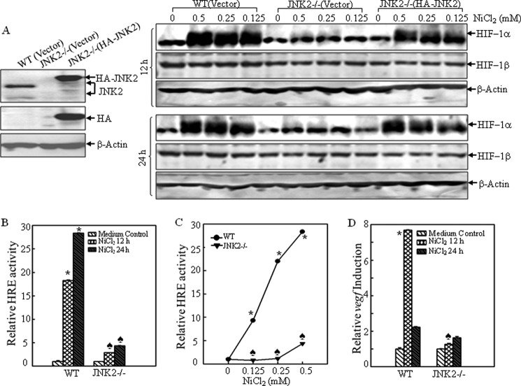
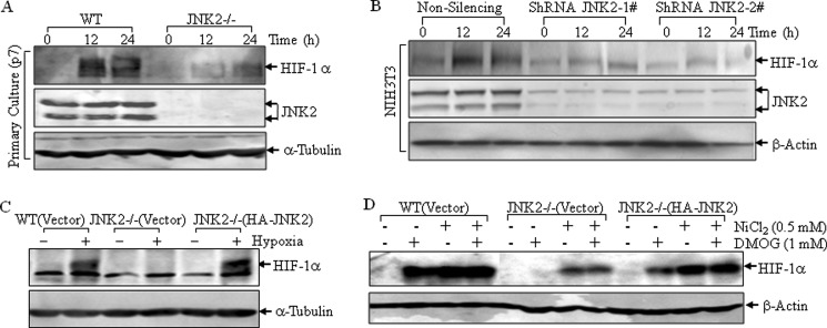
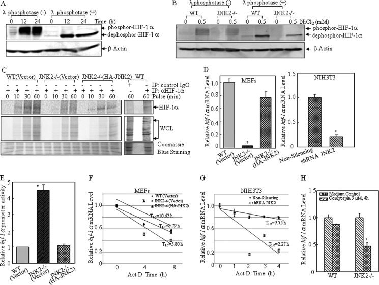
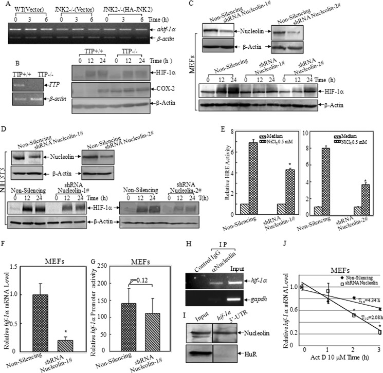
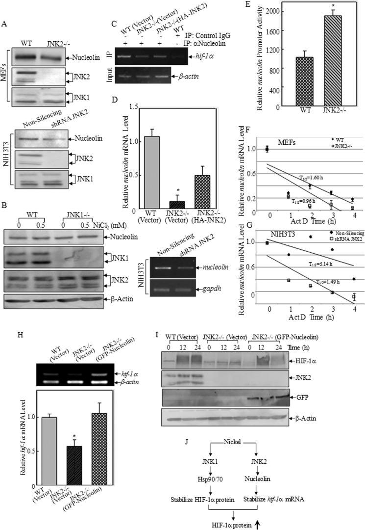
The mRNA of hif-1α is considered as being constitutively and ubiquitously expressed, regardless of the level of oxygen tension. However many recent reports have showed that hif-1α mRNA could be regulated by natural antisense transcripts, potential microRNAs, and low O(2). In this study, it was found that a deficiency of JNK2 expression reduced HIF-1α protein induction in response to nickel treatment resulting from the impaired expression of hif-1α mRNA. Both the promoter luciferase assay and mRNA degradation assay clearly showed that depletion of JNK2 affected stability of hif-1α mRNA, rather than regulated its transcription. In addition, nucleolin, a classic histone chaperone, was demonstrated to physically bind to hif-1α mRNA and maintain its stability. Further investigation indicated that JNK2 regulated nucleolin expression and might in turn stabilize hif-1α mRNA. Collectively, we provided one more piece of evidence for the oncogenic role of JNK2 and nucleolin in regulating the cancer microenvironments by controlling HIF-1α expression.


Figures

FIGURE 1.
FIGURE 1.
JNK2 deficiency impaired nickel-induced HIF-1α expression and transactivation. A, WT(Vector), JNK2−/− (Vector), and JNK2−/− (HA-JNK) MEFs were identified by Western blotting assay for expression of JNK2 (left panel). The induction of HIF-1α protein was compared after the indicated cells were exposed to nickel at doses and time points as indicated (right panel). B--D, WT and JNK2−/− cells that were stably transfected with HRE-luciferase reporter (B and C) or vegf-luciferase reporter (D) were exposed to 0.5 mm nickel for 12 h and 24 h (B and D), or different doses of nickel for 24 h (C). The luciferase activities were determined using a luminometer. The results are expressed as relative HRE activities or vegf inductions to medium control. Each bar indicates the mean and standard error of triplicate assay wells. The asterisk (*) indicates a significant increase as compared with medium control in WT cells (p < 0.05). The spade (♣) indicates a significant decrease as compared with that in the WT cells for the same treatment (p < 0.05).
FIGURE 2.
FIGURE 2.
JNK2 regulated HIF-1α protein expression in various cells. A, primarily cultured WT and JNK2−/− MEFs at passage 7 were exposed to nickel (0.5 mm) for 12 h and 24 h. B, two separate shRNAs for JNK2 or non-silencing control plasmid were transfected into NIH3T3 cells. The stable transfectants were treated with nickel (0.5 mm) for 12 h and 24 h. C and D, WT(Vector), JNK2−/− (Vector), and JNK2−/− (HA-JNK) MEFs were exposed to hypoxia (1% O2) for 8 h (C), or DMOG in combination with or without nickel for 24 h (D). The cell extracts were subjected to Western blotting assay.
FIGURE 3.
FIGURE 3.
JNK2 regulated hif-1α mRNA stability. A and B, WT (A) or WT and JNK2−/− MEFs (B) (1 × 10⁶/well) were seeded into 10-mm dish and exposed to 0.5 mm nickel for 12 h and 24 h (A), or 24 h (B). The whole cell lysate was incubated with λ phosphatase for 40 min at 30 °C. Proteins were resolved in SDS-PAGE and revealed by Western blotting assay. C, WT(Vector), JNK2−/− (Vector), and JNK2−/− (HA-JNK) MEFs were treated with nickel (0.5 mm) for 12 h in complete medium. Cells were then accommodated in methionine- and cysteine-free DMEM for 1 h. 35S-labeled methionine and cysteine was then added for the indicated times for pulse assay. Cell extracts were immunoprecipitated with anti-HIF-1α antibody or control IgG, and subjected to SDS-PAGE. Autoradiography was used to visualize 35S-labeled HIF-1α. D, hif-1α mRNA levels in the individual cells were determined by real-time PCR. The asterisk (*) indicates a significant decrease as compared with those in WT(Vector) and JNK−/− (HA-JNK2) MEFs or non-silencing control cells (p < 0.05). E, basal levels of hif-1α promoter activity were evaluated by transfecting the indicated cells with a construct containing hif-1α promoter-driven luciferase. pRL-TK vector was used as an internal control. The results are expressed as the ratios of firefly to Renilla lucifease activity, as means ± S.D. (n = 3). The asterisk (*) indicates a significant increase as compared with that of WT(Vector) cells or JNK−/− (HA-JNK2) cells (p < 0.05). F and G, mRNA degradation rate of hif-1α was detected following treatment with actinomycin D (5 μm) for the indicated time. The PCR products were separated over 2% agarose gels, stained with ethidium bromide. The densitometric analyses of the product bands were conducted using the software of ImageQuant 5.2 (GE Healthcare). The results were shown as means ± S.D. (n = 3). H, indicated cells were treated with cordycepin (5 μm) for 4 h. Real-time PCR was conducted to detect the hif-1α mRNA expression. The asterisk (*) indicates a significant decrease as compared with that in WT cells under the same treatment (p < 0.05).
FIGURE 4.
FIGURE 4.
Nucleolin bound to hif-1α mRNA and increased its stability. A, expression of a natural antisense transcript of hif-1α (ahif) was determined by RT-PCR. B, WT and TTP−/− MEFs were treated with 0.5 mm nickel for 12 and 24 h. The cell extracts were subjected to Western blotting assay. C and D, shRNA-nucleolin was stably transfected into WT MEFs (C) or NIH3T3 (D). The transfectants were identified by Western blotting. HIF-1α protein induction by nickel exposure (0.5 mm) was compared between non-silencing and shRNA-nucleolin transfectants. E, HRE-luciferase reporter and pRL-TK vector were transiently transfected into non-silencing and shRNA-nucleolin MEFs. The HRE induction was determined by luciferase assay. The results were presented as relative HRE activities by normalizing the ratios of firefly to Renilla lucifease activity, as means ± S.D. (n = 3). The asterisk (*) indicates a significant decrease as compared with that in non-silencing cells (p < 0.05). F, hif-1α mRNA expression levels were detected by real-time PCR in the indicated MEFs. The asterisk (*) indicates a significant decrease as compared with that in non-silencing cells (p < 0.05). G, hif-1α transcription was determined using hif-1α promoter-driven firefly luciferase and pRL-TK vector. The results were expressed relative hif-1α promoter activities by normalizing the ratios of firefly to Renilla lucifease activity, as means ± S.D. (n = 3). H, RNA-IP was performed to examine the binding of nucleolin to hif-1α mRNA in 293T cells. I, RNA-pull down was performed using an in vitro transcribed RNA containing mouse hif-1α 3′-UTR labeled with biotin. Magnetic streptavidin beads were used to capture the proteins bound with hif-1α 3′-UTR after incubating with cell lysate. Western blotting was carried out to detect the presence of nucleolin in pull-down complex. HuR was used as a negative control. J, hif-1α mRNA degradation rates were compared between the indicated MEFs by real-time PCR following treatment with actinomycin D (10 μm). The data were shown as means ± S.D. (n = 3). The asterisk (*) indicates a significant decrease as compared with that in non-silencing cells (p < 0.05).
FIGURE 5.
FIGURE 5.
Nucleolin was regulated by JNK2, but not JNK1. A and B, expression of nucleolin protein in the indicated cell lines was analyzed by Western blotting assay. C, RNA-IP was performed to compare the binding with nucleolin to hif-1α mRNA in the indicated cell lines using anti-nucleolin antibody. D, comparison of nucleolin mRNA expression in the transfectants as indicated by real-time PCR (top panel and RT-PCR (bottom panel). The asterisk (*) indicates a significant decrease as compared with that in WT(Vector) or JNK2−/− (HA-JNK2) cells (p < 0.05). E, nucleolin transcription levels were determined using nucleolin promoter-driven firefly luciferase and pRL-TK vector. The results were expressed relative nucleolin promoter activities by normalizing the ratios of firefly to Renilla lucifease activity, as means ± S.D. (n = 3). The asterisk (*) indicates a significant increase as compared with that in WT cells (p < 0.05). F and G, mRNA degradation rate of nucleolin was determined by reverse transcript-PCR following treatment with actinomycin D (5 μm) as indicated. The densitometric analyses of the product bands were conducted using the software of ImageQuant 5.2 (GE Healthcare). The results were shown as means ± S.D. (n = 3). H, hif-1α mRNA expression levels were assessed by RT-PCR in the indicated cells. The figures shown were representative of three independent experiments. The densitometric analyses of the product bands were conducted using the software of ImageQuant 5.2 (GE Healthcare). The results were shown as means ± S.D. (n = 3). I, WT(Vector), JNK2−/− (Vector), and JNK2−/− (GFP-Nucleolin) MEFs were exposed to nickel (0.5 mm) for 12 and 24 h. The cell extracts were subjected to Western blotting as indicated. J, scheme showing the molecular mechanisms underlying JNK1 and JNK2 regulation of HIF-1α expression following nickel exposure.

Similar articles

Cited by

References

    1. Grandjean P. (1984) Human exposure to nickel. IARC. Sci. Publ. 53, 469–485 - PubMed
    1. Furst A. (1984) Mechanism of action of nickel as a carcinogen: needed information. IARC. Sci. Publ. 53, 245–252 - PubMed
    1. Salnikow K., Zhitkovich A. (2008) Genetic and epigenetic mechanisms in metal carcinogenesis and cocarcinogenesis: nickel, arsenic, and chromium. Chem. Res. Toxicol. 21, 28–44 - PMC - PubMed
    1. Lu H., Shi X., Costa M., Huang C. (2005) Carcinogenic effect of nickel compounds. Mol. Cell. Biochem. 279, 45–67 - PubMed
    1. Ke Q., Costa M. (2006) Hypoxia-inducible factor-1 (HIF-1). Mol. Pharmacol. 70, 1469–1480 - PubMed

Publication types

MeSH terms