. 2012 Oct 12;287(42):35506-35515.
doi: 10.1074/jbc.M112.394924. Epub 2012 Aug 21.

Neuroimmunoendocrine regulation of the prion protein in neutrophils

Affiliations

Neuroimmunoendocrine regulation of the prion protein in neutrophils

Rafael M Mariante et al. J Biol Chem. .

Abstract

The prion protein (PrP(C)) is a cell surface protein expressed mainly in the nervous system. In addition to the role of its abnormal conformer in transmissible spongiform encephalopathies, normal PrP(C) may be implicated in other degenerative conditions often associated with inflammation. PrP(C) is also present in cells of hematopoietic origin, including T cells, dendritic cells, and macrophages, and it has been shown to modulate their functions. Here, we investigated the impact of inflammation and stress on the expression and function of PrP(C) in neutrophils, a cell type critically involved in both acute and chronic inflammation. We found that systemic injection of LPS induced transcription and translation of PrP(C) in mouse neutrophils. Up-regulation of PrP(C) was dependent on the serum content of TGF-β and glucocorticoids (GC), which, in turn, are contingent on the activation of the hypothalamic-pituitary-adrenal axis in response to systemic inflammation. GC and TGF-β, either alone or in combination, directly up-regulated PrP(C) in neutrophils, and accordingly, the blockade of GC receptors in vivo curtailed the LPS-induced increase in the content of PrP(C). Moreover, GC also mediated up-regulation of PrP(C) in neutrophils following noninflammatory restraint stress. Finally, neutrophils with up-regulated PrP(C) presented enhanced peroxide-dependent cytotoxicity to endothelial cells. The data demonstrate a novel interplay of the nervous, endocrine, and immune systems upon both the expression and function of PrP(C) in neutrophils, which may have a broad impact upon the physiology and pathology of various organs and systems.


Figures

FIGURE 1.
FIGURE 1.
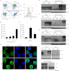
Systemic administration of LPS regulates PrPC in bone narrow neutrophils. A, mice were injected intraperitoneally with 50 μg of LPS, and 24 h later BMC were collected, and PrPC content at the surface of Gr-1+ cells was analyzed by flow cytometry. Control animals (CTR) were injected with sterile saline alone. NIS, nonimmune serum. B, mice were injected intraperitoneally with varying doses of LPS, and BMC were analyzed after 24 h. C, animals were injected intraperitoneally with 50 μg of LPS, and BMC were collected at various time points after injection, and PrPC content at the surface of Gr-1+ cells was analyzed. D, neutrophils purified from the bone marrow of animals either treated or not with 50 μg of LPS for 24 h were fixed, permeabilized, labeled with anti-PrPC, counterstained with TO-PRO-3, and examined by confocal microscopy. Scale bar, 5 μm. E, BMC from untreated animals were cultured in vitro for 24 h either with varying doses of LPS (left panel) or with 10% serum from animals previously treated at various intervals with 50 μg of LPS (LPS-serum; right panel). The relative increase of surface PrPC in bone marrow neutrophils was estimated by flow cytometry. F, immunoblots showing PrPC content of lysates from cerebral cortex or bone marrow neutrophils from animals either treated (+) or not (−) with 50 μg of LPS for 24 h. Erk2 was used as a loading control. Molecular mass standards are indicated at the left in kDa. G, BMC were cultured in vitro for 24 h with either 10% control or 10% LPS-serum obtained 12 h after administration of PBS or LPS (50 μg). Immunoblots of the respective lysates were stained for PrPC. *, p < 0.01 (B); *, p < 0.05, and **, p < 0.01 (C and E) compared with untreated groups; n ≥ 3.
FIGURE 2.
FIGURE 2.
LPS-induced PrPC regulation is not dependent on major proinflammatory cytokines. A, BMC were cultured in vitro for 24 h with CM prepared from thioglycolate-elicited peritoneal macrophages stimulated for 24 or 48 h with 0.1 or 1 μg/ml LPS. The relative increase of surface PrPC in Gr-1+ cells was evaluated by flow cytometry. B, BMC were cultured for 30 min in the presence of 1 μg/ml neutralizing antibody against TNF-α (TNF-α nAb) or IL-1β (IL-1β nAb) before stimulation with CM derived from macrophages. C, BMC were cultured in the presence of various doses (ng/ml) TNF-α, and analyzed as before. D, BMC were cultured for 30 min in the presence of 10 μg/ml TNF-α nAb before treatment for 24 h with different doses of LPS-serum. E, TNFR1 null mice were injected intraperitoneally with 50 μg of LPS; 24 h later their BMC were collected and analyzed by flow cytometry. Control animals (CTR) received intraperitoneal injections of sterile saline alone. F, BMC obtained from TNFR1 null mice were cultured in vitro for 24 h with increasing doses of TNF-α (gray bars) or in the presence of 10% control or 10% LPS-serum (black bars) and examined as before. G, wild-type BMC were cultured in vitro for 24 h in the presence of 10% serum obtained from TNFR1 null mice previously treated with either LPS or sterile saline alone (CTR). *, p < 0.05 (A and B); *, p < 0.01 (C and E); **, p < 0.001 (A, F, and G), and **, p < 0.01 (B) compared with untreated groups, and #, p < 0.05 compared with CM LPS alone; n ≥ 3.
FIGURE 3.
FIGURE 3.
TGF-β and glucocorticoids concur to regulate PrPCin vitro. Bone marrow cells were cultured in vitro for 24 h with various concentrations of either TGF-β (A) or DEX (B) and analyzed by flow cytometry for PrPC content at the surface of Gr-1+ cells. In some cases, the glucocorticoid receptor antagonist RU486 was added to the cultures 30 min before DEX treatment (B). *, p < 0.01, and **, p < 0.001 compared with untreated groups, and #, p < 0.01 compared with each dose of DEX alone; n ≥ 5 (A) and n ≥ 4 (B).
FIGURE 4.
FIGURE 4.
Glucocorticoid up-regulates PrPCin vivo. A, mice received intraperitoneal injections of 2 mg of DEX, and 24 h later their BMC were collected and analyzed by flow cytometry. Control animals (CTR) received intraperitoneal injection of sterile saline alone. NIS, nonimmune serum. B, mice were injected intraperitoneally with varying doses of DEX, and BMC were analyzed as above. C, mice were injected intraperitoneally with 2 mg of DEX and had their BMC collected at various time points for analysis. D, neutrophils purified from the bone marrow of animals either treated or not with 2 mg of dexamethasone for 24 h were fixed, permeabilized, labeled with anti-PrPC, counterstained with TO-PRO-3, and examined by confocal microscopy. Scale bar, 5 μm. E–H, immunoblots showing PrPC content of lysates from brain and bone marrow cells (E–G and I) or isolated neutrophils (H) from C57/BL6 (E, H, and I) or C57/BL10 (F and G) mice either treated or not with DEX as before. A polyclonal antiserum raised in Prnp0/0 mice was used in E and F, and a monoclonal anti-PrPC SAF32 was used in GI. Erk2 was used as a loading control. Molecular mass standards are indicated at the left in kDa. In I we show the analysis of deglycosylated PrPC. BMC obtained from DEX-treated C57/BL6 mice were treated or not with N-glycosidase F (PNGase) and probed for PrPC. *, p < 0.001 (B); *, p < 0.05, and **, p < 0.001 (C) compared with untreated groups; n ≥ 3.
FIGURE 5.
FIGURE 5.
LPS-induced PrPC up-regulation is dependent on both glucocorticoids and TGF-β. A, BMC were cultured for 30 min in the presence or absence of 2 μm RU486 and/or 1 μg/ml neutralizing antibody to TGF-β (TGF-β nAb) before adding either 10% control (CTR) or 10% LPS-serum from animals previously treated for 12 h. PrPC at the surface of neutrophils was estimated by flow cytometry. B, BMC were cultured in vitro for 24 h with 10 ng/ml TGF-β and/or various concentrations of DEX and analyzed by flow cytometry. The inset shows comparatively the combined action (CA) and the sum of the individual actions (IA) of both compounds. C, mice were treated or not by oral gavage with 500 μg of RU486 for 2 h before intraperitoneal injection of 50 μg of LPS. After 24 h, their BMC were collected and analyzed as before. *, p < 0.001 (A and C), and **, p < 0.001 (B) compared with untreated groups, and #, p < 0.001 compared with LPS-serum or LPS alone; n ≥ 5 (A) and n ≥ 4 (B and C).
FIGURE 6.
FIGURE 6.
Behavioral stress increases PrPC in neutrophils through increased levels of glucocorticoids in the serum. A, mice pretreated for 2 h by oral gavage with either 500 μg of RU486 or vehicle alone were subject to restraint stress (RS) for 15 h, as described under “Experimental Procedures.” Then their BMC were collected and analyzed by flow cytometry for PrPC content at the surface of Gr-1+ cells. Control animals (CTR) received either no pretreatment or 500 μg of RU486 by oral gavage. B, BMC were cultured in vitro for 24 h in the presence of 10% serum from mice either subject (RS serum) or not (CTR serum) to restraint stress for 15 h, as before. Control cultures (CTR) received medium alone. PrPC at the surface of neutrophils was assessed by flow cytometry. C, serum corticosterone levels were measured in mice either subject to restraint for 15 h (RS) or at 24 h after injection of 50 μg of LPS. *, p < 0.05 (A and C); *, p < 0.0001 (B), and **, p < 0.01 (C) compared with untreated groups; #, p < 0.01 compared with restraint stress alone; n ≥ 3.
FIGURE 7.
FIGURE 7.
Up-regulation of PrPC involves enhanced gene expression and has functional consequences. A, increased PrPC is associated with augmented transcription. Analysis of the induction of the transcript levels of PrPC was done after LPS or DEX treatment or restraint stress (RS). Total RNA from bone marrow was isolated, and real time RT-PCR measurements were done as described under “Experimental Procedures” and normalized against GAPDH. B, cytotoxicity of neutrophils following peripheral stress. bAEC were cultured in the presence of CM from neutrophils harvested from WT or KO mice either treated or not with DEX and incubated for 30 min with 1 μg/ml PMA. In some cases, catalase (CAT) (1 μg/ml) was added to the CM 15 min before the treatment of the bAEC. Cytotoxicity was measured by the neutral red uptake assay as described under “Experimental Procedures.” C, peroxide-induced neutrophil cell death. BMC obtained from WT or KO mice previously treated or not with DEX were cultured for 24 h in RPMI without serum, containing H2O2 at various concentrations. Cells were stained with 1 μg/ml propidium iodide (PI) and analyzed by flow cytometry. *, p < 0.05, and **, p < 0.001 compared with untreated group; n ≥ 4 (A) and n = 3 (B and C).

References

    1. Linden R., Martins V. R., Prado M. A., Cammarota M., Izquierdo I., Brentani R. R. (2008) Physiology of the prion protein. Physiol. Rev. 88, 673–728 - PubMed
    1. Weissmann C., Büeler H., Sailer A., Fischer M., Aguet M., Aguzzi A. (1993) Role of PrP in prion diseases. Br. Med. Bull. 49, 995–1011 - PubMed
    1. Prusiner S. B. (1998) Prions. Proc. Natl. Acad. Sci. U.S.A. 95, 13363–13383 - PMC - PubMed
    1. Gunther E. C., Strittmatter S. M. (2010) β-Amyloid oligomers and cellular prion protein in Alzheimer disease. J. Mol. Med. 88, 331–338 - PMC - PubMed
    1. Kessels H. W., Nguyen L. N., Nabavi S., Malinow R. (2010) The prion protein as a receptor for amyloid-β. Nature 466, E3–E4 - PMC - PubMed

Publication types

MeSH terms