. 2012;7(7):e39715.
doi: 10.1371/journal.pone.0039715. Epub 2012 Jul 20.

Highly pure and expandable PSA-NCAM-positive neural precursors from human ESC and iPSC-derived neural rosettes

Affiliations

Highly pure and expandable PSA-NCAM-positive neural precursors from human ESC and iPSC-derived neural rosettes

Dae-Sung Kim et al. PLoS One. 2012.

Abstract

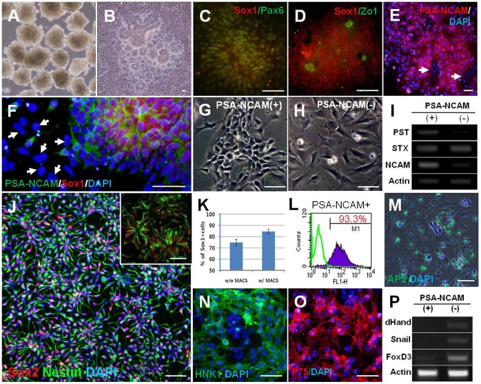
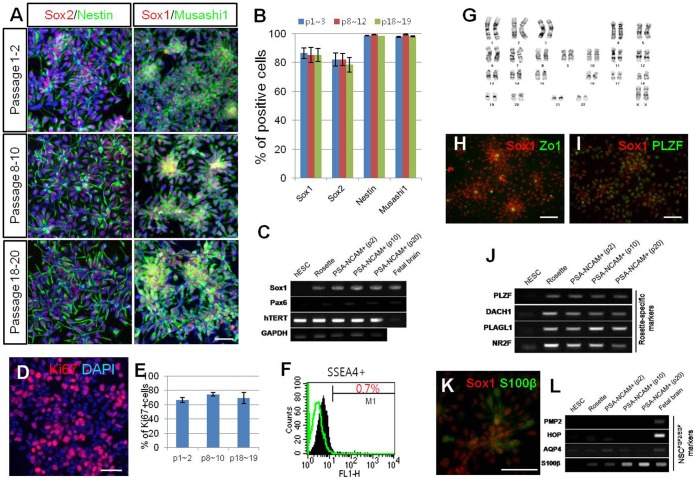
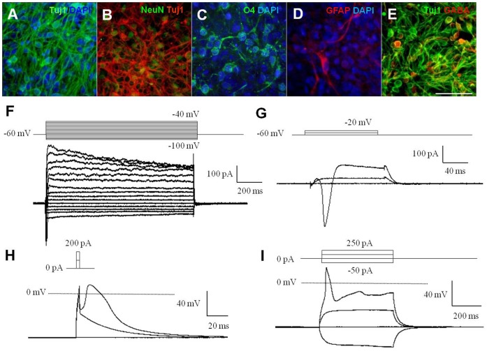
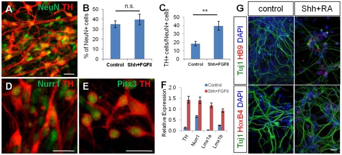
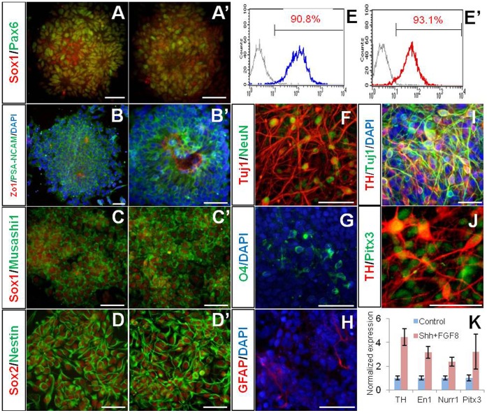
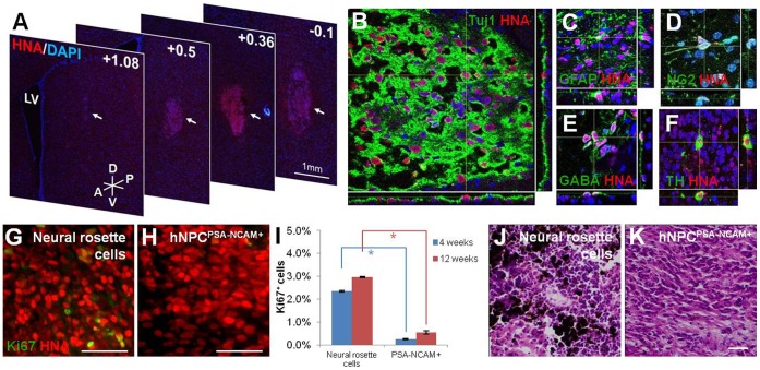
Homogeneous culture of neural precursor cells (NPCs) derived from human pluripotent stem cells (hPSCs) would provide a powerful tool for biomedical applications. However, previous efforts to expand mechanically dissected neural rosettes for cultivation of NPCs remain concerns regarding non-neural cell contamination. In addition, several attempts to purify NPCs using cell surface markers have not demonstrated the expansion capability of the sorted cells. In the present study, we show that polysialic acid-neural cell adhesion molecule (PSA-NCAM) is detected in neural rosette cells derived from hPSCs, and employ PSA-NCAM as a marker for purifying expandable primitive NPCs from the neural rosettes. PSA-NCAM-positive NPCs (termed hNPC(PSA-NCAM+)) were isolated from the heterogeneous cell population of mechanically harvested neural rosettes using magnetic-based cell sorting. The hNPC(PSA-NCAM+) extensively expressed neural markers such as Sox1, Sox2, Nestin, and Musashi-1 (80∼98% of the total cells) and were propagated for multiple passages while retaining their primitive characteristics in our culture condition. Interestingly, PSA-NCAM-negative cells largely exhibited characteristics of neural crest cells. The hNPC(PSA-NCAM+) showed multipotency and responsiveness to instructive cues towards region-specific neuronal subtypes in vitro. When transplanted into the rat striatum, hNPC(PSA-NCAM+) differentiated into neurons, astrocytes, and oligodendrocytes without particular signs of tumorigenesis. Furthermore, Ki67-positive proliferating cells and non-neural lineage cells were rarely detected in the grafts of hNPC(PSA-NCAM+) compared to those of neural rosette cells. Our results suggest that PSA-NCAM-mediated cell isolation provides a highly expandable population of pure primitive NPCs from hPSCs that will lend themselves as a promising strategy for drug screening and cell therapy for neurodegenerative disorders.


Conflict of interest statement

Competing Interests: The authors have declared that no competing interests exist.

Figures

Figure 1
Figure 1. Efficient induction of neural rosette cells and isolation of hNPCPSA-NCAM+.
(A) hESC derived-EBs treated with dorsomorphin (DM) and SB431542 (SB), (B) When EBs treated with DM and SB were attached onto the Matrigel-coated dish, a large number of rosette structures formed in the center of the colonies after 4–5 days. (C–D) Typical neural markers such as Sox1 and Pax6 were strongly expressed in neural rosettes (C) with strong expression of Zo-1 in the central lumens of rosettes (D). (E–F) PSA-NCAM was highly expressed on the surface of neural rosette cells, but not on the cells out-migrating from the rosette clump (indicated with white arrows in E and F). (G–H) PSA-NCAM-positive cells showed typical morphology of NPCs (G), whereas PSA-NCAM-negative cells had flat and large cell bodies, similar to neural crest cells (H). (I) NCAM and PST, a polysialylating enzyme, were more abundantly expressed in PSA-NCAM-positive fraction after cell sorting. (J) A representative image for the culture of hNPCPSA-NCAM+ . After cell sorting, most of the cells were Sox2/Nestin double-positive, indicating a highly homogeneous culture of NPCs (J, inset) hNPCPSA-NCAM+ spontaneously formed the neural rosette structure. (K–L) After sorting, the proportion of Sox1-positive cells and PSA-NCAM-positive cells were enriched up to ∼85% and ∼93% of the total cells, respectively. (M-P) Cells in PSA-NCAM negative-fraction were predominantly positive for AP2 (M), HNK1 (N), and P75 (O) which were used to identify neural crest cells, and enhanced the expression of several genes implicated in neural crest development, such as dHand, Snail, and FoxD3 (P). Scale bars: 50 µm.
Figure 2
Figure 2. Maintenance and characterization of hNPCPSA-NCAM+.
(A–B) Neural markers (Sox1, Sox2, Nestin, and Musashi1) were extensively and consistently expressed throughout multiple passages. (C) RT-PCR analysis shows that the expression of Sox1 and Pax6 were sustained from the neural rosette stage to hNPCPSA-NCAM+; furthermore, hTERT was continuously expressed throughout the maintenance culture. (D–E) During multiple passages, about 70% of cultured cells were positive for Ki67, supporting their self-renewing potential. (F) At passage 9, SSEA4-positive cells were rarely detected. (G) hNPCPSA-NCAM+ exhibited a normal karyotype at passage 20. (H–J) Along with Zo-1 expression in the lumen-side of the rosette structure (H), co-expression of PLZF with Sox1 (I) as well as prominent expression of “neural rosette-specific” genes (J) indicate that hNPCPSA-NCAM+ exhibited the characteristics of typical neural rosette cells. (K–L) Although NPs did not express either PMP2 or HOP, they did express AQP4 and S100β to some extent, which were proposed to be specifically expressed in FGF/EGF-expanded neural cells (NPCFGF/EGF). Scale bars: 50 µm.
Figure 3
Figure 3. hNPCPSA-NCAM+ exhibited multipotency and differentiated neurons were electrophysiologically functional.
(A–D) hNPCPSA-NCAM+ were spontaneously differentiated into neurons (A­B), oligodendrocytes (C), and astrocytes (D) in the absence of mitogens. (E) GABA-immunoreactivity was frequently observed among Tuj1-positive cells. (F–G) Voltage-dependent membrane currents: depolarizing voltage steps elicited outward K+ currents (F) and fast inward Na+ currents (G). (H–I) In current-clamp recordings, short (3 ms) or prolonged (500 ms) depolarizing current injections above supra-threshold elicited the single action potential (H), and the fast action potentials (I). Scale bars: 50 µm.
Figure 4
Figure 4. hNPCPSA-NCAM+ were able to differentiate into specific neuronal subtypes using regionalizing cues.
(A) Neurons exhibiting dopaminergic phenotypes were derived from hNPCPSA-NCAM+. (B–C) The cells treated with Shh and FGF8 for 8 days, followed by treatment with BDNF, GDNF, and ascorbic acid gave rise to a larger number of TH-positive neurons than control cells (non-treated group) without a significant change in total neuronal differentiation. (D–E) TH-positive cells co-expressed with Nurr1 and Pitx3. (F) Quantitative RT-PCR analysis confirmed the enhanced expression of several transcription factors involved in dopaminergic differentiation after treatment with Shh and FGF8 to hNPCPSA-NCAM+. (G) RA/Shh-exposed hNPCPSA-NCAM+ showed induction of HoxB4 and HB9, a posterior gene and a motor neuron-specific gene, respectively. Scale bars: 20 µm, n.s., not significant, **, p<0.01.
Figure 5
Figure 5. Isolation and differentiation of hNPCPSA-NCAM+ from iPSCs; spontaneous and directed differentiation from them.
(A–E) Data from WT-iPSC3 and (A’–E’) from PD-iPSC4. (A, A’ and B, B’) Induction of neural rosettes from iPSCs. Neural markers (Sox1, Pax6, and PSA-NCAM) were prominently expressed in rosette structure with distinct Zo-1 expression in lumen. (C–E and C’–E’) hNPCPSA-NCAM+ were successfully isolated from two hiPSC lines and maintained in adherent culture with the comparative efficiency to that of hESCs. (F–H) hNPCPSA-NCAM+ derived from PD-iPSC were able to differentiate into neurons, oligodendrocytes and astrocytes. (I–K) PD-specific hNPCPSA-NCAM+ primed with Shh and FGF8 efficiently gave rise to neurons exhibiting DA neuron phenotypes. Scale bars: 50 µm.
Figure 6
Figure 6. Integration and survival of hNPCPSA-NCAM+ in adult rat striatum.
(A) A series of coronal sections from +1.08 to −0.1 shows survival and integration of hNPCPSA-NCAM+ grafts cells in the host striatum 4 weeks after transplantation. (B) Dual-label confocal immunofluorescence microscopy with DAPI confirms a large population of HNA- and Tuj1-positive cells within the graft of hNPCPSA-NCAM+. (C–D) In addition to Tuj1-positive cells, GFAP- and NG2-positive cells within HNA-positive graft sites show the potential of hNPCPSA-NCAM+ in differentiating into all three neural lineages in vivo. (E–F) Further dual immunohistochemistry for HNA:GABA and HNA:TH indicates the ability of grafted cells to differentiate into various neuronal subtypes. (G–H) A representative area of a neural rosette cell graft indicates a significantly higher number of Ki67-positive cells and clusters whereas only a few Ki67-positive cells with no clusters are observed for hNPCPSA-NCAM+. (I) Bars indicate the proportions of Ki67-positive cells in the grafts of neural rosette cells and hNPCPSA-NCAM+ at 4 weeks (blue bars) and 12 weeks (red bars) post-transplantation as mean ± s.e.m. (J–K) Representative images of H/E-stained brain tissues grafted with unsorted neural rosette cells (J) and hNPCPSA-NCAM+ (K). Melanocyte-like cells stained in dark-brown color were shown only in the grafts of unsorted neural rosette cells (J). Scale bar: 50 µm. Abbreviations: LV, lateral ventricle; A, anterior; P, posterior; D, dorsal; V, ventral. *, p<0.05.

Similar articles

Cited by

References

    1. Zhang SC, Wernig M, Duncan ID, Brüstle O, Thomson JA. In vitro differentiation of transplantable neural precursors from human embryonic stem cells. Nat Biotechnol. 2001;19:1129–1133. - PubMed
    1. Chambers SM, Fasano CA, Papapetrou EP, Tomishima M, Sadelain M, et al. Highly efficient neural conversion of human ES and iPS cells by dual inhibition of SMAD signaling. Nat Biotechnol. 2009;27:275–280. - PMC - PubMed
    1. Perrier AL, Tabar V, Barberi T, Rubio ME, Bruses J, et al. Derivation of midbrain dopamine neurons from human embryonic stem cells. Proc Natl Acad Sci U S A. 2004;101:12543–12548. - PMC - PubMed
    1. Conti L, Pollard SM, Gorba T, Reitano E, Toselli M, et al. Niche-independent symmetrical self-renewal of a mammalian tissue stem cell. PLoS Biol. 2005;3:e283. - PMC - PubMed
    1. Koch P, Opitz T, Steinbeck JA, Ladewig J, Brüstle O. A rosette-type, self-renewing human ES cell-derived neural stem cell with potential for in vitro instruction and synaptic integration. Proc Nat Acad Sci U S A. 2009;106:3225–3230. - PMC - PubMed

Publication types

MeSH terms