. 2012;7(7):e41077.
doi: 10.1371/journal.pone.0041077. Epub 2012 Jul 20.

A Wnt/beta-catenin pathway antagonist Chibby binds Cenexin at the distal end of mother centrioles and functions in primary cilia formation

Affiliations

A Wnt/beta-catenin pathway antagonist Chibby binds Cenexin at the distal end of mother centrioles and functions in primary cilia formation

Nathan Steere et al. PLoS One. 2012.

Abstract

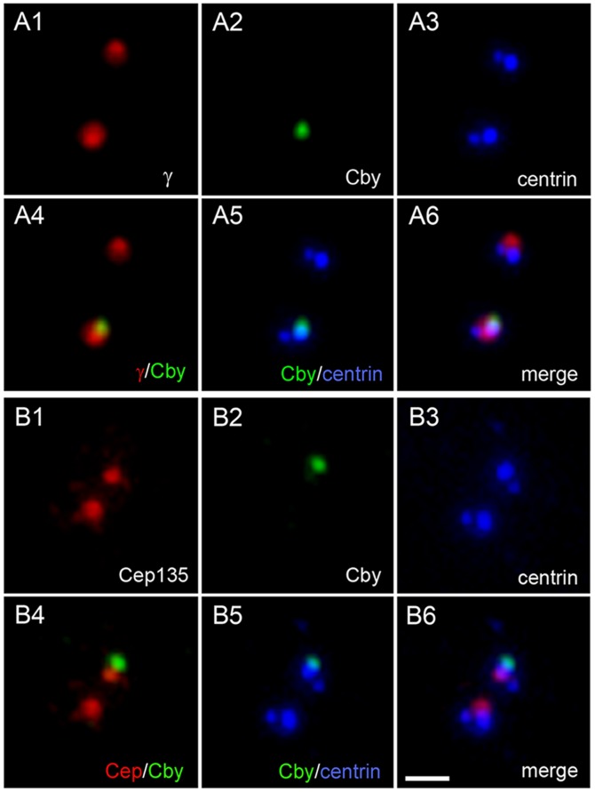
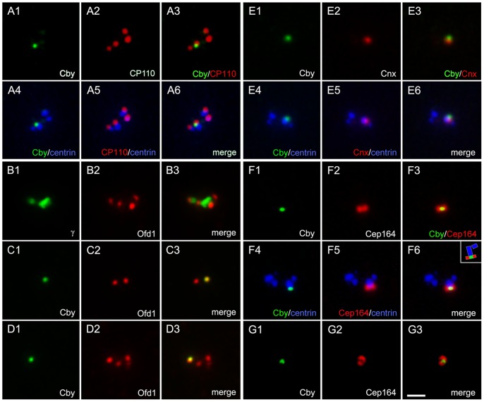
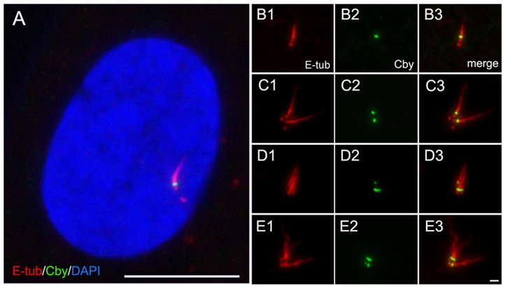
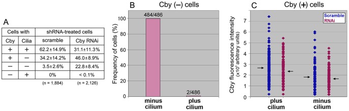
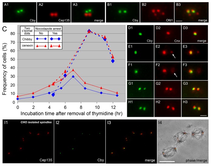
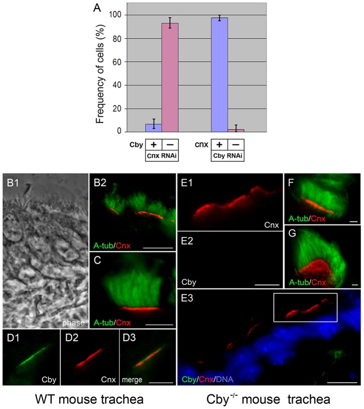
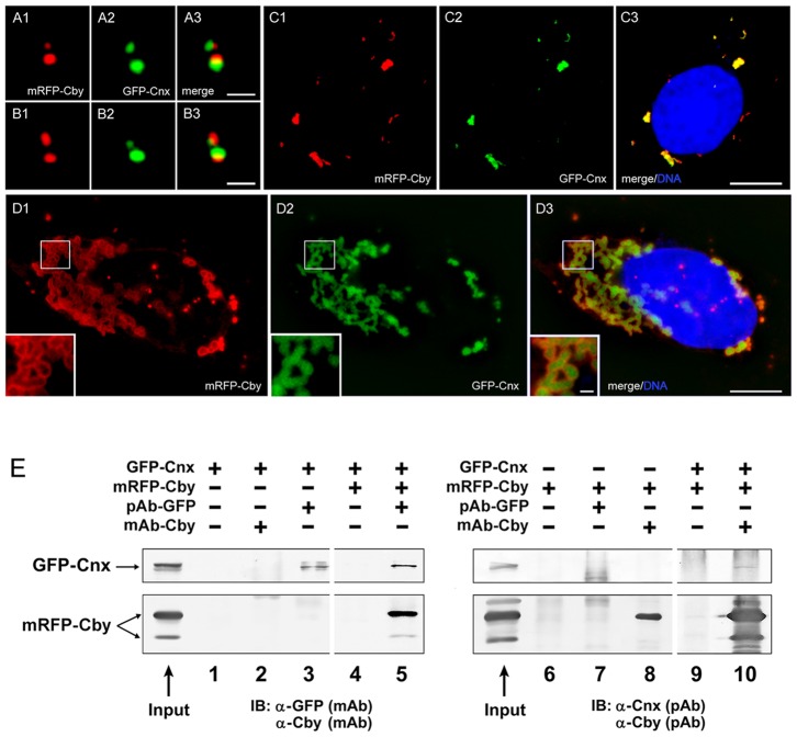
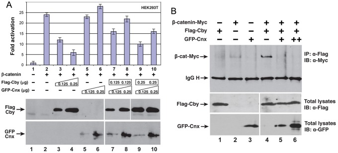
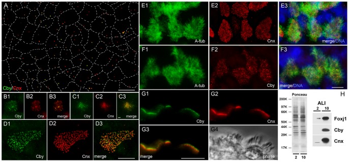
The mother centriole of the centrosome is distinguished from immature daughter centrioles by the presence of accessory structures (distal and subdistal appendages), which play an important role in the organization of the primary cilium in quiescent cells. Primary cilia serve as sensory organelles, thus have been implicated in mediating intracellular signal transduction pathways. Here we report that Chibby (Cby), a highly conserved antagonist of the Wnt/β-catenin pathway, is a centriolar component specifically located at the distal end of the mother centriole and essential for assembly of the primary cilium. Cby appeared as a discrete dot in the middle of a ring-like structure revealed by staining with a distal appendage component of Cep164. Cby interacted with one of the appendage components, Cenexin (Cnx), which thereby abrogated the inhibitory effect of Cby on β-catenin-mediated transcriptional activation in a dose-dependent manner. Cby and Cnx did not precisely align, as Cby was detected at a more distal position than Cnx. Cnx emerged earlier than Cby during the cell cycle and was required for recruitment of Cby to the mother centriole. However, Cby was dispensable for Cnx localization to the centriole. During massive centriogenesis in in vitro cultured mouse tracheal epithelial cells, Cby and Cnx were expressed in a similar pattern, which was coincident with the expression of Foxj1. Our results suggest that Cby plays an important role in organization of both primary and motile cilia in collaboration with Cnx.


Conflict of interest statement

Competing Interests: The authors have declared that no competing interests exist.

Figures

Figure 1
Figure 1. Localization of Cby at one of the centrioles in cultured mammalian cells.
HeLa cells expressing GFP-tagged centrin1 were immunostained with Cby and γ-tubulin (A) or Cep135 (B). Cby is detected at one of two centrioles revealed by GFP-centrin. Bar, 1 µm.
Figure 2
Figure 2. Localization of Cby at the distal end of the mother centriole.
RPE1 (A, F–G), CHO (B–D), and HeLa (E) cells were double immunostained with Cby-CP110 (A), Cby-Ofd1 (B–D), Cby-Cnx (E), and Cby-Cep164 (F–G) to show Cby at the distal end of the mother centriole. Both RPE1 and HeLa cells stably expressed GFP-tagged centrin1. The inset (F6) shows a schematic diagram of Cby/Cep164/centrin localization. Bar, 1 µm.
Figure 3
Figure 3. Localization of Cby at the base of primary cilia.
Primary cilia induced in quiescent RPE1 cells were stained with Cby and non-tryosinated α-tubulin (E-tub) antibodies. A is a merged image with DAPI, and B–E show multiple cilia induced by cytochalasin treatment. Bars, 1 µm (B–E) and 10 µm (A).
Figure 4
Figure 4. Cilia formation in RPE1 cells expressing different amounts of Cby at the centriole. A:
Number of cells with and without Cby and cilia after transfection with scrambled and Cby shRNA. Cells were cultured for 48 hr in a serum-free medium before fixation. Total 1,884 (scramble) and 2,126 (Cby RNAi) cells were examined in repeated six experiments. B: Number of Cby-lacking cells with and without cilia after Cby RNAi. No cilia were formed in nearly 100% (484/486) cells expressing non-detectable levels of Cby. C: Quantification of the fluorescence intensity of Cby remaining at the centriole in ciliated and non-ciliated cells after transfection with scramble and Cby shRNA. Arrows indicate the position of average fluorescence intensities (266 vs.182 in control and 237 vs. 167 in RNAi cells in arbitrary units with and without cilia, respectively). Cells lacking cilia contained Cby in consistently lower amounts than cells with cilia in both control and RNAi samples.
Figure 5
Figure 5. Emergence of Cby at the second site of the centrosome during the cell cycle. A, B:
CHO cells double stained with Cby and Cep135 (A) or Ofd1 (B) antibodies show the presence of two Cby-containing centrioles. C–H: Time course analysis of Cby/Cnx emergence in partially synchronized CHO cells arrested at G1/S or S phase by thymidine treatment. Two Cby (blue) or Cnx (red) dots were counted after removal of thymidine at time zero (C). Dotted lines indicate cells treated with nocodazole that was added at 4.25 h after washing out thymidine. D–H show double staining of centrioles with Cby and Cnx antibodies in cells fixed at 0 (D), 6 (E–F), and 7 (G–H) hr after removal of thymidine. Arrows indicate the position of Cnx-specific centrioles. I shows isolated CHO spindles stained with Cep135 and Cby antibodies and a merged image with phase-contrast is shown in I4. Cby is seen at one of the two centrioles at each spindle pole. Bars, 1 µm (A–B, D–H) and 10 µm (I).
Figure 6
Figure 6. Localization of Cby/Cnx at centrioles/basal bodies in the absence of Cnx/Cby.
A: Numbers of HeLa cells with (blue) and without (pink) Cby (two columns at left side) and Cnx (two columns at right side) at the centriole after depletion of Cnx and Cby, respectively. B–G: Tracheal epithelial tissues of wild type (B–D) and Cby−/− (E–G) mice stained with acetylated α-tubulin (A-tub), Cnx, and Cby antibodies. B1 is a phase image and E3 is a merged image with DAPI. A high magnification of the outlined area of E3 is shown in E1 and E2. In G, Cnx-containing basal bodies are seen as individual dots. Bars, 10 µm (B, E3), 5 µm (C–D, E1-E2), and 1 µm (F–G).
Figure 7
Figure 7. Interaction of Cby with Cnx.
A–D: U2OS cells expressing mRFP-Cby and GFP-Cnx at the centriole (A–B) and cytoplasmic foci (C–D). Insets of D show a high magnification of the outlined area. Bars, 1 µm (A–B, insets of D) and 10 µm (C–D). E: Co-immunoprecipitation of mRFP-Cby and GFP-Cnx expressed in HEK293T cells. Proteins were pulled down with polyclonal GFP or monoclonal Cby antibodies and blotted with monoclonal GFP/Cby and polyclonal Cnx/Cby antibodies.
Figure 8
Figure 8. Effect of Cby and Cnx on β-catenin-mediated transcriptional activation. A:
HEK293T cells were transfected with a TopFlash luciferase reporter along with stabilized β-catenin, Flag-Cby and GFP-Cnx plasmids. The basal TopFlash value was set as 1. Western blot analysis indicates that Cby and Cnx were stably expressed. B: Cnx competes with β-catenin for Cby binding. HEK293T cells were cotransfected with constant amounts of stabilized β-catenin-Myc (1.0 µg) and Flag-Cby (0.5 µg) and increasing amounts of GFP-Cnx (0.5 µg and 1.0 µg). Cell lysates were immunoprecipitated using anti-Flag antibody and detected with anti-Myc antibody. IgG H denotes the IgG heavy chain.
Figure 9
Figure 9. Cby and Cnx in differentiating mouse tracheal epithelial cells (mTECs).
A–G: mTECs of ALI-0 (A), ALI-2 (B–C), and ALI-10 (D to G) stained with Cby, Cnx, and acetylated α-tubulin antibodies. Merged images with DAPI are shown in E3 and F3, and G4 is a phase image. Dotted lines in A represent borders between adjacent cells. Bars, 1 µm (B–C) and 10 µm (A, D, E–G). H: Immunoblot analysis of ALI-2 and ALI-10 mTECs probed with Foxj1, Cby, and Cnx.

References

    1. Kuriyama R, Borisy GG. Centriole cycle in Chinese hamster ovary cells as determined by whole-mount electron microscopy. J Cell Biol. 1981;91:814–821. - PMC - PubMed
    1. Kleylein-Sohn J, Westendorf J, Le Clech M, Habedanck R, Stierhof YD, et al. Plk4-induced centriole biogenesis in human cells. Dev. Cell. 2007;13:190–202. - PubMed
    1. Singla V, Romaguera-Ros M, Garcia-Verdugo JM, Reiter JF. Ofd1, a human disease gene, regulates the length and distal structure of centrioles. Dev Cell. 2010;18:410–424. - PMC - PubMed
    1. Schmidt TI, Kleylein-Sohn J, Westendorf J, Le Clech M, Lavoie SB, et al. Control of centriole length by CPAP and CP110. Curr. Biol. 2009;19:1005–1011. - PubMed
    1. Vorobjev IA, Chentsov YS. Centrioles in the cell cycle I. Epithelial cells. J Cell Biol. 1982;93:938–949. - PMC - PubMed

Publication types